Corresponding author: Atilla KAZANCI E-mail: [email protected]
Original Investigation
Published Online: 12.08.2016
Atilla KAZANCI
1, Oktay GuRCAN
1, Ahmet Gurhan GuRCAY
1, Evrim ONDER
2, Burak KAZANCI
3,
Mesut Emre YAMAN
4, Murad BAVBEK
11Ataturk Research and Education Hospital, Department of Neurosurgery, Ankara, Turkey
2Diskapi Yildirim Beyazit Training and Research Hospital, Department of Pathology, Ankara, Turkey 3Ufuk University, Department of Neurosurgery, Ankara, Turkey
4Memorial Hospital, Department of Neurosurgery, Ankara, Turkey
Effects of Topical Cova
™
, Tisseel
®
and Adcon
®
Gel
Application on the Development of Spinal Peridural Fibrosis:
An Experimental Study in Rats
ABSTRACT
pain. The FBSS rate is reported as 5-40% in the literature (5,6,12,14,29-31). Peridural fibrosis (PF), which commonly causes compression and/or stretching of the nerve roots or the dura mater, is a common reason for “post-laminectomy syndrome”. To some extent, peridural fibrosis is a healing process after spinal surgery (5,7,12,14,26,33). Excessive PF causes a clinical situation similar to that of LDH by the way █
INTRODUCTION
L
umbar disc herniation (LDH) is one of the most common causes of sciatica and surgery is one of the most effective treatment methods of LDH. The failed back surgery syndrome (FBSS) also known as “post-laminectomy syndrome” (PLS) following laminectomy is the major cause of postoperative persisting low back pain and/or radicularAIm: Leptomeningeal adhesions and fibrosis in the spinal peridural space are the most common causes of post-laminectomy syndrome. Fibrin sealant agents and membrane barriers are commonly used for hemostasis and sealing purposes in spinal surgery. Peridural fibrosis may be a risk of the usage of these topical agents. In this study, we aimed to compare the effects of Cova™,
Tisseel® and Adcon®Gel on the development of spinal peridural fibrosis in the experimental rat model.
mATERIAl and mEThODS: Thirty-two Sprague Dawley female rats were randomly divided into 4 groups. Groups were constituted as group 1; Cova™ group (laminectomy+CovaTM), group 2; Tisseel® group (laminectomy+Tisseel®), group 3; Adcon®Gel group
(laminectomy + Adcon®Gel), group 4; control group (laminectomy only). Six weeks after laminectomy, spinal columns were removed
en bloc between L1 and L4 vertebrae. Peridural fibrosis was evaluated histologically and the results were compared statistically. RESUlTS: Statistically significant reduction of peridural fibrosis was achieved in groups 1, 2, and 3 when compared with the control group (p<0.05). Our data revealed a statistically significant difference between group 1 and group 3 (p<0.05). When we compared with group 2 and 3, the fibrosis grades were not different between these two groups (p>0.05).
CONClUSION: Fibrin sealant agent Tisseel® and membrane barrier Cova™ do not enhance peridural fibrosis following laminectomy.
Cova™ and Tisseel® may be appropriate for hemostasis and leakage prevention during the spinal surgery and it is safe to leave these
materials on the operation surface.
of adhesions, tension and compression on the nerve roots. The multi-factorial process that causes PF involves increased extracellular matrix components such as fibronectin, dermatan sulfate and collagen fibers and the accumulation of fibroblasts with cellular reduction (20,21,28,34). FBSS generally leads to revision surgeries with higher rates of complications mostly due to the disrupted anatomical plane. Many biological and non-biological materials for the prevention of peridural fibrosis such as autologous fat grafts, hemostatic polysaccharide agents, polytetrafluoroethylene membrane, antineoplastic agents, CO2 laser, hyperbaric oxygen treatment, fibrinolytic
agents, anti-inflammatory agents and dural adhesion barriers have been studied in animals (4,5,7,8,11,14,26,28 29,31,33). Hematoma and dural lacerations ending in cerebrospinal fluid (CSF) leakage are the most frequent undesirable complications following spinal surgery. Many operative techniques, devices and agents have been used to minimize these complications. Electro-coagulation, compression, suture ligation, and haemostatic agents are generally used to achieve haemostasis (32). Direct surgical repair techniques with sutures, muscle or other patch grafts, fibrin glues or fibrin sealants and soft or suturable microfibrillar collagen membranes are used for repairing dural lacerations (10).
Cova™ (Biom’Up SA Saint Priest, France) is a membrane,
which is composed of reticulated acellular porcine collagen. Cova™ forms a barrier that guides the healing along distinct
anatomical planes, thereby enabling the preservation of cleavage planes and the prevention of adhesions and fibrosis. It is completely resorbable within a period that is compatible with healing. In spinal surgery, this membrane may act as a natural barrier dura mater and adjacent tissues (1-3).
Tisseel® (Baxer AG,Vienna, Austria) is a biological
two-component fibrin sealant composed of sealer protein solution (Total protein: 96 – 125 mg/mL, fibrinogen: 67 – 106 mg/ mL, synthetic aprotinin: 2250 – 3750 KIU/mL) and thrombin solution (human thrombin 400 – 625 units/mL, calcium chloride: 36 – 44 μmol/mL). Tisseel® is a fibrin sealant that is
used as an adjunct to hemostasis when control of bleeding by conventional surgical techniques, such as suture, ligature, and cautery, is ineffective or impractical (19,24,32). It received Food and Drug Administration (FDA) approval for hemostasis at 1998 (32). It is also used for preventing leakage from anastomoses or tissue lacerations as an adjunct to standard surgical techniques such as suture and ligature (10).
Adcon®Gel (European Medical Contract Manufacturing B.V.
Nijmegen, The Netherlands) is a resorbable carbohydrate polymer gel composed of gelatin (porcine) and a polyglycan ester in phosphate buffered saline (15,17,22). It has FDA approval for preventing peridural fibrosis. It must be kept in mind that some case reports have stated increased risk of CSF leakage after the use of Adcon®Gel (9,16,18). Although
discussions continue on whether Adcon®Gel reduces scar
formation and adhesions, neurosurgeons frequently use Adcon®Gel during recurrent spine surgery to reduce peridural
scarring and adhesions (13,17,27).
Fibrin sealant agents and membrane barriers are frequently and abundantly used for hemostasis and leakage preven-tion. Neurosurgeons generally avoid leaving extra-surgical materials on the surgical field due to the anxiety of causing additional morbidities. However, for both fibrin sealant agents and membrane barriers, there is not enough evidence in the current English literature about the relationship with peridural fibrosis.
The purpose of this study was to investigate the effects of topical application of Cova™ and Tisseel® in the epidural space
after laminectomy in an experimental rat model. █
mATERIAl and mEThODS
All experimental procedures were approved by the ethical committee of Kobay Experimental Animals Laboratory (Kobay DHL). The procedures were carried out at Kobay Experimental Animals Incorporated Company under veterinarian surveil-lance. Thirty-two adult Sprague-Dawley female rats weighing on average 250-350 g were randomly allocated into 4 groups. All animals were housed individually in a climate-controlled room at constant 23ºC temperature and there was no water or food deprivation. Experimental groups are summarized in Table I. The groups and procedures were as follows:
Group 1 (Cova™ group, n=8): After laminectomy, acellular
collagen membrane COVA™ was placed on the dura mater at the laminectomy site.
Group 2 (Tisseel® group, n=8): After laminectomy, Tisseel® was
applied on the dura mater at the laminectomy site.
Group 3 (Adcon®Gel group, n=8): After laminectomy,
Adcon®Gel was applied on the dura mater at the laminectomy
site.
Group 4 (Laminectomy group, n=8): Only laminectomy was performed in this control group.
Surgical Procedure
For general anesthesia, the rats underwent intramuscular (i.m.) injection mixture of Ketamin (Ketalar, Pfizer, Istanbul, Turkey) 60 mg/kg and xylazine (Rompun, Bayer, Istanbul, Turkey) 10 mg/kg. Vital parameters such as arterial saturation, cardiac rate, and rectal temperatures were monitorized and the body temperature of the animals was fixed (37°C± 0.5°C) by the help of a heating pad. Under aseptic circumstances, the rats were placed on the fixation board in prone position, and the thoracolumbar region was shaved and cleansed with povidone iodine. A posterior midline incision at T12-L4 level was performed. After splitting the paravertebral muscles, total laminectomy was performed with the help of a microscope at the L2-L4 levels with uttermost caution not to traumatize the spinal cord. The site of laminectomy was marked with a nylon suture at the neighboring tissue. After visualizing the dura mater, Cova™, Tisseel® and Adcon®Gel were applied
on the dural surface according the related group. At the end of the laminectomy, paraspinal fascia, muscles were closed with 4-0 absorbable sutures. Skin incisions were closed with staples. The rats were kept in separate cages for 6 weeks at
23ºC temperature with free access to water and food with the body temperature 35-37°C. Postoperative care included regular bladder excretion was established. At the end of 6 weeks, the rats were sacrificed by using a lethal dose (60 mg/ kg) of pentobarbital (IE Ulagay, Istanbul, Turkey). The previous operation site was incised and the spinal cord at the level of laminectomy was harvested along with one vertebra level above and below the surgical site. The specimens were then placed in 10% buffered formalin.
histological Analysis
A blind observer evaluated the histological sections microscopically. Pathological sampling was carried out after formalin fixation, decalcification and rehydration. All tissue layers of vertebral column including the spinal cord, epidural space, bony structures and muscles were sampled en bloc. Paraffin-embedded tissue blocks were prepared and 2 sections were obtained from each block. Primary microscopical examination was based on the Haematoxylin and Eosin (HE) stained sections. Presence or absence of fibrosis was further confirmed by Masson’s Trichrome (MT) stain. In sections with MT stain muscle fibers were observed as red, collagen tissues and bone gets blue or green, cytoplasm light gets red or pink and cell nuclei is observed as dark brown to black (23). The fibrotic process in the epidural space was graded under the light microscope according to the method proposed by He et al. and summarized in Table II (8).
Grade 0: Absence of fibrosis
Grade 1: Thin fibrous bands between dura mater and scar tissue (Figure 1).
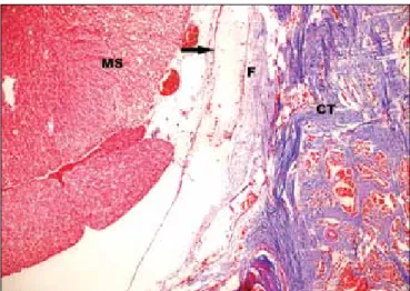
Grade 2: Continuous adherence in less than two-thirds of the laminectomy defect (Figure 2).
Grade 3: Scar tissue affecting more than two-thirds of laminectomy defect.
Table I: Summary of Experimental Groups
Groups Surgical Procedures
Cova™ Group 1 Laminectomy + Cova™ Application
Tisseel® Group 2 Laminectomy + Tisseel® Application
Adcon® Gel Group 3 Laminectomy + Adcon® Gel Application
Laminectomy Group 4 Laminectomy only
Table II: Histopathological Fibrosis Grades (8)
Grade 0 No scar tissue over dura mater
Grade 1 Only thin fibrous bands were observed between the scar tissue and dura mater
Grade 2 Continuous adherence was observed in two-thirds of the laminectomy defect
Grade 3 Scar tissue adherence was large, affecting more than two-thirds of the laminectomy defect or the adherence extended to the nerve roots
Figure 2: Grade 2 fibrosis. (Masson’s Trichrome, x 200). mS: Medulla spinalis, F: fibrosis, CT: connective tissue.
Black arrow shows dura mater.
Figure 1: Grade 1 fibrosis with thin fibrous bands (Hematoxylin- Eosin, x100). mS: Medulla Spinalis, F: fibrosis, CT: connective tissue. Black arrow shows dura mater.
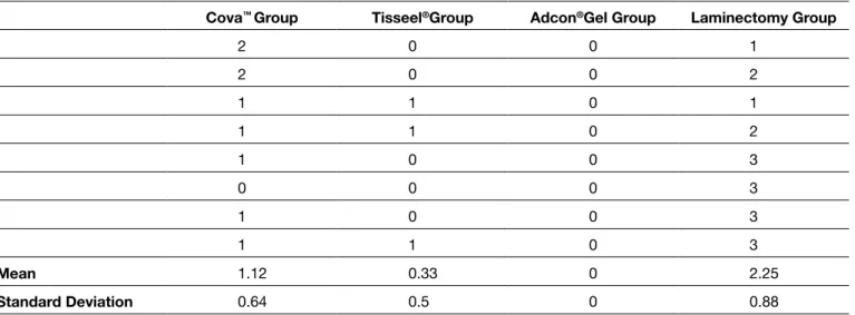
standard deviations are summarized in Figure 3. There was no grade 0 in the laminectomy group. All animals’ fibrosis grades were grade 0 in Adcon®Gel group. There was no grade 3 in
Cova™ and Tisseel® groups.
Based on this grading system, the results revealed that statistically significant less peridural fibrosis developed in Cova™, Tisseel® and Adcon®Gel groups when compared to
the laminectomy group (p=0.001). This data was interpreted as showing that these 3 bio-usable agents prevent peridural fibrosis.
Tisseel® and Adcon®Gel groups were compared individually
by post hoc analysis. There was no statistically significant difference between the Tisseel® and Adcon®Gel groups
(p=0.608). Our data revealed that both Tisseel® and Adcon®Gel
prevent peridural fibrosis to a similar extent.
We compared by post hoc analysis and there was a statistically significant difference between the Cova™ and Adcon®Gel
groups (p=0.001). This data was interpreted that although Cova™ prevents peridural fibrosis, Adcon®Gel prevents
peridural fibrosis statistically more effectively than Cova™.
Briefly, Cova™, Tisseel® and Adcon®Gel reduced PF when
compared with laminectomy group based on the examinations of PF grades. When the results of the Cova™, Tisseel® groups
were compared with the Adcon®Gel group, both Tisseel® and
Adcon®Gel prevented peridural fibrosis statistically to a similar
extent and Adcon®Gel prevented peridural fibrosis statistically
more effectively than Cova™.
█
DISCUSSION
FBSS is a well-known complication of spinal surgery mostly results in inadequate pain relief after LDH operations. FBSS sometimes needs revision surgeries, which have higher rates of complications such as neural tissue injuries and dural lacerations due to disrupted anatomical structures. The most important cause of FBSS is PF following laminectomy (4,6,8,11,12,14,20,29-31). Actually, PF is a healing process after surgery and it occurs to some degree in response to Samples of histopathological sections for grade 1 (HE stain
sample of grade 1 fibrosis) and grade 2 (MT stain sample of grade 2 fibrosis) are demonstrated in Figure 1 and Figure 2.
Statistical Analysis
Data analysis was performed using SPSS for Windows version 15.0 (SPSS Inc., Chicago, IL, USA). One-way ANOVA test was performed for the analysis of the difference between experimental groups and groups were compared individually by Tukey-HSD post hoc analysis test. P<0.05 was considered as statistically significant.
█
RESUlTS
There was no mortality and morbidity related to the procedure. No complication such as wound infection, hematoma or CSF leakage, was observed. All animals were healthy and mobile before specimens were obtained.
Based on the scar tissue level, fibrosis grades of each rat were determined separately by the pathologist (Table III). The mean values of histopathological fibrosis grades of groups with
Figure 3: Mean values of histopathological fibrosis grades of groups with standard deviations.
Table III: Histopathological Fibrosis Grades of Groups
Cova™ Group Tisseel®Group Adcon®Gel Group laminectomy Group
2 0 0 1 2 0 0 2 1 1 0 1 1 1 0 2 1 0 0 3 0 0 0 3 1 0 0 3 1 1 0 3 mean 1.12 0.33 0 2.25 Standard Deviation 0.64 0.5 0 0.88
adhesions following sternotomy (2). In spinal surgery, this membrane may act as a natural barrier between duramater and adjacent tissues. To our knowledge, this is the first study that investigates the effect of Cova™ on PF. The present study
revealed that Cova™ reduced PF formation significantly when
compared with the only laminectomy group (p=0.001). Tisseel® is fibrin sealant composed of sealer protein
and thrombin solution. Tisseel® is used as an adjunct to
hemostasis when control of bleeding by conventional surgical techniques such as suture, ligature, and cautery is ineffective or impractical. It received FDA approval for hemostasis in 1998. Yao et al. reported the effective usage of Tisseel® in
neurosurgery practice (32). It is also used for preventing leakage from anastomoses or tissue lacerations as an adjunct to standard surgical techniques such as suture and ligature. Hutchinson et al. stated that Tisseel® is an effective
fibrin sealant in achieving and maintaining a watertight seal of the dura (10). In our study the data revealed that Tisseel®
reduces PF formation significantly when compared with only laminectomy group (p=0.001).
Adcon®Gel is a resorbable carbohydrate polymer gel
composed of gelatin and a polyglycan ester. It has FDA approval for preventing PF. It is suggested that Adcon®Gel
prevents PF by covering nervous structures until fibrosis occurs (15,17). In present study, the Adcon®Gel group was
used as a positive control group. McKinley and Shaffer stated that is cost effective in means of pain relief, time of returning to work, and the need for additional pain medicine (22). Although Richter et al. reported that Adcon®Gel is not effective
in preventing fibrosis, our data revealed that Adcon®Gel
reduces PF formation significantly when compared with the only laminectomy group (p=0.001) (25). In the present study, Tisseel® and Adcon®Gel groups were compared individually.
There was no statistically significant difference between these two groups (p=0.608). Our result revealed that both Tisseel®
and Adcon®Gel prevent PF to a similar extent.
█
CONClUSION
Peridural fibrosis following spinal surgery is a well-known and described situation among neurosurgeons. Besides improved minimally invasive surgery techniques, many agents and mechanical barriers have been used to prevent PF. In the present study, we aimed to investigate the effects of Cova™ and Tisseel® on PF and to compare this effect with
Adcon®Gel. Our results revealed that Cova™, Tisseel® and
Adcon®Gel reduced PF when compared with the laminectomy
group. When the results of Cova™ and Tisseel® compared with
Adcon®Gel, Tisseel® and Adcon®Gel prevented PF statistically
to a similar extent but Adcon®Gel prevented PF statistically
more effectively than Cova™.
Neurosurgeons generally hesitate to leave these materials on the laminectomy field with the anxiety of additional morbidities. Cova™, Tisseel®, which are used for both hemostasis and
preventing CSF leakage, could be applied safely during spinal surgeries. These agents do not increase PF and conversely reduce PF after laminectomy. We suggest that they can be manipulations in the peridural space. Excessive formation of
PF causes the clinical situation known as FBSS. Many factors play a role in amount of peridural scar formation such as postoperative hematoma, postoperative infection and surgical techniques. Improved minimally invasive surgery techniques, many agents and mechanical barriers have been used to accomplish this bothering problem (5,6,12,14,15,17,19,26, 29-31,33).
The basic pathophysiological mechanism of excessive PF formation has not been explained sufficiently clearly yet. Many mechanisms play a role in the wound healing process. Inflammation is the first step of scar formation. Lymphocytes, fibroblasts, macrophages migrate and accumulate in peridural space. Scar tissue is composed of mainly extracellular matrix components such as fibronectin, collagen fibers rather than a cellular component. Pro-inflammatory cytokines are secreted and trigger PF formation. Formation of PF also involves the transformation of fibroblasts to myofibroblasts. Transforming Growth Factor-1β (TGF-1β) plays an important role in transformation of fibroblasts into myofibroblasts (20,21,34). Hemostasis is essential for both obtaining intraoperative adequate visualization of underlying structures and prevent-ing post-operative complications of microneurosurgical procedures. Damage to small blood vessels and capillaries frequently occurs during surgery. Following vascular damage, there is an immediate reflex that promotes vasoconstric-tion, thus diminishing blood loss. Intraoperative bleeding control can be achieved generally via standard methods such as mechanical compression with patties, irrigation with warm saline, bipolar coagulation, oxidized regenerated cel-lulose and various absorbable gelation sponges (32). Novel hemostatic agents such as fibrin sealants, fibrin patches and gelatin-thrombin matrix sealants that work independently of the coagulation cascade are available for neurosurgical pro-cedures. Fibrin sealants and fibrin patches seem to have more of a role within neurosurgery in treating or preventing CSF leak (10,19,24).
Any tear or laceration in the dura mater may result in CSF leakage. These leaks may occur after diagnostic or therapeutic lumbar puncture, spinal surgery or trauma. CSF leakage may cause serious problems such as persistent headaches, meningitis and intracranial subdural hematomas. Surgical management of leakage is commonly closure of the dural tear by means of direct surgical repair techniques with sutures, muscle, fascial grafts, other patch grafts, fibrin glues, fibrin sealants and soft or suturable microfibrillar collagen membranes used as a barrier (19,24).
Cova™ is composed of reticulated acellular porcine collagen. It
is an elastic resorbable and suturable membrane and is cross-linked with an oxidized polysaccharide. Cova™ forms a barrier
that guides healing along distinct anatomical structures; thereby it may prevent adhesions and fibrosis. It is completely resorbable within a period that is compatible with healing without cellular toxicity (1-3). Armoiry et al. reported that this absorbable collagen membrane is effective in the prevention of adhesions in pediatric cardiac surgery (1). Bel et al. stated that Cova™ is an effective membrane in reducing postoperative
13. Kasimcan MO, Bakar B, Aktas S, Alhan A, Yilmaz M: Effectiveness of the biophysical barriers on the peridural fibrosis of a postlaminectomy rat model: An experimental research. Injury 42(8):778-781, 2011
14. Kato T, Haro H, Komori H, Shinomiya K: Evaluation of hyaluronic acid sheet for the prevention of postlaminectomy adhesions. Spine J 5(5):479-488, 2005
15. Kaya B, Esen HH, Ustun ME: A comparison of the effects of Adcon-L, Mitomycin C and Sodium Hyaluronate in experimental epidural fibrosis in rabbits. J Neurol Sci (Turk) 28(2):236-242, 2011
16. Kim SB, Lim YJ: Delayed detected unexpected complication of ADCON-L® Gel in lumbar surgery. J Korean Neurosurg Soc 48(3):268-271, 2010
17. Kurt G, Aytar MH, Doğulu F, Cemil B, Erdem O, Baykaner MK, Ceviker N: A comparison of the local effectiveness of mitomycin C, aprotinin, and Adcon-L in experimental peridural fibrosis. Surg Neurol 70(6):608-613, 2008
18. Le AX, Rogers DE, Dawson EG, Kropf MA, De Grange DA, Delamarter RB: Unrecognized durotomy after lumbar discectomy: A report of four cases associated with the use of ADCON-L. Spine (Phila Pa 1976) 26(1):115-117, 2001 19. Lee JY, Ebel H, Friese M, Schillinger G, Schröder R, Klug N:
Influence of TachoComb in comparison to local hemostyptic agents on epidural fibrosis in a rat laminectomy model. Minim Invasive Neurosurg 46(2):106-109, 2003
20. Liu J, Ni B, Zhu L, Yang J, Cao X, Zhou W: Mitomycin C-polyethylene glycol controlled-release film inhibits collagen secretion and induces apoptosis of fibroblasts in the early wound of a postlaminectomy rat model. Spine J 10(5):441-447, 2010
21. Lv P, Zhao J, Su W, Liang X, Zhang K: An experimental novel study: Hyperbaric oxygen treatment on reduction of epidural fibrosis via down-regulation of collagen deposition, IL-6, and TGF-β1. Eur J Orthop Surg Traumatol 25 Suppl 1:S53-58, 2015
22. McKinley DS, Shaffer LM: Cost effectiveness evaluation of Adcon-L adhesion control gel in lumbar surgery. Neurol Res 21 Suppl 1:67-71, 1999
23. Pereira P, Avelino A, Monteiro P, Vaz R, Castro-Lopes JM: New insights from immunohistochemistry for the characterization of epidural scar tissue. Pain Physician 17:465-474, 2014 24. Richards PJ, Turner AS, Gisler SM, Kraft S, Nuss K, Mark
S, Seim HB 3rd, Schense J: Reduction in postlaminectomy epidural adhesions in sheep using a fibrin sealant-based medicated adhesion barrier. J Biomed Mater Res B Appl Biomater 92(2):439-446, 2010
25. Richter HP, Kast E, Tomczak R, Besenfelder W, Gaus W: Results of applying ADCON-L gel after lumbar discectomy: The German ADCON-L study. J Neurosurg 95(2 Suppl):179-189, 2001
26. Rodgers KE, Robertson JT, Espinoza T, Oppelt W, Cortese S, diZerega GS, Berg RA: Reduction of epidural fibrosis in lumbar surgery with Oxiplex adhesion barriers of carboxymethylcellulose and polyethylene oxide. Spine J 3(4):277-283, discussion 284, 2003
safely left in the operation field. To our knowledge, this is the first study that investigates the effect of Cova™ on PF. More
investigations are needed to reveal the exact effects of Cova™
and Tisseel® on PF.
█
REFERENCES
1. Armoiry X, Viprey M, Constant H, Aulagner G, Roux AS, Huot L, Roubertie F, Ninet J, Henaine R: Potential interest of a new absorbable collagen membrane in the prevention of adhesions in paediatric cardiac surgery: A feasibility study. Arch Cardiovasc Dis 106(8-9):433-439, 2013
2. Bel A, Kachatryan L, Bruneval P, Peyrard S, Gagnieu C, Fabiani JN, Menasché P: A new absorbable collagen membrane to reduce adhesions in cardiac surgery. Interact Cardiovasc Thorac Surg 10(2):213-216, 2010
3. Bel A, Ricci M, Piquet J, Bruneval P, Perier MC, Gagnieu C, Fabiani JN, Menasché P: Prevention of postcardiopulmonary bypass pericardial adhesions by a new resorbable collagen membrane. Interact Cardiovasc Thorac Surg 14(4):469-473, 2012
4. Chen F, Zuo Z, Wang K, Zhang C, Gong H, Ye F, Ji A, Tao H: Study on salvianolic acid B in the reduction of epidural fibrosis in laminectomy rats. BMC Musculoskelet Disord 15:337, 2014 5. Esposito F, Cappabianca P, Fusco M, Cavallo LM, Bani GG, Biroli F, Sparano A, de Divitiis O, Signorelli A: Collagen-only biomatrix as a novel dural substitute. Examination of the efficacy, safety and outcome: Clinical experience on a series of 208 patients. Clin Neurol Neurosurg 110(4):343-351, 2008 6. Farrokhi MR, Lotfi M, Masoudi MS, Gholami M: Effects
of methylene blue on postoperative low-back pain and functional outcomes after lumbar open discectomy: A triple-blind, randomized placebo-controlled trial. J Neurosurg Spine 11:1-9, 2015
7. Gunaldi O, Guclu G, Postalci L, Eseoglu M, Yilmaz I, Ofluoglu E, Erdogan S: Can royal jelly prevent epidural fibrosis development after laminectomy? An experimental study. J Neurol Sci (Turk) 31(2): 257-265, 2014
8. He Y, Revel M, Loty B: A quantitative model of post-laminectomy scar formation. Effects of a nonsteroidal anti-inflammatory drug. Spine (PhilaPa 1976) 20:557–563, 1995 9. Hieb LD, Stevens DL: Spontaneous postoperative
cerebrospi-nal fluid leaks following application of anti-adhesion barrier gel: Case report and review of the literature. Spine (Phila Pa 1976) 26(7):748-751, 2001
10. Hutchinson RW, Mendenhall V, Abutin RM, Muench T, Hart J: Evaluation of fibrin sealants for central nervous system sealing in the mongrel dog durotomy model. Neurosurgery 69(4):921-928, 2011
11. Ismailoglu O, Albayrak B, Gulsen I, Tanriover G, Demir N: Topical application of tacrolimus prevents epidural fibrosis in a rat postlaminectomy model: Histopathological and ultrastructural analysis. Turk Neurosurg 21(4):630-633, 2011 12. Ivanic GM, Pink PT, Schneider F, Stuecker M, Homann
NC, Preidler KW: Prevention of epidural scarring after microdiscectomy: A randomized clinical trial comparing gel and expanded polytetrafluoroethylene membrane. Eur Spine J 15(9):1360-1366, 2006
31. Turkoglu E, Dinc C, Tuncer C, Oktay M, Serbes G, Sekerci Z: Use of decorin to prevent epidural fibrosis in a post-laminectomy rat model. Eur J Pharmacol 724:86-91, 2014 32. Yao HH, Hong MK, Drummond KJ: Haemostasis in
neurosurgery: What is the evidence for gelatin-thrombin matrix sealant? J Clin Neurosci 20(3):349-356, 2013
33. Yu CH, Lee JH, Baek HR, Nam H: The effectiveness of poloxamer 407-based new anti-adhesive material in a laminectomy model in rats. Eur Spine J 21(5):971-979, 2012 34. Zhang C, Kong X, Ning G, Liang Z, Qu T, Chen F, Cao D,
Wang T, Sharma HS, Feng S: All-trans retinoic acid prevents epidural fibrosis through NF-κB signaling pathway in post-laminectomy rats. Neuropharmacology 79: 275-281, 2014 27. Rönnberg K, Lind B, Zoega B, Gadeholt-Göthlin G, Halldin
K, Gellerstedt M, Brisby H: Peridural scar and its relation to clinical outcome: A randomised study on surgically treated lumbar disc herniation patients. Eur Spine J 17(12):1714-1720, 2008
28. Schimizzi AL, Massie JB, Murphy M, Perry A, Kim CW, Garfin SR, Akeson WH: High-molecular-weight hyaluronan inhibits macrophage proliferation and cytokine release in the early wound of a preclinical postlaminectomy rat model. Spine J 6(5):550-556, 2006
29. Temiz C, Temiz P, Sayin M, Ucar K: Effect of cepea extract-heparin and allantoin mixture on epidural fibrosis in a rat hemilaminectomy model. Turk Neurosurg 19(4):387-392, 2009 30. Tural Emon S, Somay H, Orakdogen M, Uslu S, Somay A:
Effects of hemostatic polysaccharide agent on epidural fibrosis formation after lumbar laminectomy in rats. Spine J 16(3):414-419,2016