case report
1 3
S626 Meandering right pulmonary vein associated with retrocaval ureter and vertebral fusion anomaliesSummary We present multimodality imaging of a
meandering right pulmonary vein in a 29-year-old female patient. It was associated with right retrocaval ureter causing hydronephrosis and stone formation, ver-tebral fusion anomalies and corrected cardiac anomalies (patent ductus arteriosus and ventricular septal defect). To the best of our knowledge, only a few meandering right pulmonary vein have been presented in the litera-ture until now and this is the first reported case that is associated with retrocaval ureter and vertebral fusion anomalies.
Keywords Meandering right pulmonary vein ·
Retroca-val ureter · Vertebral fusion · CT · MR
Gleichzeitiges Vorliegen einer mäandernden rechten Pulmonalvene mit einem retrocavalem Ureter und Wirbelfusionsanomalien
Zusammenfassung Wir berichten über eine
multimo-dale Bildgebung einer mäandernden rechten Pulmo-nalvene in einer 29 Jahre alten weiblichen Patientin. Gleichzeitig lag der rechte Ureter retrocaval, was zu Hyd-ronephrosis und Steinbildung geführt hatte. Außerdem bestanden Wirbelfusionsanomalien und korrigierte
Herzanomalien (offener Ductus arteriosus und Ventri-kelseptumdefekt). Soweit wir wissen, wurden bisher nur wenige mäandernde Pulmonalvenen in der Literatur beschrieben. Dieser Fall ist außerdem der erste, bei dem gleichzeitig ein retrocavaler Ureter und Wirbelfusionsan-omalien vorliegen.
Schlüsselwörter Mäandernde rechte Pulmonalvene ·
Retrocavaler Ureter · Wirbelfusion · Computertomogra-phie (CT) · Magnetresonanz (MR)
Introduction
Meandering right pulmonary vein (MRPV) also known as pseudo-scimitar syndrome is a rare pulmonary vein anomaly in which a winding and widened right pulmo-nary vein traveling through the right lung and finally draining normally to the left atrium in contrast to the inferior vena cava (IVC) as seen in scimitar syndrome [1]. MRPV is an innocent vascular anomaly and sometimes associated with other anomalies [1, 2]. In this report, we present a MRPV associated with right retrocaval ure-ter, vertebral fusion anomalies and corrected cardiac anomalies. To the best of our knowledge, this is the first reported case of a MRPV with retrocaval ureter and ver-tebral fusion anomalies.
Case
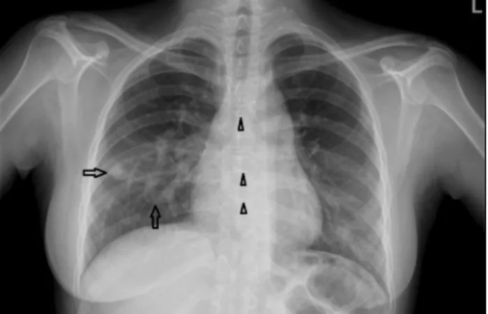
A 29-year-old female patient who had operated for pat-ent ductus arteriosus and vpat-entricular septal defect 22 years ago was admitted to our hospital with the com-plaint of coughing, palpitation, and weakness. Tubular structures in the right perihilar region was seen on chest radiograph but no marked volume difference between the two lungs was detected (Fig. 1). A contrast enhanced
Assoc. Prof. C. Erol, MD () · O. Baskan, MD · F. Z. Gungoren, MD · R. Kılıçarslan, MD
Faculty of Medicine, Department of Radiology, Medipol Mega Hospital, Istanbul Medipol University,
TEM Avrupa Otoyolu Göztepe Çıkışı No: 1, 34214 Bağcılar, Istanbul, Turkey
e-mail: [email protected] V. Guzelburc, MD
Faculty of Medicine, Department of Urology, Istanbul Medipol University,
34214 Bağcılar, Istanbul, Turkey
Received: 5 January 2014 / Accepted: 1 December 2014 / Published online: 14 January 2015 © Springer-Verlag Wien 2014
Wien Klin Wochenschr (2016) 128 [Suppl 8]:S626–S629 DOI 10.1007/s00508-014-0695-1
Meandering right pulmonary vein associated with
retrocaval ureter and vertebral fusion anomalies
case report
Meandering right pulmonary vein associated with retrocaval ureter and vertebral fusion anomalies S627
1 3
thorax computed tomography (CT) examination was performed and showed dilated right superior and infe-rior pulmonary veins with aberrant meandering courses. The right superior pulmonary vein coursing inferolater-ally toward the diaphragm before turning upwards and the right inferior pulmonary vein coursing superolater-ally and then traveling anteromedisuperolater-ally were joining in the pulmonary hilus to form a common right pulmo-nary vein before draining into the left atrium (Fig. 2). No connection was seen between the right pulmonary vein and systemic venous circulation. There was calcifica-tions in the interventricular septum and cardiac apex, and an incidental right hydronephrotic kidney contain-ing calculus was also detected. She had no abdominal symptoms. Cardiac magnetic resonance imaging (MRI) and abdominal MR urography showed MRPV (Fig. 3a), regional wall thinning, myocardial scarring and focal aneurysmatic dilatation of the inferoseptum secondary to operation or chronic infarction. The right ureter was coursing posterior to the IVC causing high grade hydro-nephrosis (Fig. 3b). It was also noted that there was lower lumbar and sacral vertebral body fusion (Fig. 3c). Patient was operated for retrocaval ureter. Retrocaval segment of the ureter was identified (Fig. 3d) and excised by lapa-roscopic transperitoneal approach. After a double-J stent insertion, Anderson-Hynes pyeloplasty was performed.
Discussion
Congenital pulmonary venous return anomalies are usu-ally classified into three categories as venous diameter abnormalities (varices, stenosis, and atresia), anoma-lous pulmonary venous course within the lung without abnormal drainage (MRPV), and anomalous pulmonary venous return with abnormal drainage (scimitar syn-drome) [2]. MRPV is charecterised by a dilated varicose right pulmonary vein that travels within the lung in a cir-cuitous route before draining into the left atrium. Only
a few MRPV cases have been reported in the literature since its description by Goodman et al. in 1972 [1, 2]. It is hemodynamically normal, produce no symptoms and do not require any intervention [3, 4]. On the other hand, its appearance on chest radiograph is similar with the scimitar sign (Turkish sword) described for scimi-tar syndrome in which there are hypoplastic right lung, abnormal pulmonary venous drainage to the systemic venous circulation and systemic arterial supply of some right lung parts from the aorta [1–3]. Scimitar syndrome is hemodynamically abnormal because of left to right shunt, can lead to cyanosis and may require intervention [1]. Another subtype named as scimitar variant is used to describe connection of an anomalous right pulmonary vein to both the IVC and the left atrium [3]. That is why, these entities should be distinguished from each other by the way of imaging modalities [1, 2].
Fig. 2 Axial thick slice maximum intensity projection image (a)
and three-dimensional volume-rendered posterior view com-puted tomography (CT) image (b) demonstrate that the right
superior pulmonary vein (asterisk) courses inferolaterally to-ward the diaphragm and then turns upto-wards, and the right in-ferior pulmonary vein (arrow) courses superolaterally and then travels anteromedially. Meandering right pulmonary veins join in the pulmonary hilus to form a common right pulmonary vein before draining into the left atrium
Fig. 1 Posteroanterior chest X-ray shows two dilated and curved tubular structures (arrows) coursing and converging toward the right pulmonary hilus, anomalously. Notice midline sternal wires (arrow heads) indicating previous cardiothoracic surgery
case report
1 3
S628 Meandering right pulmonary vein associated with retrocaval ureter and vertebral fusion anomaliesIn conclusion, MRPV is a rare and hemodynamically normal pulmonary vein anomaly that does not require any treatment and should be differentiated from scimitar syndrome. It can be revealed strictly by CT or MR angi-ography. Although, MRPV is a benign anomaly, it may be associated with serious cardiovascular, urologic and vertebral anomalies.
Conflict of interest
The authors declare that there is no actual or potential conflict of interest in relation to this article.
References
1. Rodrigues MA, Ritchie G, Murchison JT. Incidental mean-dering right pulmonary vein, literature review and pro-posed nomenclature revision. World J Radiol. 2013;5:215–9. 2. Médart L, Giordano V, Ghaye B. Meandering pulmonary
vein associated with left thoracic isomerism. JBR-BTR. 2009;92:164–5.
3. Legras A, Guinet C, Alifano M, Lepilliez A, Régnard JF. A case of variant scimitar syndrome. Chest. 2012;142:1039–41. 4. Tortoriello TA, Vick GW III, Chung T, Bezold LI, Vincent JA. Meandering right pulmonary vein to the left atrium and inferior vena cava: the first case with associated anomalies. Tex Heart Inst J. 2002;29:319–23.
Reported retrocaval ureter is around 200 cases since the first case described in 1893 in the literature [5]. It is a rare congenital anomaly and is found in 0.06–0.17 % of the autopsy series [5]. External compression of the proxi-mal ureter coursing behind the IVC leads to obstruction of the right ureter and causes hydronephrosis and usually becomes symptomatic by the third to fourth decades of life [5, 6]. It is fortunate for our patient that thorax CT and meandering right pulmonary vein facilitated the diagno-sis of retrocaval ureter by showing right hydronephrodiagno-sis in the last CT slices. It is easy to diagnose this anomaly by the typical appearance of “S” or “J” shaped upper ureter and renal pelvis with intravenous urography and contrast enhanced CT or MR urography [5].
Retrocaval ureter is not usually associated with urinary obstruction. Patients who have moderate to high grade hydronephrosis with symptoms such as stone formation, infection, and worsening of renal function are candidates for laparoscopic pyeloplasty [5, 6]. Our patient was oper-ated for high grade hydronephrosis and stone formation. Reported associated anomalies with MRPV are right lung hypoplasia, pulmonary artery hypoplasia, systemic arterial supply to the lung, dextroposition and left tho-racic isomerism [1, 2]. To the best of our knowledge, this is the first case of MRPV associated with retrocaval ure-ter, vertebral fusion anomalies, and operated cardiac anomalies.
Fig. 3 Coronal view magnetic resonance (MR) angiogra-phy (a) shows dilated right
superior and inferior pulmo-nary veins (white arrows) with aberrant meandering courses. Coronal view MR urography (b) demonstrates high grade
right hydronephrosis and S shaped proximal right ureter (black arrow) which is typical for retrocaval course. Sagittal T1 weighted lumbar vertebra image (c) shows lower lumbar
and sacral vertebral body fusion (arrows), anteriorly. Laparoscopic view (d) shows
course of the right proximal ureter behind the inferior vena cava. White asterisk inferior vena cava, black asterisk aorta
case report
Meandering right pulmonary vein associated with retrocaval ureter and vertebral fusion anomalies S629
1 3
6. Singh V, Sinha RJ. Laparoscopic dismembered pyeloplasty and pyelolithotomy in a patient with a retrocaval ureter: our experience and review of literature. Urol J. 2011;8:231–5. 5. Gupta P, Khullar M, Sharma R, Singh R. A rare
presenta-tion of the double inferior vena cava with an anomalous retrocaval right ureter: embryogenesis and clinical impli-cations. J Clin Diagn Res. 2013;7:518–21.