Oktay OLMUŞÇELİK1,a Mesut YILMAZ 2,b
Ömür Gökmen SEVİNDİK3,c
Ayşe TOSUN İSTANBULLU 4,d Ecem Cemre CEYLAN 5,e Ayşenur GÜLER 5,f Büşra VARDAR 5,g 1 Medipol University, Faculty of Medicine, Department of Internal Medicine, İstanbul, TURKEY 2 Medipol University, Faculty of Medicine, Department of Infectious Diseases, İstanbul, TURKEY 3 Medipol University, Faculty of Medicine, Department of Hematology, İstanbul, TURKEY 4 Medipol University, Faculty of Medicine, Department of Microbiology, İstanbul, TURKEY 5 Medipol University, Faculty of Medicine, Undergraduate Student, İstanbul, TURKEY a ORCİD: 0000-0002-9815-1848 b ORCİD: 0000-0001-8022-7325 c ORCİD: 0000-0001-9636-4113 d ORCİD: 0000-0003-3952-1914 e ORCİD: 0000-0001-9501-1340 f ORCİD: 0000-0001-9242-0659 g ORCİD: 0000-0001-9698-5411 Received : 15.05.2019 Accepted : 29.07.2019
Can Procalcitonin Levels Differentiate Gram-Negative
Bacteremia from Gram-Positive Bacteremia?
Objective: Patients with sepsis constitute a significant proportion of critically ill patients. Although
outcomes have significantly improved in the last decade, mortality still remains high 25-30% and 40-50% when shock is present. Procalcitonin has been studied in sepsis for diagnosis, and guidance of antibiotic therapy. We investigated the levels of C-reactive protein (CRP) and procalcitonin in patients with bacteremia seen in various departments of the hospital and to evaluate the possible discriminative role of procalcitonin in differentiating bacterial etiology of infection.
Materials and methods: Serum procalcitonin and CRP levels of patients with Gram negative and
positive bacteremia were recorded retrospectively. CRP and procalcitonin levels of 407 consecutive patients with 636 positive blood cultures were included in the study. The blood cultures were first grouped as Gram-positive bacteremia (GPB) and Gram-negative bacteremia (GNB). 316 (49.7%) of 636 positive blood culture were in GPB group. GNB group was further divided into Enterobacteriaceae bacteremia (GNEB) and Non-Enterobacteriaceae (GNNEB).
Results: Procalcitonin levels of GNB and GPB groups were significantly different (P= 0.0001).
Procalcitonin and CRP levels of GNEB and GNNEB groups were also significantly different (P=0.015 and P= 0.003, respectively).
Conclusion: Our findings suggest that procalcitonin levels may be discriminative between GNB
and GPB groups and levels >4.055 ng/mL may be evidence of GNB.
Key words: Procalcitonin, bacteremia, gram-negative, gram-positive
Prokalsitonin Düzeyleri Gram Negatif Bakteriyemiyi Gram Pozitif Bakteriyemiden Ayırt Edebilir mi?
Amaç: Sepsis hastaları, kritik durumdaki hastaların önemli bir bölümünü oluşturmaktadır. Sonuçlar
bakımından son on yılda önemli ölçüde gelişme kaydedilse de, mortalite oranı halen %25-30 düzeylerinde seyretmekte ve şok eşliğinde %40-50’ye kadar yükselebilmektedir. Prokalsitonin, tanı amacıyla ve antibiyotik tedavisinin yönetimi bakımından sepsiste üzerinde çalışılmış bir peptiddir. Çalışmada bakteriyemi hastalarında hastanemizin çeşitli bölümlerinde C-reaktiv protein (CRP) ve prokalsitonin düzeylerini araştırdık ve prokalsitoninin bakteriyel enfeksiyon etiyolojisini ayırt etme bakımından ne tür bir rolü olabileceğini değerlendirildi.
Gereç ve Yöntem: Gram negatif ve pozitif bakteriyemisi olan hastaların serum prokalsitonin ve
CRP düzeyleri, retrospektif olarak kayıt altına alındı. Çalışmaya, 636 pozitif kan kültürü olan 407 hastanın CRP ve prokalsitonin düzeyleri dahil edildi. Kan kültürlerine göre bakteriyemik ataklar, önce Gram-pozitif bakteriyemi (GPB) ve Gram-negatif bakteriyemi (GNB) olarak gruplandırıldı. GPB grubunda 636 kültür arasından 316 (%49.7) pozitif kan kültürü tespit edildi. GNB grubu ayrıca Enterobacteriaceae bakteriyemi (GNEB) ve Non-Enterobacteriaceae (GNNEB) olarak ikiye ayrıldı.
Bulgular: GNB ve GPB gruplarının prokalsitonin düzeyleri anlamlı olarak farklıydı (P= 0.0001).
GNEB ve GNNEB gruplarının prokalsitonin ve CRP düzeyleri de anlamlı şekilde farklıydı (sırasıyla P= 0.015 ve P= 0.003).
Sonuç: Bulgularımız, prokalsitonin düzeylerinin, GNB ve GPB grupları arasında ayırt edici
olabileceğini ve 4.055 ng/mL’nin üzerindeki düzeylerin GNB'nin kanıtı olabileceğini düşündürmektedir.
Anahtar Kelimeler: Prokalsitonin, bakteriyemi, gram negatif, gram pozitif
Introduction
Procalcitonin, a prohormone of 116 amino acids, is the precursor of the calcium homeostasis hormone, calcitonin. This prohormone circulates at very low concentrations in normal serum and is thought to be produced in physiological conditions by the neuroendocrine cells in the thyroid gland and the lungs (1). Procalcitonin is massively synthetized by various types of cells during sepsis, which is defined as a systemic inflammatory response to infection (2, 3). Its synthesis is up-regulated in bacterial infections and down-regulated in viral infections. In a landmark study, procalcitonin levels were shown to differ significantly between systemic bacterial infections and noninfectious inflammatory states in intensive care unit patients (4). Consequently, with the increased incidence of antibiotic resistance worldwide, interest has focused on the ability of this marker not only to diagnose infection but also to tailor antibiotic treatment and help to reduce the development of antibiotic resistance. A key clinical challenge
Correspondence Yazışma Adresi Oktay OLMUŞÇELİK Medipol University, Faculty of Medicine, Department of Internal Medicine, İstanbul – TURKEY [email protected]
associated with antibiotic therapy is the overuse and misuse of antibiotics —including continuing antibiotics longer than necessary— can result in adverse effects and add to the increasing problem of antibiotic resistance. However, the duration of antibiotic therapy that is appropriate for these patient populations is often undefined, and clinical features are of limited help in guiding discontinuation of the therapy (5). There is evidence that procalcitonin is more specific for bacterial infections when compared to other markers, with regard to the increased serum levels at the onset of infection and falling rapidly as the infection resolves (6-10). Thereby, it can guide to manage local or systemic bacterial infections in association with clinical impression, which can result in reduction of antibacterial resistance. A second question arose after identifying its potential role in managing bacterial infections, whether it is capable of differentiating between bacterial etiologies of the infection. Some pivotal studies have shown that higher procalcitonin levels are associated with bloodstream infections due to Gram-negative bacteria (BSIGN) rather than bloodstream infections due to Gram-positive bacteria (BSIGP) or fungemia (11). Varying cut-off limits are defined in literature with varying sensitivity and specificity in order to discriminate BSIGN. Herein, we present a retrospective analysis in order to investigate the levels of two biomarkers, C-reactive protein (CRP) and procalcitonin in patients with positive blood culture bloodstream infections. We aimed to evaluate the possible discriminative role of these biomarkers in differentiating bacterial etiology of infection with a reasonably high number of infectious episodes.
Materials and Methods
Following the approval of the institutional review board, medical records of the patients with positive blood cultures were reviewed. The patients, who were enrolled to the study, were followed up at different clinics of our center such as ICU, internal medicine inpatient clinics, infectious diseases and surgery. Out of 920 patients with at least one set of positive blood
cultures, 872 patients with clinically relevant
bloodstream infections were included in the study. Afterwards 407 patients in whom the CRP and procalcitonin levels were studied at the time of detection of positive cultures (n=636) were selected for further analysis. CRP and procalcitonin levels were evaluated using the immunoturbidometric system (Cobas® 6000 Analyzer Systems, Roche Diagnostics, Indianapolis, IN, USA) and the ECLIA (electro-chemiluminescence immunoassay) method (Cobas®
6000 Analyzer Systems, Roche Diagnostics,
Indianapolis, IN, USA), respectively. The cutoff values were 5 mg/L and 0.5 ng/mL for CRP and procalcitonin, respectively. Patients were divided into two groups: Group 1 included BSIGP (N:320, 50.3%) and group 2 included BSIGN patients (N:316, 49.7%). Blood stream infection due to Gram negative bacteria were further categorized into two groups: bloodstream infections due to Enterobacteriaceae (BSIE) and bloodstream infections due to non-Enterobacteriaceae (BSInE). The
levels and numerical variations of the markers were further evaluated in order to detect a possible role in differing the etiologies in the subgroups.
Quantitative variables were expressed as median (range). Frequency and percentage were used for categorical variables. Mann-Whitney U and Kruskal Wallis tests were used to compare median biomarker values among different patient groups. Categorical variables were compared by the Chi-Square test. Correlation analyses were performed using Pearson’s coefficients. Spearman’s test was used when necessary. If any significant discriminative ability of a
biomarker was detected, Receiver Operating
Characteristics (ROC) curve was drawn to assess sensitivity and specificity of certain biomarker among two different groups. All tests of significance were two tailed. P values less than 0.05 were considered
statistically significant. Statistical analysis was
performed using Statistical Package of Social Sciences (SPSS Inc, Chicago, IL, USA) version 11.0 for Windows.
Results
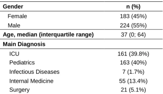
Basic demographic characteristics of analyzed cohorts were shown in Table 1. Among the demographic subgroups (male/female, age over and below median, diagnostic subgroups) there were no statistically significant difference regarding the levels of both crp and procalcitonin between aforementioned groups.
Of 636 positive blood stream infection of 407 patients, 316 (49.7%) had BSIGN and 320 (50.3%) had BSIGP. There was no statistically significant difference regarding CRP levels between gram positive and negative groups. Instead, procalcitonin levels were significantly higher in BSIGN group when compared with BSIGP group (Table 2).
Table 1. Basic demographic characteristics of
analyzed cohorts
Gender n (%)
Female 183 (45%)
Male 224 (55%)
Age, median (interquartile range) 37 (0; 64)
Main Diagnosis ICU 161 (39.8%) Pediatrics 163 (40%) Infectious Diseases 7 (1.7%) Internal Medicine 55 (13.4%) Surgery 21 (5.1%)
Table 2. CRP and procalcitonin levels of BSIGP and
BSIGN groups
BSIGP BSIGN P value
C-reactive protein 123 (0-552) 121 (0-544) 0.872 Procalcitonin 1.51 (0.07-100) 5.59 (0.07-749) 0.0001 BSIGP: bloodstream infection due to Gram-positive bacteria BSIGN: bloodstream infection due to Gram-negative bacteria
When groups within BSIGN were compared to each other; CRP levels were significantly lower in BSInE group, however, procalcitonin levels were significantly higher in BSIE group (Table 3).
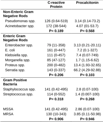
When analyzed between cause specific groups, CRP and Procalcitonin levels did not differ among any subgroup (Table 4).
ROC curves demonstrated a 4.055 cut-off value of procalcitonin should significantly predicted blood stream infection due to negative or Gram-positive bacteria with a sensitivity of 58.8% and specificity of 69.9% (AUC: 0.650, 95% CI: 0.594-0.705, P<0.001) (Figure 1). Also, a cut-off a 35.21 cut-off value of procalcitonin was able to distinguish blood stream infection due to Enterobacteriaceae or non- Enterobacteriaceae in patients who were known to be infected with Gram-negative bacteria with a sensitivity of 25% and specificity of 93.9 % (AUC: 0.609 95% CI: 0.542-0.676, P= 0.003) (Figure 2).
Table 3. CRP and Procalcitonin Levels of BSInE and
BSIE
BSInE BSIE P value
C-reactive protein 141 (0.64-544) 112 (0-482) 0.015 Procalcitonin 3.6 (0.1-73.2) 6.49 (0.07-749) 0.003 BSInE, blood stream infection due to non-Enterobacteriaceae BSIE, blood stream infection due to Enterobacteriaceae
Table 4. CRP and Procalcitonin Levels of Cause
Specific Subgroups C-reactive Protein Procalcitonin Non-Enteric Gram Negative Rods Pseudomonas spp. 126 (0.64-519) 3.14 (0.14-73.2) Acinetobacter spp. 172 (38-544) 4.07 (01-53.7) P= 0.189 P= 0.568 Enteric Gram Negative Rods Enterobacter spp. 79 (11-358) 3.13 (0.21-20.11) E. coli 161 (0-447) 7.2 (0.1-327) Klebsiella spp. 111 (0-457) 7.4 (0.07-749) Morganella spp. 85 (47-127) 1.7 (1.15-6.62) Proteus spp. 200 (0-482) 13.4 (1.93-32.65) Serratia spp. 143 (0-337) 66.2 (4.29-92.88) P= 0.206 P= 0.103 Gram Positive Bacteria Staphylococcus spp. 141 (0.42-495) 2.8 (0.07-100) Streptococcus spp. 114 (0-552) 1.4 (0.007-100) P= 0.318 P= 0.268 MSSA 141 (0.42-495) 2.86 (0.07-100) MRSA 130 (10-343) 3.85 (0.11-50.98) P= 0.906 P= 0.946
Figure 1. ROC Curve to demonstrate the ability of
Procalcitonin to distinguish gram negative bacteremia from gram positive bacteremia
Figure 2. ROC Curve to demonstrate the ability of
Procalcitonin to distinguish fermentative vs non-fermentative gram negative bacteremia
Discussion
In this study, we aimed to search for a possible role of two different biomarkers in predicting the
microbiological agent which is responsible of
bacteremia with a reasonably high number of episodes. Our data indicates that a procalcitonin level, which was drawn simultaneously with blood culture, higher than 4.055 was significantly associated with BSIGN with a sensitivity of 58.8% and a specificity of 69.9%. Furthermore, a procalcitonin level of 35.21 or higher was significantly associated with BSIE. While waiting for blood culture results it would be reasonable to put the patients, who has a high procalcitonin level, on antibiotics which have high Gram-negative coverage if clinically indicated.
Previous studies showed that procalcitonin is highly effective in diagnosing sepsis. Procalcitonin was also a significant biomarker to exclude bloodstream infections regardless of pathogen categories (12).
While being a useful tool in diagnosing sepsis and excluding blood stream infections, a certain cut off
value was not established considering the
heterogeneity and variations in recent studies. A recent meta-analysis showed that procalcitonin had a mean sensitivity and specificity of 0.77 and 0.79, respectively, for the diagnosis of sepsis (13). Oussalah et al. (13) reported an optimal procalcitonin thresholds ranging from 0.4 to 0.75 ng/mL had very high diagnostic accuracies for excluding all pathogen categories with the following negative predictive values; Gram-negative bacteria (98.9%) (including Enterobacteriacae (99.2%), non-fermentative Gram-negative bacilli (99.7%), and anaerobic bacteria (99.9%)), Gram-positive bacteria (98.4%), and fungi (99.6%). In their paper, they have reported different median procalcitonin levels among different pathogen groups. Procalcitonin levels above 10 ng/mL were associated with a high risk of BSIGN and BSIGP but also dramatically reduced the risk of potential contaminants found in blood cultures (PCBCs) or fungemia. Median procalcitonin levels were 2.2 (0.6-12.2), 2.5 13.5), 1.1 (0.3-8.1), 2.7
(0.7-13.0) and 1.3 (0.3-6.9) for Gram-negative,
Enterobacteriacae, Non-Enterobacteriacae, anaerobic bacteria, Gram-positive pathogen groups respectively. In our study the median procalcitonin levels were 5.59 (0.07-749), 6.49 (0.07-749), 3.6 (0.1-73.2) and 1.51 (0.07-100) for Gram-negative, Enterobacteriacae, non-Enterobacteriacae and Gram-positive pathogen groups respectively. Recently, Guo et al. (14) also have tried
to investigate the usefulness of measuring
procalcitonin to predict blood culture in septic patients.
According to blood culture classifications, a
significantly higher value of procalcitonin was observed in BSIGN bacteria (26.7 ng/mL, 0.09-188.3) than that in BSIGP bacteria (0.84 ng/mL, 0.05-18.79) or Candida spp. (1.12 ng/mL, 0.07-49.68). A cutoff value of ≥ 3.39 ng/mL for PCT showed a sensitivity of 80%, a specificity of 71%, a positive predictive value of 35%, a negative predictive value of 91% and an area under the
curve of 0.73 for gram-negative bacteremia
identification by BC. Among the 122 diagnostic episodes with positive blood culture results, a cutoff value of ≥ 6.47 ng/mL for PCT yielded a sensitivity of 74%, a specificity of 81%, a positive predictive value of 82%, a negative predictive value of 75% and an area under the curve of 0.81 for BSIGN identification. There have been reports indicating some different thresholds
regarding procalcitonin level in order to distinguish gram negative bacteremia from gram positive bacteremia, which included smaller sample sizes in the literature (15-17). It has been shown that Gram-negative bacteria tend to induce higher levels of procalcitonin when compared with Gram-positive bacteria. Gram-negative infections probably increase proinflammatory cytokines like TNF alfa, IL 1, IL 6, IL 10 and IL 8, which may be explained by lipopolysaccharide, the major component of outer membrane, pattern activate neutrophils through the Toll-like receptor (TLR)-4. TLR activation triggers inflammatory cascades leading to synthesis of proinflammatory cytokines and acute phase proteins. Previous clinical and animal studies also have confirmed that a straight lipopolysaccharide injection induced production of procalcitonin. Our results also documented Gram-negative bacteria were associated with significantly higher procalcitonin levels when compared with Gram-positive bacteria.
Our study had several limitations. First, clinical outcome was not an endpoint regarding the study design. Benefit of implementing a clinical strategy based on certain thresholds of procalcitonin levels in managing bacteremia was not assessed. So, it is not possible to address if a strategy based on procalcitonin
levels should decrease unnecessary antibiotic
prescription rates or should decrease antibiotic resistance. Retrospective study design was another limitation. Also, we did not group patients according to the severity of infectious episode using a disease severity index. We have collected data of patients with BSI, sepsis and septic shock. We also did not include patients with fungemia. It would be reasonable to
compare procalcitonin levels of patients with
bacteremia and fungemia. A prospective study
addressing all these limitations could further
demonstrate the importance of procalcitonin on clinical judgment.
As a conclusion our findings suggest that level of PCT may predict whether Gram-negative bacteremia or a Gram-positive bacteremia. In this study PCT levels >4.055 ng/mL may be evidence of BSIGN and a cue for starting antibiotic treatment for Gram-negative bacteria. PCT levels >35.21 ng/mL likely indicates blood stream infection due to Enterobacteriaceae.
References
1. Becker KL, Snider R, Nylen ES. Procalcitonin in sepsis and systemic inflammation: A harmful biomarker and a therapeutic target. Br J Pharmacol 2010;159: 253-264. 2. Russell JA. Management of sepsis. N Engl J Med 2006;
355:1699-1713.
3. Angus DC, van der Poll T. Severe sepsis and septic shock. N Engl J Med 2013; 369: 840-851.
4. Assicot M, Gendrel D, Carsin H, et al. High serum procalcitonin concentrations in patients with sepsis and infection. Lancet 1993; 341: 515-518.
5. Bouadma L, Luyt CE, Tubach F, et al. Use of procalcitonin to reduce patients' exposure to antibiotics in intensive care units (PRORATA trial): A multicentre randomised controlled trial. Lancet 2010; 375: 463-474. 6. Muller B, Becker KL, Schachinger H, et al. Calcitonin
precursors are reliable markers of sepsis in a medical intensive care unit. Crit Care Med 2000; 28: 977-983. 7. Harbarth S, Holeckova K, Froidevaux C, et al. Diagnostic
value of procalcitonin, interleukin-6, and interleukin-8 in critically ill patients admitted with suspected sepsis. Am J Respir Crit Care Med 2001; 164: 396-402.
8. Duflo F, Debon R, Monneret G, et al. Alveolar and serum procalcitonin: diagnostic and prognostic value in ventilator-associated pneumonia. Anesthesiology 2002; 96: 74-79.
9. Luyt CE, Combes A, Reynaud C, et al. Usefulness of procalcitonin for the diagnosis of ventilator-associated pneumonia. Intensive Care Med 2008; 34: 1434-1440. 10. Mofidi R, Suttie SA, Patil PV, Ogston S, Parks RW. The
value of procalcitonin at predicting the severity of acute pancreatitis and development of infected pancreatic necrosis: Systematic review. Surgery 2009; 146: 72-81. 11. Brodska H, Malickova K, Adamkova V, et al. Significantly
higher procalcitonin levels could differentiate Gram-negative sepsis from Gram-positive and fungal sepsis. Clin Exp Med 2013; 13: 165-170.
12. Wang H, Yin F, Shen DX, et al. Predictive value of procalcitonin for excluding bloodstream infection: Results of a retrospective study and utility of a rapid, quantitative test for procalcitonin. J Int Med Res 2013; 41: 1671-1681. 13. Oussalah A, Ferrand J, Filhine-Tresarrieu P, et al.
Diagnostic accuracy of procalcitonin for predicting blood
culture results in patients with suspected bloodstream infection: An observational study of 35,343 consecutive patients (A STROBE-Compliant Article). Medicine (Baltimore) 2015; 94: e1774.
14. Guo SY, Zhou Y, Hu QF, Yao J, Wang H. Procalcitonin is a marker of gram-negative bacteremia in patients with sepsis. Am J Med Sci 2015; 349: 499-504.
15. Li S, Rong H, Guo Q, et al. Serum procalcitonin levels distinguish negative bacterial sepsis from Gram-positive bacterial and fungal sepsis. J Res Med Sci 2016; 21: 39.
16. Daef EA, Elsherbiny NM, Agban MN, Riad KF, Mohammed LF. Bloodstream infections in febrile neutropenic pediatric cancer patients: Microbiological and sepsis biomarkers insight. Egypt J Immunol 2018; 25: 21-34.
17. Bilgili B, Haliloglu M, Aslan MS, et al. Diagnostic accuracy of procalcitonin for differentiating bacteraemic Gram-negative sepsis from Gram-positive sepsis. Turk J Anaesthesiol Reanim 2018; 46: 38-43.