Long-term outcomes of Absorb bioresorbable vascular
scaffold using predilation, sizing, and postdilation
protocol in a real-world patient population
Predilatasyon, uygun stent çapı, postdilatasyon protokolü ile takılan Absorb
eriyebilen vasküler çatının uzun dönem gerçek yaşam takip sonuçları
Department of Cardiology, İstanbul Medipol University Faculty of Medicine, İstanbul, Turkey
Sinem Çakal, M.D., Beytullah Çakal, M.D., Oğuz Karaca, M.D., Bilal Boztosun, M.D.
Objective: Bioresorbable vascular scaffolds (BVSs) have been a disappointment in the evolution of drug-eluting stents used in percutaneous coronary intervention because an excessive num-ber of thrombotic complications have been reported. The aim of this study was to evaluate long-term clinical outcomes of the Absorb BVS in patients treated using a predilation, proper sizing, and post-dilation implantation technique.
Methods: The records of 110 patients who had a total of 150 Absorb BVSs implanted were retrospectively analyzed. The rate of major adverse cardiovascular events (MACEs), de-fined as the composite of cardiac death, target vessel myocar-dial infarction (MI), and target-lesion revascularization were studied using quantitative coronary angiography.
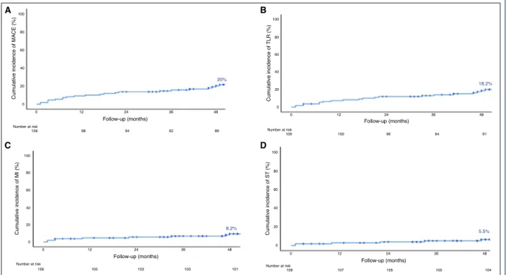
Results: Of the study population, 80% were male and the mean age was 60±11.3 years. The most common diagnosis was stable angina (84%). The median length of follow-up was 53 months (range: 46–59 months). The rate of predilation and postdilation was 100%, and 95%, respectively. The 4-year rate of MACEs was 20%: cardiac death in 3 patients (2.7%), target vessel MI in 9 (8.2%), and target lesion revascularization in 20 (18.2%). Definite device thrombosis occurred in 6 of 110 patients (5.5%). One case of very late scaffold thrombosis was observed at 47 months. A small BVS diameter (2.5 mm) was found to be the most powerful independent predictor of a MACE (p=0.05). Conclusion: The Absorb BVS was associated with an in-creased risk of adverse events, including late and very late device thrombosis, despite the use of a good implementation protocol.
Amaç: Eriyebilen vasküler çatı (EVÇ), ilaç kaplı stent tek-nolojisinde en heyecan verici gelişme olarak son yıllarda ön plana çıkmış fakat artmış tromboz komplikasyonları nede-niyle hayal kırıklığı yaşatmıştır. Bu çalışmada, çok büyük oranda predilatasyon-uygun çap postdilatasyon protokolü’ne uyarak Absorb EVÇ yerleştirilen hasta grubunda uzun dö-nem klinik sonuçlar araştırılmıştır.
Yöntemler: Bu geriye dönük çalışmaya 150 Absorb EVÇ yer-leştirilen toplam 110 hasta dahil edildi. Uzun dönem takipte kardiyak ölüm, hedef damar miyokart enfarktüsü (ME), hedef lezyon revaskülarizasyonu olarak tanımlanan majör kardiyo-vasküler olaylar (MKO) değerlendirildi.
Bulgular: Çalışmaya katılan hastaların %80’i erkek, ortalama yaş 60±11.3 yıldı. En sık tanı %84 kararlı anjinaydı. Ortanca takip süresi 53 aydı (aralık 46–59 ay). Hastaların predilatas-yon, postdilatasyon oranları sırasıyla %100, %95’ti. Dört yıllık takipte MKO oranı %20 bulundu. Hastaların 3’ünde (%2.7) kardiyak ölüm, 9’unda (%8.2) hedef damar ME ve 20’sinde (%18.2) hedef lezyon revaskülarizasyonu mevcuttu. Kesin çatı trombozu 6/110 (%5.5) hastada gözlemlendi. Bir hastada 47. ayda çok geç dönem çatı trombozu izlendi. Küçük BVS çapı (2.5 mm) MKO’ların en güçlü öngördürücüsü saptandı (p=0.05).
Sonuç: Absorb EVÇ, uygun yerleştirme protokolüne rağmen, artmış geç ve çok geç dönem çatı trombozunu da içeren olumsuz olaylarla ilişkilidir.
Received:June 01, 2020 Accepted:August 14, 2020
Correspondence: Dr. Sinem Çakal. İstanbul Medipol Üniversitesi Tıp Fakültesi, Kardiyoloji Anabilim Dalı, İstanbul, Turkey.
Tel: +90 212 - 460 77 77 e-mail: [email protected]
© 2021 Turkish Society of Cardiology
A
bioresorbable vascular scaffold (BVS) represent-ed a revolutionary alternative option to overcome the shortcomings of drug-eluting stents (DESs) in percutaneous coronary intervention (PCI).[1,2]The initial analyses of the first commercially avail-able everolimus-eluting BVS (Absorb; Abbott Vascu-lar Inc., Santa CVascu-lara, CA, USA) used in de novo sim-ple lesions showed non-inferior outcomes to metallic DESs in patients with stable coronary artery disease in short-term follow-up.[3] However, 3-year data from
the Absorb II cohort raised questions regarding the long-term safety of an Absorb BVS due to a high rate of device-related thrombosis compared with DESs.[4]
The mid- and long-term data of the AIDA (Amster-dam Investigator-initiateD Absorb Strategy All-com-ers Trial) were also disappointing, reporting a higher rate of late scaffold thrombosis compared with the XIENCE everolimus-eluting stent (EES) (Abbott Vascular, Inc., Santa Clara, CA, USA) (3.5% vs 0.9%; hazard risk [HR]: 3.87; 95% confidence interval [CI]: 1.78–8.42; p<.001).[5,6] A European medical device
advisory task force recommended additional testing and study,[7] and in September 2017, the manufacturer
halted sales of the first-generation Absorb BVS. The aim of the present study was to analyze long-term clinical outcomes of Absorb BVS implantation performed with the predilation, sizing, and post-dilation (PSP) implantation technique in a single high-volume PCI center.[8]
METHODS Study design and population
This was an observational, retrospective, single-cen-ter study of consecutive patients treated for coronary artery disease at Istanbul Medipol University hospi-tal, Turkey, between May 2014 and December 2016 with the Absorb BVS. The use of an Absorb BVS was at the discretion of the operator in charge. Clinical and procedural characteristics were assembled ret-rospectively from hospital medical records and fol-low-up data was collected through hospital visits and telephone consultations. This study was approved by the İstanbul Medipol University Faculty of Medicine Ethics Committee (Approval Date: 08/11/2019 Num-ber: 10840098-604.01.01-E.60925). A total of 110 pa-tients treated with 150 Absorb BVSs were included in the analysis.
Patients who were >18 years of age with ev-idence of myo-cardial ischemia, including those with stable cor-onary artery dis-ease and acute coronary syn-drome, with a reference vessel diameter (RVD) ≥2.50 mm were enrolled in the study. Stenosis of >50% was
evident in the native coronary arteries of all of the treated lesions. The exclusion criteria were a left main coronary artery lesion, a saphenous vein graft lesion, or the presence of a lesion requiring stents >4.0 mm or <2.5 mm. No restrictions were applied for the num-ber of lesions and vessels treated, lesion length, or the number of implanted stents.
Absorb bioresorbable vascular scaffold implantation
The implantation of an Absorb BVS according the principles of the PSP technique was not manda-tory, but was highly recommended given the cir-cumstances at the time. Angiographic assessments of BVS size and position were based on visual as-sessment using a guiding catheter as a reference for calibration, the length of opaque wire sections, and balloon length. Predilation was performed with compliant or non-compliant balloons. Generally, for more calcified lesions, a Scoreflex balloon (Or-busNeich Medical Co. Ltd., Hong Kong, China) or an AngioSculpt PTA scoring balloon (AngioScore Inc., Fremont, CA, USA) was preferred. The im-plantation of a scaffold was performed with a grad-ual increase of 1 atm of pressure every 5 seconds, without exceeding the rated burst pressure. The bal-loon was then rapidly deflated, reinflated, and kept at nominal pressure for 15–30 seconds. Finally, an-other angiogram was performed to evaluate BVS ex-pansion. Postdilation was performed with non-com-pliant balloons with the same size BVS or 0.25–0.5 mm larger. Long-segment lesions (>28 mm) that could not be covered with a single BVS therefore
Abbreviations:
BVS Bioresorbable vascular scaffold CI Confidence interval DAPT Dual anti-platelet therapy DES Drug-eluting stent EES Everolimus-eluting stent HR Hazard risk IQR Interquartile range IVUS Intravascular imaging MACE Major adverse cardiovascular event MI Myocardial infarction MLD Minimal lumen diameter P2Y12 Adenosine diphosphate chemoreceptor PCI Percutaneous coronary intervention PSP Predilation, sizing, and postdilation QCA Quantitative coronary angiography RVD Reference vessel diameter TLF Target lesion failure TLR Target lesion revascularization TVR Target vessel revascularization
required a BVS-BVS or DES-BVS combination. An overlapping BVS-BVS was used in prognosti-cally significant segments and vessels, such as the left atrial descending artery, to enable future surgi-cal vessel grafting options. Overlapping DES-BVS was typically preferred if a BVS longer than 28 mm was not available or there were special lesion char-acteristics of calcification, tortuosity, or bifurcation. In some cases, easier insurance reimbursement was also a factor. All of the patients were anticoagulated with unfractionated heparin to achieve an activated clotting time of 250 seconds. All of the patients were treated with dual anti-platelet therapy (DAPT) for at least 12 months after the procedure. The need for specific treatment strategies, such as additional stent implantation after postdilation were left to the oper-ator’s discretion.
Quantitative coronary angiography (QCA) was performed offline using standard techniques with au-tomated edge detection algorithms (CAAS 5.7.1, Pie Medical Imaging BV, Masstricht, The Netherlands) in the hospital’s angiographic analysis center. RVD, minimal lumen diameter (MLD), stenosis percent-age, MLD after stent implantation, and acute gain (defined as the difference between MLD postproce-dure and MLD preprocepostproce-dure) were measured. Binary angiographic restenosis was defined as stenosis of >50% of the luminal diameter in the target segment. A bend of >45° proximal to the lesion was defined as tortuosity. A single bend of 45–90° proximal to the lesion was defined as mild tortuosity, while 3 or more of 45–90° or one or more >90° was defined as severe tortuosity. Bends not meeting these criteria (mild and severe tortuosity) were defined as moderate tortuos-ity.[9] Calcification was defined as overt radiopacity
of the vessel wall across the lesion site. It was clas-sified as moderate (radiopacity noted only during the cardiac cycle before contrast injection) or severe (ra-diopacity noted across both sides of the vessel wall before contrast injection and independent of cardiac motion).[10]
Angiographic success was defined as <30% resid-ual diameter stenosis as determined by QCA with a Thrombolysis In Myocardial Infarction grade 3 flow in the treated target vessel. Procedural success was defined as angiographic success in the absence of in-hospital death, myocardial infarction (MI), or revas-cularization.
Outcomes and definitons
The primary outcomes of the study were a major ad-verse cardiovascular event (MACE), which was a composite of cardiac death, target vessel MI, and clin-ically-driven target lesion revascularization (TLR). TLR was defined as any revascularization within 5 mm of the scaffold. Target vessel revascularization (TVR) was defined as repeat PCI or coronary artery bypass graft in the target vessel. Deaths were consid-ered cardiac-related unless a non-cardiac cause was identified. All components of the composite endpoint, and definite stent thrombosis was determined accord-ing to the Academic Research Consortium.[11]
Statistical analysis
Continuous variables were expressed as mean±SD or median and interquartile range (IQR), as appropriate. Categorical variables were expressed as number and percentage. The Kolmogorov-Smirnov test was used to test the normality of distribution of continuous variables. A chi-squared test or Fisher’s exact test was used to compare binary variables, and Student’s t-test or a non-parametric test was used to compare contin-uous variables. Cumulative event rates were estimat-ed using the Kaplan-Meier method. Cox regression analysis was used to identify the factors affecting the occurrence of a MACE. IBM SPSS Statistics for Win-dows, Version 23.0 software (IBM Corp., Armonk, NY, USA) was used to perform the statistical analysis.
RESULTS Baseline characteristics
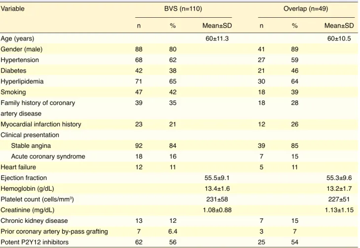
The demographic characteristics of the overall cohort and the groups with overlapping stents are presented in Table 1. A total of 110 patients with 150 scaffolds were enrolled in the study. Overlapping techniques with a DES or BVS were used in 49 patients (BVS-BVS: 19 patients, DES-(BVS-BVS: 30 patients). The pa-tients were predominantly male (80%), with a mean age of 60±11.3 years, and there was a high prevalence of stable angina (84%). Diabetes mellitus was present in 38% of the patients, while hypertension, hyperlip-idemia, and a current smoking history were recorded in 62%, 65%, and 42%, respectively.
Procedural and angiographic characteristics of the bioresorbable vascular scaffold implantation
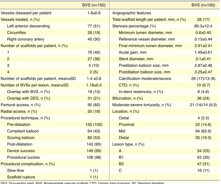
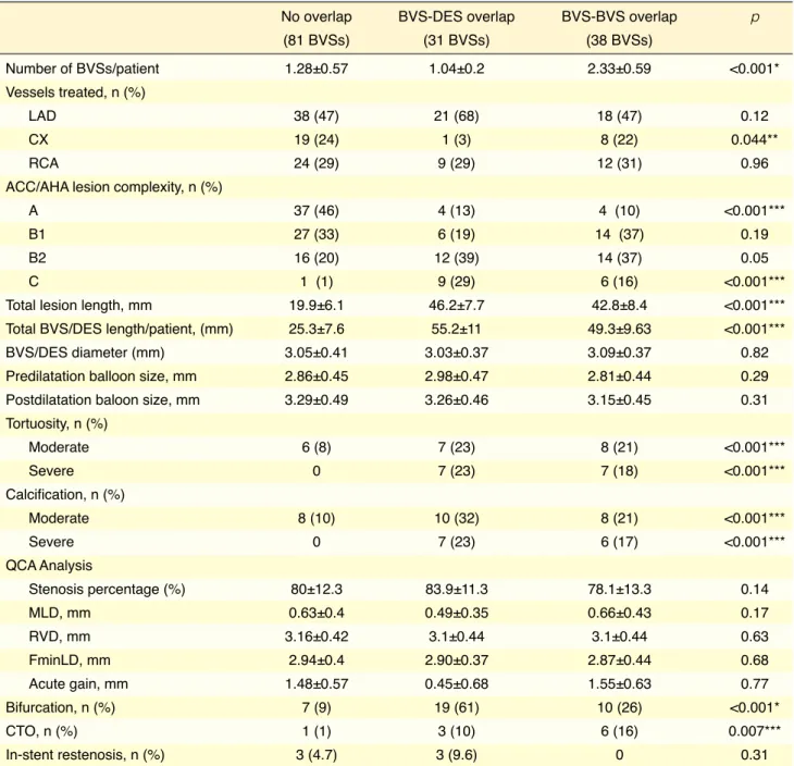
gies compared with the non-overlap group in terms of tortuosity, lesion calcification, and C-type lesions. Bi-furcation lesions were more frequent in the DES-BVS group (Table 3). The implanted stent/scaffold length did not differ significantly between the DES-BVS group (55.2 mm±11 mm) and the BVS-BVS group (49.3 mm±9.6 mm). One patient had a periprocedur-al MI, and another patient suffered scaffold rupture, which was managed with prolonged balloon inflata-tion. More than half (57%) of the patients used po-tent adenosine diphosphate chemoreceptor (P2Y12) inhibitor treatment during first year (Table 1).
Clinical outcomes
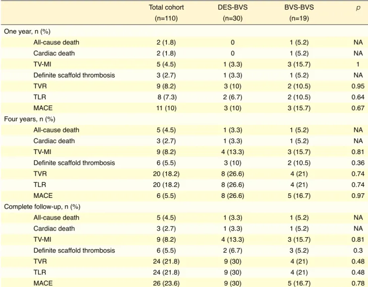
Outcomes of the overall cohort and the overlap groups are provided in Table 4. The BVS-BVS over-lap and DES-BVS overover-lap groups had similar clinical outcomes. The median length of follow-up was 53 months (IQR: 46–59 months). The MACE rate was 10% at the 12-month follow-up, 20% at the 4-year characteristics of the lesions treated. Most were in the
anterior descending coronary artery (51%), followed by the right coronary (30%) and circumflex (19%) ar-teries. The mean number of Absorb BVSs implanted per patient was 1.4±0.6. Predilation was performed in all lesions and postdilatation in 95% of the treated lesions. The clinical success of the device was 98%, as well as the procedural clinical success (n=108/110, 98%). Offline QCA indicated that the mean grade of stenosis was 80.3±12.4%, the lesions had a RVD of 3.13±0.44 mm and that the median length of the scaf-fold per patient was 28 mm (IQR: 17 mm). Most of the lesions (58%) treated with an Absorb BVS were classified as type A or B1 (American Heart Associa-tion/American College of Cardiology classification).
Approximately half (51%) of the patients had at least 2 scaffolds implanted. Two BVSs were over-lapped in 19 lesions, and an overlapping of BVS and a DES was performed in 31 lesions (Table 3). The overlap patients had more complex lesion
morpholo-Table 1. Patient characteristics
Variable BVS (n=110) Overlap (n=49) n % Mean±SD n % Mean±SD Age (years) 60±11.3 60±10.5 Gender (male) 88 80 41 89 Hypertension 68 62 27 59 Diabetes 42 38 21 46 Hyperlipidemia 71 65 30 64 Smoking 47 42 18 39
Family history of coronary 39 35 18 28
artery disease
Myocardial infarction history 23 21 12 26
Clinical presentation
Stable angina 92 84 39 85
Acute coronary syndrome 18 16 7 15
Heart failure 12 11 5 11
Ejection fraction 55.5±9.1 55.3±9.6
Hemoglobin (g/dL) 13.4±1.6 13.2±1.7
Platelet count (cells/mm3) 231±58 227±51
Creatinine (mg/dL) 1.08±0.88 1.13±1.15
Chronic kidney disease 13 12 7 15
Prior coronary artery by-pass grafting 7 6.4 3 7
Potent P2Y12 inhibitors 62 56 25 54
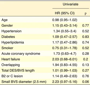
follow-up, and 23.6% at the end of the study fol-low-up period. The 4-year Kaplan-Meier estimate of MACE was 20% (Fig. 1). There were 3 (2.7%) cases of cardiac death, 20 (18.2%) cases of target lesion revascularization, and 9 (8.2%) cases of target ves-sel MI. Two non-cardiac deaths were due to prostate cancer at 23 and 56 months, and a third patient died of gastric cancer. The rate of early and late device thrombosis was 1.8% and 2.7%, respectively, and very late events continued to accrue beyond 1 year (5.5%). Table 5 provides a detailed description of the cases with definite stent thrombosis. Univariate Cox regression analysis indicated that only a small BVS diameter (2.5 mm) was a risk factor for the develop-ment of a MACE during follow-up (HR: 2.23; 95% CI: 0.97–2.23; p=0.05) (Table 6).
DISCUSSION
To the best of our knowledge, this is the largest long-term analysis of Absorb BVS real-world outcomes from a single high-volume center in Turkey. We found that the long-term incidence of MACE was primarily driven by a higher rate of TLR and MI, as well as early, late, and very late scaffold thrombosis events. A higher rate of a MACE was observed despite the greater use of a PSP implantation strategy and more frequent use of P2Y12 inhibitors compared with other Absorb BVS studies.[12]
After initial enthusiasm for the use of a BVS, poor clinical outcomes in terms of TLR and scaffold thrombosis of the Absorb BVS, the most
comprehen-Table 2. Angiographic and procedural characteristics
BVS (n=150)
Vessels diseased per patient 1.6±0.6 Vessels treated, n (%)
Left anterior descending 77 (51)
Circumflex 28 (19)
Right coronary artery 45 (30)
Number of scaffolds per patient, n (%)
1 76 (49)
2 27 (36)
3 5 (10)
4 2 (5)
Number of scaffolds per patient, mean±SD 1.4 ±0.6 Number of BVSs per lesion, mean±SD 1.18±0.4 Overlap with BVS, n (%) 19 (13) Overlap with DES, n (%) 31 (21)
Femoral access, n (%) 90 (82) Radial access, n (%) 20 (18) Procedural technique, n (%) Pre-dilatation 150 (100) Compliant balloon 64 (43) Scoring balloon 80 (53) Post-dilatation 142 (95) Device success 149 (99) Procedural sucess 108 (98) Procedural complication, n (%) Slow-flow 1 (1) Scaffold rupture 1 (1) BVS (n=150) Angiographic features
Total scaffold length per patient, mm, n (%) 28 (17) Stenosis percentage (%) 80.3±12.4 Minimum lumen diameter, mm 0.6±0.40 Reference vessel diameter, mm 3.13±0.44 Final minimum lumen diameter, mm 2.91±0.41
Acute gain, mm 1.49±0.61
Stent diameter, mm 3.1±0.41
Predilation balloon size, mm 2.87±0.46 Postdilation balloon size, mm 3.25±0.47 Calcification moderate/severe 26 (17)/13 (8) CTO, n (%) 10 (6.7) In-stent restenosis, n (%) 6 (4.6) Bifurcation, n (%) 36 (24) Moderate-severe tortuosity, n (%) 21 (14)/14 (9.3) Location, n (%) Ostial 4 (2.5) Proximal 22 (14.6) Mid 94 (62.6) Distal 30 (19.3) Lesion type, n (%) A 34 (23) B1 53 (35) B2 47 (31) C 16 (11)
withdrew the Absorb BVS from the market, and the implementation of BVS was given a class III indi-cation in clinical practice outside of studies in the current European Society of Cardiology guidelines.
[15,16] Scaffold discontinuities as a result of
intralumi-nal scaffold dismantling can precipitate thrombosis sively studied BVS, have hampered the clinical use
of BVSs.[13] A meta-analysis of 4 randomized BVS
trials assigning patients to an Absorb BVS (n=2164) or a DES (n=1225) resulted in higher 3-year rates of target lesion failure (TLF) in the Absorb BVS group (11.7% versus 8.1%; p=0.006).[14] The manufacturer
Table 3. Angiographic and lesion features of overlap and non-overlap groups
No overlap BVS-DES overlap BVS-BVS overlap p
(81 BVSs) (31 BVSs) (38 BVSs) Number of BVSs/patient 1.28±0.57 1.04±0.2 2.33±0.59 <0.001* Vessels treated, n (%) LAD 38 (47) 21 (68) 18 (47) 0.12 CX 19 (24) 1 (3) 8 (22) 0.044** RCA 24 (29) 9 (29) 12 (31) 0.96
ACC/AHA lesion complexity, n (%)
A 37 (46) 4 (13) 4 (10) <0.001***
B1 27 (33) 6 (19) 14 (37) 0.19
B2 16 (20) 12 (39) 14 (37) 0.05
C 1 (1) 9 (29) 6 (16) <0.001***
Total lesion length, mm 19.9±6.1 46.2±7.7 42.8±8.4 <0.001***
Total BVS/DES length/patient, (mm) 25.3±7.6 55.2±11 49.3±9.63 <0.001***
BVS/DES diameter (mm) 3.05±0.41 3.03±0.37 3.09±0.37 0.82
Predilatation balloon size, mm 2.86±0.45 2.98±0.47 2.81±0.44 0.29
Postdilatation baloon size, mm 3.29±0.49 3.26±0.46 3.15±0.45 0.31
Tortuosity, n (%) Moderate 6 (8) 7 (23) 8 (21) <0.001*** Severe 0 7 (23) 7 (18) <0.001*** Calcification, n (%) Moderate 8 (10) 10 (32) 8 (21) <0.001*** Severe 0 7 (23) 6 (17) <0.001*** QCA Analysis Stenosis percentage (%) 80±12.3 83.9±11.3 78.1±13.3 0.14 MLD, mm 0.63±0.4 0.49±0.35 0.66±0.43 0.17 RVD, mm 3.16±0.42 3.1±0.44 3.1±0.44 0.63 FminLD, mm 2.94±0.4 2.90±0.37 2.87±0.44 0.68 Acute gain, mm 1.48±0.57 0.45±0.68 1.55±0.63 0.77 Bifurcation, n (%) 7 (9) 19 (61) 10 (26) <0.001* CTO, n (%) 1 (1) 3 (10) 6 (16) 0.007*** In-stent restenosis, n (%) 3 (4.7) 3 (9.6) 0 0.31
ACC/AHA: American College of Cardiology/American Heart Association; BVS: Bioresorbable vascular scaffold; CTO: Chronic total occlusion; CX: Circum-flex; DES: Drug-eluting stent; FminLD: Final minimum lumen diameter; LAD: Left anterior descending; MLD: Minimum lumen diameter; QCA: Quantitative coronary angiography; RCA: Right coronary artery; RVD: Reference vessel diameter.
*Post-hoc analysis showed significant differences between all of the subgroups.
**Post-hoc analysis showed significant differences between the BVS-DES group and the other 2 subgroups in CX vessels. *** Post-hoc analysis showed significant differences between the no-overlap group and the other 2 subgroups.
outcomes is a suboptimal implantation technique. Though a precise optimal definition of PSP implanta-tion remains unknown, the importance of adherence to PSP is frequently emphasized.[8,17] Postdilatation
rates of <50% and varying PSP protocols have been reported in previous large-scale, randomized clinical trials and cohort studies.[18] Puricel et al.[13] noted that
in a multicenter registry of 1305 patients implanted with an Absorb BVS, the rate of scaffold thrombosis declined significantly in patients when a strategy op-timized for BVS was applied rather than a DES-ori-ented implantation strategy. However, despite optimal implantation practice, in the COMPARE-ABSORB trial, a substantially increased risk of TLF was found in complex de novo target lesions, defined as at least 28 mm in length, located in a small vessel, and with and/or restenosis more than a year after implantation.
An appropriate implantation technique with careful device sizing, appropriate expansion through long periods of balloon inflation, extensive use of post-dilation, and intracoronary imaging guidance in or-der to accurately assess the dimensions of the vessel and detect the presence of possible calcification were considered to be particularly important during BVS implantation in contrast to current-generation DESs because of the larger strut thickness and less radial strength. Special attention to intravascular imaging (IVUS) is called for when a BVS is implanted, espe-cially in long, complex lesions requiring overlapping, since the stacked struts of BVSs can reach a thickness of ~300 μm in overlap areas. One of the major factors hypothesized to be a determining factor in adverse
Table 4. Clinical outcomes
Total cohort DES-BVS BVS-BVS p
(n=110) (n=30) (n=19)
One year, n (%)
All-cause death 2 (1.8) 0 1 (5.2) NA
Cardiac death 2 (1.8) 0 1 (5.2) NA
TV-MI 5 (4.5) 1 (3.3) 3 (15.7) 1
Definite scaffold thrombosis 3 (2.7) 1 (3.3) 1 (5.2) NA
TVR 9 (8.2) 3 (10) 2 (10.5) 0.95 TLR 8 (7.3) 2 (6.7) 2 (10.5) 0.64 MACE 11 (10) 3 (10) 3 (15.7) 0.67 Four years, n (%) All-cause death 5 (4.5) 1 (3.3) 1 (5.2) NA Cardiac death 3 (2.7) 1 (3.3) 1 (5.2) NA TV-MI 9 (8.2) 4 (13.3) 3 (15.7) 0.81
Definite scaffold thrombosis 6 (5.5) 3 (10) 2 (10.5) 0.36
TVR 20 (18.2) 8 (26.6) 4 (21) 0.74 TLR 20 (18.2) 8 (26.6) 4 (21) 0.74 MACE 6 (5.5) 8 (26.6) 5 (16.7) 0.97 Complete follow-up, n (%) All-cause death 5 (4.5) 1 (3.3) 1 (5.2) NA Cardiac death 3 (2.7) 1 (3.3) 1 (5.2) NA TV-MI 9 (8.2) 4 (13.3) 3 (15.7) 0.81
Definite scaffold thrombosis 6 (5.5) 2 (6.7) 3 (5.2) 0.3
TVR 24 (21.8) 9 (30) 4 (21) 0.48
TLR 24 (21.8) 9 (30) 4 (21) 0.48
MACE 26 (23.6) 9 (30) 5 (16.7) 0.78
BVS: Bioresorbable vascular scaffold; DES: Drug-eluting stent; MACE: Major adverse cardiac event; TLR: Target lesion revascularization; TV-MI: Target vessel myocardial infarction; TVR: Target vessel revascularization.
Figure 1. (A-D) Cumulative event curves for major adverse cardiac events calculated using the Kaplan-Meier method. TLR: Target lesion revascularization; MACE: Major adverse coronary event; MI: Myocardial infarction; ST: Stent thrombosis.
A C B D 100 100 100 100
Cumulative incidence of MACE (%) Cumulative incidence of
TLR (%)
Cumulative incidence of MI (%) Cumulative incidence of ST
(%)
Follow-up (months) Follow-up (months)
Follow-up (months) Follow-up (months)
80 80 80 80 60 60 60 60 40 40 40 40 20 20 20 20 0 0 0 0 0 0 0 0 109 109 109 109 98 100 105 107 94 96 103 105 92 94 103 105 89 20% 8.2% 18.2% 5.5% 91 101 104 Number at risk Number at risk
Number at risk Number at risk
12 12 12 12 24 24 24 24 36 36 36 36 48 48 48 48
thrombosis or TLR rates. The estimated 2-year event rate for TLF (a composite of cardiac death, MI and TVR) was 11.0% for the Absorb BVS and 9.9% for the cobalt-chromium EES (p=0.003 for non-inferi-ority).[17,21] The careful attention paid to the
implan-tation technique in our study (predilaimplan-tation: 100%, a pre-existing total occlusion or bifurcation.[19] In our
study, a small BVS diameter (2.5 mm) was the only predictor of a MACE, which is consistent with the results of previous studies.[13,20] In the prospective
AIDA trial, scaffold implantation according to an optimised PSP protocol did not result in lower stent
Table 5. Detailed description of the cases of scaffold thrombosis
Case Initial PCI Treated Lesion Calcification Postdilation Scaffold size New Thrombosis Treatment
indication vessel type P2Y12 (months)
1 SAP LAD C No Yes 2.5x28 No 1 Thrombus aspiration 2 SAP RCA C Yes Yes 2.5x18, 3.5x18 Yes 1 Thrombus aspiration
overlapped with 3x33 Xience
3 SAP LAD B1 No Yes 2.5x28 No 10 Multiple balloon inflations GIIbIIIa inhibitor 4 SAP RCA A No Yes 3.0x28 Yes 22 3x28 Xience
GIIbIIIa inhibitor 5 ACS RCA B2 Yes Yes 3.5x18 overlapped Yes 33 3.0x38 Resolute Integrity
with 3.5x18 Resolute Integrity
6 SAP RCA B2 Yes Yes 3.0x28 overlapped No 47 Thrombus aspiration with 3.5x38 Xience 3.0x38 Ultimaster Tansei ACS: Acute coronary syndrome; LAD: Left anterior descending; P2Y12: Adenosine diphosphate receptor; PCI: Percutaneous coronary intervention; RCA: Right coronary artery; SAP: Stable angina pectoris.
um EES.[15,23] Whether these events are linked to
inter-ruption of dual antiplatelet therapy remains uncertain. Data from the INVEST registry showed that at the time of very late scaffold thrombosis, the majority of pa-tients (83%) were receiving aspirin monotherapy and that a minority were still receiving DAPT.[24]
Corrob-orating previous results, we also observed early, late, and very late scaffold thrombosis events. In our series, there were 3 cases of late thrombosis, 1 of which oc-curred 47 months after device implantation with on-going aspirin therapy. We had a higher rate of potent P2Y12 inhibitor use (57%) compared with random-ized Absorb BVS trials (Absorb Japan, Absorb China, Absorb III, which had rates of 21–24%).[25] Prolonged
use of P2Y12 antagonists, which have been associated with more potent antiplatelet activity than clopidogrel, has been advocated, however, whether this strategy leads to a reduction of very late scaffold thrombosis remains unproven. Moreover, prolongation of dual an-tiplatelet therapy increases the risk of bleeding events.
Limitations
A small sample size and the lack of randomization or blinding constitute significant limitations in the de-sign of this study. The decision to implant an Absorb BVS was left to the operator; therefore, selection bias cannot be ruled out, and the lack of IVUS guidance is an additional limitation. The absence of IVUS could have, at least in part, had an impact on the adverse events observed in the overlap groups. IVUS was used in only some cases in published BVS trials.[8] Further studies are required to determine whether improved outcomes can be achieved with routine IVUS. Some of the adverse events seen in the current study could be related to DES-BVS overlapping. Since routine an-giography and imaging modalities during follow-up were not performed, we may have missed non-clinical BVS stenosis.
Conclusion
Despite greater use of P2Y12 inhibitors and very strict adherence to PSP protocol, we found that the MACE rate was high and that cases of very late stent throm-bosis continued to accrue during long-term follow-up after Absorb BVS implantation in a patient population reflecting routine clinical practice.
Funding: This research did not receive any specific grant
from any funding agency.
Ethical statement: This study was approved by the postdilatation: 95%) is consistent with this concept.
Nonetheless, while optimization of the implantation technique appears to be of great importance, we still detected a higher MACE rate in our study. Therefore, the effect of an optimal implantation technique on BVS outcomes remains controversial.
Conflicting results from clinical studies have chal-lenged the expectation that complete scaffold degra-dation occurred within 3–4 years after BVS implan-tation. Sotomi et al.[22] evaluated the causes of acute
and subacute scaffold thrombosis and determined that the most frequent causes of thrombosis were malap-position (23.5%), uncovered struts (17.6%), strut un-der-deployment (11.8%), acute scaffold disruption (5.9%), overlapping stents (5.9%), and acute scaffold recoil (5.9%). Malapposition and late scaffold discon-tinuity (34.6% and 30.8%, respectively) were the most common mechanism of thrombosis in late and very late phases. Scaffold thrombosis could be prevented with an appropriate PSP protocol, however, even when optimal PSP technique was applied, extensive scaf-fold discontinuity with struts protruding into the lu-men might cause late scaffold thrombosis. The AIDA and ABSORB III trials and subsequent meta-analyses clearly showed that very late scaffold thrombosis was significantly more frequent after 1 year of implantation with the ABSORB BVS than with the
cobalt-chromi-Table 6. Factors associated with major adverse cardiac events at 4 years Univariate HR (95% CI) p Age 0.98 (0.95–1.02) Gender 1.15 (0.43–3.14) 0.77 Hypertension 1.34 (0.55–3.4) 0.52 Diabetes 1.09 (0.47–2.57) 0.83 Hyperlipidemia 1.17 (0.47–2.86) 0.74 Smoker 0.75 (0.31–1.78) 0.52
Acute coronary syndrome 1.73 (0.63–4.7) 0.28 Heart failure 2.03 (0.68–6.01) 0.2 Overlapping 1.94 (0.83–4.55) 0.13 Total DES/BVS length 1.01 (0.99–1.03) 0.27 B2 or C lesion 1.14 (0.49–2.63) 0.76 Small BVS diameter (2.5 mm) 2.23 (0.97–5.16) 0.06
BVS: Biovascular scaffold; DES: Drug-eluting stent; HR: Hazard risk; CI: Confidence interval.
fold implantation for prediction of cardiac adverse events: development and internal validation of the PSP score. EuroIn-tervention 2017;12:2110−7.
9. Ho HH, Jafary FH, Loh KK, Tan JK, Ooi YW, Ong PJ. Deliv-erability of integrity coronary stents in severely tortuous cor-onary arteries: a preliminary experience. J Invasive Cardiol 2012;24:650−4.
10. Mintz GS, Popma JJ, Pichard AD, Kent KM, Satler LF, Chuang YC, et al. Patterns of calcification in coronary artery disease. A statistical analysis of intravascular ultrasound and coronary angiography in 1155 lesions. Circulation 1995;91:1959−65. 11. Cutlip DE, Windecker S, Mehran R, Boam A, Cohen DJ, van
Es GA, et al. Clinical end points in coronary stent trials: a case for standardized definitions. Circulation 2007;115:2344−51. 12. Stone GW, Abizaid A, Onuma Y, Seth A, Gao R, Ormiston
J, et al. Effect of Technique on Outcomes Following Biore-sorbable Vascular Scaffold Implantation: Analysis From the ABSORB Trials. J Am Coll Cardiol 2017;70:2863−74. 13. Puricel S, Cuculi F, Weissner M, Schmermund A,
Jamshi-di P, Nyffenegger T, et al. Bioresorbable Coronary Scaffold Thrombosis: Multicenter Comprehensive Analysis of Clinical Presentation, Mechanisms, and Predictors. J Am Coll Cardiol 2016;67:921−31.
14. Ali ZA, Gao R, Kimura T, Onuma Y, Kereiakes DJ, Ellis SG, et al. Three-Year Outcomes With the Absorb Bioresorbable Scaffold: Individual-Patient-Data Meta-Analysis From the ABSORB Randomized Trials. Circulation 2018;137:464−79. 15. Ali ZA, Serruys PW, Kimura T, Gao R, Ellis SG, Kereiakes
DJ, et al. 2-year outcomes with the Absorb bioresorbable scaffold for treatment of coronary artery disease: a systematic review and meta-analysis of seven randomised trials with an individual patient data substudy. Lancet 2017;390:760−72. 16. Neumann FJ, Sousa-Uva M, Ahlsson A, Alfonso F, Banning
AP, Benedetto U, et al. 2018 ESC/EACTS Guidelines on myocardial revascularization. Eur Heart J 2019;40:87−165. 17. Tijssen RYG, Kraak RP, Elias J, van Dongen IM, Kalkman
DN, Nassif M, et al. Implantation techniques (predilatation, sizing, and post-dilatation) and the incidence of scaffold thrombosis and revascularisation in lesions treated with an everolimus-eluting bioresorbable vascular scaffold: insights from the AIDA trial. EuroIntervention 2018;14:e434−e42. 18. Gori T, Weissner M, Gonner S, Wendling F, Ullrich H,
El-lis S, et al. Characteristics, Predictors, and Mechanisms of Thrombosis in Coronary Bioresorbable Scaffolds: Differenc-es Between Early and Late Events. JACC Cardiovasc Interv 2017;10:2363−71.
19. Danzi GB, Bernelli C, Cerrato E. Outcomes of Optimised Im-plantation Technique with Bioresorbable scaffolds: A Pooled Analysis of ABSORB-IV and COMPARE-ABSORB Trials. Cardiovasc Revasc Med 2020;21:559−61.
20. Ishibashi Y, Nakatani S, Sotomi Y, Suwannasom P, Grunde-ken MJ, Garcia-Garcia HM, et al. Relation Between Biore-sorbable Scaffold Sizing Using QCA-Dmax and Clinical
İstanbul Medipol University Faculty of Medicine Ethics Committee (Approval Date: 08/11/2019 Num-ber: 10840098-604.01.01-E.60925).
Peer-review: Externally peer-reviewed. Conflict-of-interest: None.
Funding: No funding to declare.
Authors’ Contribution: All authors provided critical
feed-back and helped shape the research, analysis and manu-script.
Authorship contributions: Concept: B.Ç., S.Ç.; Design:
B.B., O.K.; Supervision: B.B., B.Ç.; Materials: B.Ç., S.Ç.; Data: B.Ç.; Analysis: B.B., O.K.; Literature search: S.Ç., B.Ç.; Writing: B.Ç., O.K.; Critical revision: B.B., O.K.
REFERENCES
1. Serruys PW, Garcia-Garcia HM, Onuma Y. From metallic cages to transient bioresorbable scaffolds: change in paradigm of coronary revascularization in the upcoming decade? Eur Heart J 2012;33:16−25b.
2. Caiazzo G, Kilic ID, Fabris E, Serdoz R, Mattesini A, Foin N, et al. Absorb bioresorbable vascular scaffold: What have we learned after 5 years of clinical experience? Int J Cardiol 2015;201:129−36.
3. Ellis SG, Kereiakes DJ, Metzger DC, Caputo RP, Rizik DG, Teirstein PS, et al. Everolimus-Eluting Bioresorb-able Scaffolds for Coronary Artery Disease. N Engl J Med 2015;373:1905−15.
4. Serruys PW, Chevalier B, Sotomi Y, Cequier A, Carrie D, Piek JJ, et al. Comparison of an everolimus-eluting bioresorbable scaffold with an everolimus-eluting metallic stent for the treatment of coronary artery stenosis (ABSORB II): a 3 year, randomised, controlled, single-blind, multicentre clinical tri-al. Lancet 2016;388:2479−91.
5. Kerkmeijer LSM, Tijssen RYG, Hofma SH, van der Schaaf RJ, Arkenbout KE, Kraak RP, et al. Comparison of an evero-limus-eluting bioresorbable scaffold with an everolimus-elut-ing metallic stent in routine PCI: three-year clinical outcomes from the AIDA trial. EuroIntervention 2019;15:603−6. 6. Wykrzykowska JJ, Kraak RP, Hofma SH, van der Schaaf
RJ, Arkenbout EK, AJ IJ, et al. Bioresorbable Scaffolds versus Metallic Stents in Routine PCI. N Engl J Med 2017;376:2319−28.
7. Byrne RA, Stefanini GG, Capodanno D, Onuma Y, Baumbach A, Escaned J, et al. Report of an ESC-EAPCI Task Force on the evaluation and use of bioresorbable scaffolds for percuta-neous coronary intervention: executive summary. Eur Heart J 2018;39:1591−601.
8. Ortega-Paz L, Capodanno D, Gori T, Nef H, Latib A, Cara-manno G, et al. Predilation, sizing and post-dilation scoring in patients undergoing everolimus-eluting bioresorbable
scaf-Keywords: Absorb bioresorbable vascular scaffold; coronary artery disease; percutaneous coronary intervention.
Anahtar sözcükler: Absorb eriyebilen vasküler çatı; koroner arter hastalığı; perküten koroner girişim.
sus Everolimus-Eluting Metallic Stent Implantation: A Me-ta-Analysis of Randomized Controlled Trials. Circulation 2017;135:2145−54.
24. Yamaji K, Raber L, Windecker S. What determines long-term outcomes using fully bioresorbable scaffolds - the device, the operator or the lesion? EuroIntervention 2017;12:1684−7. 25. Kereiakes DJ, Ellis SG, Metzger C, Caputo RP, Rizik
DG, Teirstein PS, et al. 3-Year Clinical Outcomes With Everolimus-Eluting Bioresorbable Coronary Scaffolds: The ABSORB III Trial. J Am Coll Cardiol 2017;70:2852−62. Outcomes at 1 Year in 1,232 Patients From 3 Study Cohorts
(ABSORB Cohort B, ABSORB EXTEND, and ABSORB II). JACC Cardiovasc Interv 2015;8:1715−26.
21. Tijssen RYG, Kerkmeijer LSM, Takahashi K, Kogame N, Katagiri Y, Kraak RP, et al. The influence of implantation techniques on lesion oriented-outcomes in Absorb BVS and Xience EES lesions treated in routine clinical practice at com-plete three year follow-up: AIDA trial QCA substudy. Int J Cardiovasc Imaging 2020;36:565−75.
22. Sotomi Y, Suwannasom P, Serruys PW, Onuma Y. Possible mechanical causes of scaffold thrombosis: insights from case reports with intracoronary imaging. EuroIntervention 2017;12:1747−56.
23. Montone RA, Niccoli G, De Marco F, Minelli S, D’Ascenzo F, Testa L, et al. Temporal Trends in Adverse Events After Everolimus-Eluting Bioresorbable Vascular Scaffold