To the Editor,
In this issue of Turk J Gastroenterol 2014, Şahin et al. (1) presented an informative study on the prevalence and etiologic distribution of hepatic granulomas (HGs) iden-tified in patients who were biopsied in their gastroen-terology division and reviewed the literature.
It has been reported that HGs are found in approximate-ly 5% (1.6%-15%) of patients who undergo a liver biopsy and may be caused by a variety of conditions (2). Differ-ences in the rate of HGs and the underlying systemic disorders depend on the location of the study and the patient population. HGs may result from a primary liver disease or an ongoing systemic disease. Various causes of HG may be subdivided into infections (particularly brucellosis, miliary TB, pulmonary TB, Q-fever), sarcoid-osis, primary biliary cirrhosis (PBC), and drugs. The cause of HG may remain unclear, despite careful evaluation, in approximately 20% of patients (idiopathic HG). Liver biopsies of patients with chronic hepatitis C and B virus infections may show HG in ~1% of cases (3-5). Typhoid fever may also rarely cause HG (6).
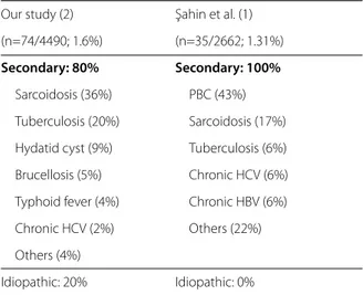
It would have been more suitable ıf the authors had cited the first article regarding etiology of HGs in Tur-key, which was published by our group in 2001 (2). Our rate of HG (≈1.5%) compares with that of the authors; however, there are differences in etiologic distribution (Table 1). The main reason for this difference may be that we had included all liver biopsy samples examined in our pathology department. The fact that sarcoidosis is the leading cause in HG in our study is actually mis-leading. Samples from another study investigating the role of hepatic involvement in sarcoidosis were also included in our study. Given these facts, TB would be the leading etiology of HG in our country. The authors claim that the etiology in up to 50% of cases with HG is PBC. The reason for this particular finding may be that
the authors studied only samples from a gastroenter-ology clinic. Moreover, our experiences in the last few decades have shown us that chronic hepatitis B and C virus infections may also cause HG (3-5).
To conclude, HG may be anticipated from liver biopsy samples of patients with PBC and various systemic dis-orders causing HG. Asymptomatic HG observed in pa-tients with chronic HBV and HCV infections generally have a viral etiology. Nevertheless, a simple diagnostic approach in order to exclude other causes of HG is war-ranted. Chest X-ray (for sarcoidosis and TB), PPD skin test, and serologic tests (Wright test, IFA for Q-fever, VDRL/RPR, IgM/AMA) may be performed.
Conflict of Interest: No conflict of interest was declared by the authors.
Ali Mert1, Mesut Yılmaz2, Bahadır Ceylan2, Ferhat Aslan2 1Department of Internal Medicine, İstanbul Medipol University Faculty
of Medicine, İstanbul, Turkey
2Department of Infectious Diseases and Clinical Microbiology, İstanbul
Medipol University Faculty of Medicine, İstanbul, Turkey Turk J Gastroenterol2014; 25: 529-30
Hepatic granulomas: Etiologic distribution
Address for Correspondence: Ali Mert, Department of Internal Medicine, İstanbul Medipol University Faculty of Medicine, İstanbul, Turkey E-mail: [email protected]
Received: 1.10.2014 Accepted: 3.10.2014
© Copyright 2014 by The Turkish Society of Gastroenterology • Available online at www.turkjgastroenterol.org • DOI: 10.5152/tjg.2014.0052
529
Editor
ial C
omment
Our study (2) Şahin et al. (1)
(n=74/4490; 1.6%) (n=35/2662; 1.31%)
Secondary: 80% Secondary: 100%
Sarcoidosis (36%) PBC (43%)
Tuberculosis (20%) Sarcoidosis (17%)
Hydatid cyst (9%) Tuberculosis (6%)
Brucellosis (5%) Chronic HCV (6%)
Typhoid fever (4%) Chronic HBV (6%)
Chronic HCV (2%) Others (22%)
Others (4%)
Idiopathic: 20% Idiopathic: 0%
REFERENCES
1. Şahin M, Yılmaz G, Arhan M, Sen İ. Hepatic granulomas in Turkey: A six year clinicopathological study of 35 cases. Turk J Gastroen-terol 2014; 25: 524-8. [CrossRef]
2. Mert A, Ozaras R, Bilir M, et al. The etiology of hepatic granulomas. J Clin Gastroenterol 2001; 32: 275-6. [CrossRef]
3. Mert A, Tabak F, Ozaras R, Tahan V, Sentürk H, Ozbay G. Hepatic granulomas in chronic hepatitis C. J Clin Gastroenterol 2001; 33: 342-3. [CrossRef]
4. Ozaras R., Tahan V, Mert A, et al. The prevalence of hepatic granu-lomas in chronic hepatitis C. J Clin Gastroenterol 2004; 38: 449-52.
[CrossRef]
5. Tahan V, Ozaras R, Lacevic N, et al. Prevalence of hepatic gran-ulomas in chronic hepatitis B. Dig Dis Sci 2004; 49: 1575-77.
[CrossRef]
6. Mert A, Tabak F, Ozaras R, Oztürk R, Akı H, Aktuglu Y. Typhoid fever as a rare cause of hepatic, splenic, and bone marrow granulomas. Intern Med 2004; 43: 436-9 [CrossRef]
530
Mert et al. Hepatic granulomas Turk J Gastroenterol 2014; 25: 529-30
Editor
ial C
![[Edirne Eski Eserleri Sevenler Cemiyeti Reisi Y. Cemil Üner tarafından Trakya Umumi Müfettişlik Yüksek Makamına gönderilen dilekçe]](data:image/gif;base64,R0lGODlhAQABAIAAAP///wAAACH5BAEAAAAALAAAAAABAAEAAAICRAEAOw==)