Olgu Sunumu
© 2010
DEÜ
TIP FAKÜLTESİ DERGİSİ CİLT 24, SAYI 1, (OCAK) 2010, S: 33 - 3633
Spontaneous Rupture Of Varicocele Due To
Strain-Defecation
ZORLU DIŞKALAMAYA BAĞLI SPONTAN VARİKOSEL RÜPTÜRÜ
Ömer DEMİR
1, Ali Kemal TEMİZKAN
21Dokuz Eylül Üniversitesi Tıp Fakültesi Üroloji Anabilim Dalı 2Şırnak Asker Hastanesi
Ömer DEMİR Dokuz Eylül Üniversitesi Tıp Fakültesi Üroloji AD 25340 İnciraltı, İZMİR Tel: Tel: Tel: Tel: (232) 4123451
eeee----posta:posta:posta: [email protected] posta:
ÖZET
Zorlamaya bağlı spontan sol varikosel rüptürü olgusu sunulmuştur. Zorlu dışkılama sonrası sol skrotumda şişlik ve ağrı yakınmasıyla gelen 21 yaşındaki erkek hastada varikosel rüptürü tanısı konuldu. Doppler ultrasonografi ile sol testiste hematom, sol testisi saran alanda kan akımı ve sol spermatik vende reflü akım izlendi. Hasta konservatif takip sonrası ameliyat edildi.
Anahtar sözcükler: Spontan varikosel rüptürü, tedavi, Doppler ultrasonografi SUMMARY
We report a case of strain-induced spontaneous rupture of left varicocele. Varicocele rupture was diagnosed in a 21-year-old man who presented with swelling and pain in the left scrotum of sudden onset after straining for defecation. Doppler ultrasound revealed a hematoma and escaping to the left peritesticular space and, reflux of blood in the left spermatic vein. The patient underwent an operation after conservative follow-up.
Key words: Spontaneous varicocele rupture, Treatment, Doppler ultrasonography
Varicocele is encountered in 15% of healthy man (1). Spontaneous rupture of varicocele is a rare condition. The algorithm for the diagnosis and management of this condition has not been defined clearly. We herein report a case of spontaneous rupture of varicocele due to constipation.
CASE PRESENTATION
A 21-year-old man presented with complaints of pain and swelling in the left scrotum following defecation. He
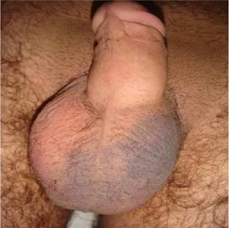
had chronic constipation and used laxatives. There was no history of trauma, previous similar complaints, blood dyscrasias or medicine intake. He had a previous abdominal operation due to hydatid cyst of the liver. Ecchymosis and swelling of the left hemiscrotum was evident (Figure 1). A soft mass was palpable around the left testis. Right hemiscrotum and testis was normal in size and consistency. Scrotal ultrasonography showed a hematoma formation around the left testis and a marked thickening of the left scrotum wall (Figure 2A and 2B).
34 Spontaneous rupture of varicocele due to strain-defecation
Doppler ultrasonography showed reflux of blood to the left spermatic veins (Figure 2C) and they were significantly dilated (Figure 2D). More than two spermatic veins of larger than 4 mm were visible, while the spermatic arterial blood flow was normal.
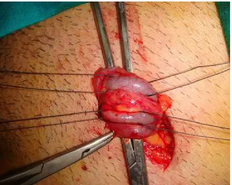
Our diagnosis was strain-defecation induced spontaneous rupture of the left varicocele. The patient was followed conservatively including antibiotics, anti-inflammatory agents, cooling of the left hemiscrotum and bed rest for five days. The scrotal ecchymosis and hematoma regressed during this period. He underwent varicocele ligation after 3 weeks for a possible recurrence of rupture (Figure 3).
DISCUSSION
Although varicocele is relatively common in the normal adult male population, spontaneous varicocele rupture is very rare (2). Review of the literature revealed less than 10 cases have been reported (3-9). Blunt trauma was the most common cause of varicocele rupture in the reported cases (3,4,6). The other causes of spontaneous rupture were sexual intercourse (8) and Valsalva’s maneuver during defecation (5,9). In the case report by Matsui et al, the cause of the rupture was associated with renal vein involvement of advanced pancreatic cancer (7). It was supposed that rise in the venous pressure due to a Valsalva’s maneuver may explain the cause of the rupture of the varix (6).
Spontaneous rupture of varicocele due to strain-defecation 35
Figure 2. A: A: A: A: Hematoma formation around the left testis (arrow). B:B:B:B: Thickening of the left scrotum wall (arrow) and significant dilatation of the left spermatic veins. C: C: C: C: Continuous reflux of blood flow from the left spermatic vein. D:D:D: Significant dilatation of D: the left spermatic veins.
As seen in other cases of varicocele rupture, scrotal swelling, pain, and ecchymosis was presented in our case. However, penile, scrotal or inguinal ecchymosis may be missing in acute cases (6). Spontaneous varicocele rupture, although rare; is commonly seen in young patients. Preoperative possible differential diagnoses include testicular torsion, strangulated hernia or scrotal hematoma. In most of the reported cases, surgery was performed since other causes of acute scrotum could not be excluded (3-6). The diagnostic procedures of spontaneous rupture of varicocele include Doppler ultrasounography, magnetic resonance imaging and computerized tomography. However, Doppler ultrasound examination is sufficient for the diagnosis in
majority of these cases.
The varicocele and hematoma around the spermatic cord easily demonstrated with Doppler ultrasound. Assessment of the scrotum by ultrasound is generally accepted as the first-line imaging technique for many common diseases (10).
We recommend the use of Doppler ultrasound to discriminate varicocele rupture from other causes of acute scrotum. Conservative treatment is a good choice for these patients as it provides resolution of the scrotal hematoma. The surgical treatment of varicocele can be performed electively for possible reccurence of rupture with a low morbidity.
36 Spontaneous rupture of varicocele due to strain-defecation
Figure 3. View of the dilated varicose veins during operation
REFERENCES
1. Pryor JL, Howards SS. Varicocele. Urol Clin N Amer 1987; 148: 499-513.
2. Hargreave TB. Varicocele—a clinical enigma. Br J Urol 1993; 72: 401-408.
3. Bisset RD. Two cases of traumatic rupture of a varicocele. J R Nav Med Serv 1945; 31: 177.
4. Redman JF, Rountree GA, Bissada NK. Injuries to scrotal contents by blunt trauma. Urology 1976; 7: 190-191.
5. Aliabadi H, Cass AS. Nontraumatic rupture of varicocele. Urology 1987; 29: 421-422.
6. Gordon JN, Aldoroty RA, Stone NN. A spermatic cord hematoma secondary to varicocele rupture from blunt
abdominal trauma: a case report and review. J Urol 1993; 149: 602-603.
7. Matsui Y, Utsunomiya N, Ichioka K, Ueda N, Yoshimura K, Terai A. Spontaneous rupture of varicocele testis associated with advanced pancreatic cancer. Int J Urol 2004; 11: 1145-1146.
8. Nishiyama Y, Nagai A, Nasu Yet al. Varicocele rupture due to sexual intercourse. Int J Urol 2005; 12: 585-587. 9. Kobayashi S, Machida T, Ishizaka K. A case of
non-traumatic rupture of varicocele. Nippon Hinyokika Gakkai Zasshi 2006; 97: 801-803.
10. Stewart VR, Sidhu PS. The testis: the unusual, the rare and the bizarre. Clin Radiol, 2007; 62: 289-302.