DOI: 10.5455/annalsmedres.2019.05.246 2019;26(8):1692-6
Evaluation of the results of pulse oximetry screening
for detection congenital heart disease in Konya region,
Turkey; Is it cost effective?
Murat Konak1, Beyza Ozcan2, Ahmet Sert3, Saime Sundus Uygun1, Hanifi Soylu1 1Selcuk University, Selcuklu Medical Faculty, Department of Division of Neonatology, Pediatrics, Konya, Turkey
2Health Science University Konya Training and Research Hospital, Department of Division of Neonatology, Pediatrics, Konya, Turkey 3Selcuk University, Selcuklu Medical Faculty, Department of Division of Pediatric Cardiology, Konya, Turkey
Copyright © 2019 by authors and Annals of Medical Research Publishing Inc.
Abstract
Aim: Congenital heart disease (CHD) is one of the most common congenital anomalies. It is known that early detection of the critical
lesions without symtomatic improves the prognosis of patients in the long term. In our study, we aimed to evaluate the efficacy and cost of pulse oximetry screening for detection of CHD in Konya region.
Material and Methods: In our study, we analyzed the results of pulse oximetry screening of infants born between 2016 and 2018
in Selcuk University and Saglik Bilimleri University Konya Training and Research Hospital. Positive screening test results and their costs was evaluated.
Results: Pulse oximetry was performed on a total of 3440 infants for screening congenital heart disease at two different centers. 2003
(58.2%) of the cases were female and 1437 (41.8%) were male. The mean timing of screening was 24.7±13.1 (4-100) hours (mean ± SD (min-max). 292 (8.48%) infants were tested positive. Echocardiographic examination was performed in the positive cases. The prevalence of CHD was found to be 27.6 ‰. The prevalence of critical CHD was 1.16 per 1000 live births. The false-positive screen rate was found to be 197 (5.7%). Among the cases who were screened, the most common CHD was the secundum ASD. The total cost of probes was 2622 ₺ ($497.91). The cost of echocardiography for 292 babies was approximately 13181 ₺ ($2503).
Conclusion: Screening for heart disease can be used effectively in the early diagnosis of serious and life-threatening diseases
in newborn and the cost is unneglectable in the early screening of patients. This study, the total cost of each infant screening is approximately 4.5 ₺ ($0.85) which appears to be a quite cheap and effective screening compared to the other countries.
Keywords: Newborn; congenital heart disease; pulse oximetry screening; cardiovascular disease.
Received: 07.05.2019 Accepted: 08.08.2019 Available online: 28.08.2019
Corresponding Author: Murat Konak,Selcuk University, Selcuklu Medical Faculty, Department of Division of Neonatology, Pediatrics,
Konya, Turkey E-mail: drmkonak@hotmail.com
INTRODUCTION
Congenital heart diseases (CHD) are one of the most common congenital malformations with a prevalence of 7-8 per 1000 live births. It is one of the leading diseases accounting for deaths in children in the first year of life (1-3). 25% of these patients present with complex congenital heart diseases, and delayed diagnosis or treatment can result in metabolic acidosis, intracranial hemorrhage, hypoxic ischemic encephalopathy, necrotizing enterocolitis, heart failure, cardiogenic shock and death (4,5). Neurological prognosis of patients is known to be poor in cases of delay or neglected diagnosis even when intervened later on (6). It is estimated that approximately 25-50% of infants with CHD are discharged from the
hospital without diagnosis (7). A murmur or weak to non-existing femoral pulses in the physical examination after delivery could be an indicator. In such cases, the physician might ask for echocardiography so that the patient can be diagnosed. These infants may not present any audible murmur due to underlying anatomical factors, decrease in pulmonary vascular resistance and decreased ventricular function, which might result in delays of the diagnosis. In recent years, many institutions, including the American Academy of Pediatrics, have recommended to perform pulse oximetry screening after delivery for detection of congenital heart diseases so that CHDs can be diagnosed in asymptomatic infants (8).
CHD screenings in our region with the aim to evaluate the feasibility, advisability, and cost-effectiveness.
MATERIAL and METHODS
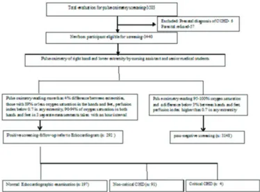
A prospective cohort study was conducted in tertiary NICU of Selçuk University and Konya Teaching and Research Hospital, Konya, between 2016 and May 2018. The local Ethics Committee approved this study. The parents were previously informed about the purpose of screening and their informed consent was obtained. The study included healthy infants beside their mothers and those who did not require hospitalization. The newborns who were suspected of having congenital heart defects upon antenatal evaluation or underwent fetal echocardiography, those who presented with dysmorphic features and genetic disorders in physical examination or those who required intensive care due to various reasons were excluded from the study. Furthermore, in our study, the births of refugee babies were excluded from the study. Refugees in our country were not included in the study because of the fact that various congenital anomalies were seen more frequently than in our society. Pulse oximetry screening methods and protocol diagram, there was shown in Figure 1.
Figure 1. Pulse oximetry screening methods, protocol diagram
and outcomes
Pre-ductal evaluation was performed by measuring the saturation and perfusion index from the right hand wrist, whereas the post-ductal evaluation was performed measuring the saturation and perfusion index from the lower extremity using the Radical 7 Masimo (Masimo Corporation, Irvine CA) device with a specific software installed for pulse oximetry screening. Masimo re-usable sensor was used for scanning. In the Training and Research Hospital, the screening was performed by a senior nurse who was experienced in neonatal infants, whereas, in the university hospital, the screening was performed by senior medical students, who were intern doctors, under the supervision of. The screening was performed 24 hours after birth, if possible, or right before discharge for the
cases who were discharged earlier. The screening results of those with 95-100% oxygen saturation and a %3 and below difference between hands and feet were determined as negative and considered to be a passed screen (7,8). The cases who had more than 4% difference between extremities, those with 89% or less oxygen saturation, a perfusion index below 0,7 in any extremity, and those who had 90-94% of oxygen saturation in both hands and feet in 3 seperate measurements taken with an hour interval were considered as positive screen and referred to cardiology for further echocardiographic examination (7,8). Cardiological examination was requested to be performed as soon as possible, but in cases where it could not be performed, the infants were hospitalized and monitored until the examination.
In echocardiographic examination, the following types of critical congenital heart defects were defined; Pulmonary atresia, truncus arteriosus, Fallot’s tetralogy, total anomalous pulmonary venous return, transposition of great arteries, tricuspid atresia, critical aortic stenosis, aortic coarctation, interruption of aortic arch and hypoplastic left heart syndrome, whereas non-critical congenital defects were defined as any ASD>5 mm, PDA>2 mm, VSD, valvular pulmonary stenosis, aortic stenosis and mild pulmonary artery stenosis. Normal variants were determined as patent foramen ovale, ASD<5 mm and PDA<2 mm (7).
For the cost analysis, the amount of pulse oximetry probes used throughout the study was calculated. In addition, cost of echocardiography which applied to positive cases was calculated as a separate outgoings. And then sum of these expenses was calculated for each patient.
Statistical evaluation was performed using SPSS 21.0 (Statistical Program in Social Sciences) software. Data were presented in number, percentage, mean and standard error of the mean (min-max). The P value less than 0.05 was considered to be statistically significant in the tests.
RESULTS
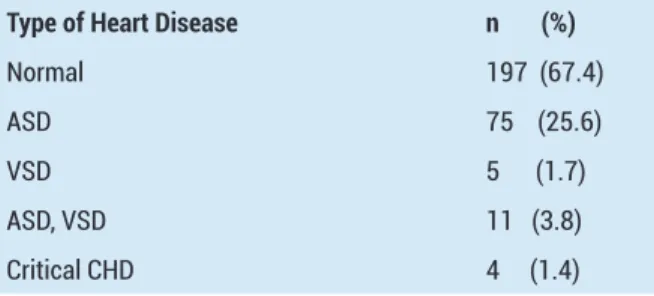
In Konya, annual birth rate is 35.000/a year. The number of annual births of the Educational Research Hospital is about 4,000 and the annual number of births of the University Hospital is around 1800. In our study, 3.440 infants were screened using pulse oximetry for congenital heart defects at two centers. 2003 (58.2%) of the cases were female and 1.437 (41.8%) were male. The average age at which screening hour of was 24.7±13.1 (4-100) (mean ± SD (min-max). 2580 infants (75%) passed the first test, 412 infants (12%) passed the second test and 156 (4.5%) passed the third test. According to the results, 292 (8.48%) infants were tested positive and had a failed test. 62% of the positive cases were female. Echocardiographic examination was performed in the positive cases. The prevalence of CHD was found to be 27.6 ‰. The prevalence of critical CHD was 1.16 per 1000 live births. The false-positive screen rate was found to be 197 (5.7%). The most common CHD among the cases was the secundum ASD (Table 1).
Table 1. Cardiac pathologies detected in patients with positive pulse oximetry screening
Type of Heart Disease n (%)
Normal 197 (67.4)
ASD 75 (25.6)
VSD 5 (1.7)
ASD, VSD 11 (3.8) Critical CHD 4 (1.4)
The sensitivity of the test was 99.9%, the specificity was 96.2%, the positive predictive value was 58.2% and the negative predictive value was 99.98%. Of the 4 cases with critical CHD, one had hypoplastic left heart syndrome, one had pulmonary atresia, one had aorticarch atresia (without hypoplastic left heart syndrome) and the other one had coarctation of the aorta. 55 (18.8%) of the cases with a positive screen were detected with a murmur during physical examination, indicating a heart problem.
If the cut-of value of the test had been taken as pre-ductal saturation of <90, 72 cases would have had a failed screen. The screening hour of the infants was 25.3±15.5 (4-100) (mean±SD (min-max). Echocardiographic examination of these 24 (33%) cases were unremarkable. ASD was detected in 46 cases (63.9%) and ASD with VSD detected in 2 cases (2.8%). 7 of the 9 cases with both pre-ductal and post-ductal saturation below 90 had ASD, and no pathology was detected in the other two.
There were 70 cases with post-ductal saturation below 90. A murmur was detected in ten of these cases (14.3%) during physical examination. The timing of screening was 23.02±11.5 (5-72) (mean ± SD (min-max). In the echocardiographic examination; there was no pathology in 21 (30%) cases while ASD was detected in 45 (64.3%), ASD with VSD was detected in 3 (4.3%) and ASD with bicuspid aortic valve in one case.
There was no statistically significant correlation between perfusion index and saturation of the same area (p>0.05). There was no statistically significant correlation between low perfusion index and the presence of CHD (p> 0.05). The recommended timing for screening test is after when the infant is 24 hours old. However, the screen came back positive in 125 cases that were screened sooner than 24 hours due to various reasons and no positive pathology was detected in 41 of them.
At the Training and Research Hospital, the cases were screened by a senior neonatal nurse after they received training on how to perform the screening test. At the University Hospital, a total of 2440 cases were screened by intern doctors under the supervision of a clinical nurse specialist. Of the 1000 infants screened at the Training and Research Hospital, 149 had referred screens and underwent cardiological examination. The results from both centers included no false positives which could affect the accuracy of the tests.
Cardiological examination of the infants who tested positive was performed on the same day. However, the examination of 6 cases was completed later on due to the unavailability of the cardiology team. Meanwhile, the cases were monitored and followed up in the hospital until the cardiologic examination. Multi-use probes were used for the screening with masimo pulse oximeter. Probes were replaced in every 50 cases. The total cost of probes was 2622 ₺ ($497.91). The cost of echocardiography for 292 babies was approximately 13181 ₺ ($2503). The overall cost revealed that the screening cost 4.5 ₺ ($0.85) for each case.
DISCUSSION
CHD is a common neonatal disease with a high morbidity and mortality rate if it is not diagnosed and intervened in an early period. As a result, several methods for antenatal diagnosis have been developed for early diagnosis of CHD over the years. Obstetricians and gynecologists have also gained more experience in parallel to the development. However, one study revealed that the antenatal diagnosis rate of CHD was 50% (9). Due to concerns about the diagnosis, routine physical examinations are performed even in asymptomatic infants during maternal hospitalization in many hospitals. The fact that congenital heart defects are asymptomatic in the early period and that only 25-50% of the findings are detected through physical examination indicate that babies are discharged from the hospital without diagnosis. It has been demonstrated that 43% of the infants discharged without diagnosis are then admitted to hospitals with symptoms of shock (6). For many years, various screening methods have been evaluated and proposed by many reputable Pediatric Cardiology Associations for early diagnosis and follow-up of infants with these defects. The most common type of these tests is pulse oximetry screening, which is used to measure the pre-ductal and post-ductal saturation of infants (8).
In our study, the prevalence of CHD was determined as 27.6 per 1000 live birth and the rate was very high compared to other studies (5,8). Only 1.16 per 1000 cases had critical CHD, which is consistent with the literature (1,2). Taksande et al. found that the incidence of CHD was 21.85 per 1000 live birth and they said that this was a high number (10). This finding was similar to our study and this may be related to the fact that the two hospitals in our study were reference hospitals.
Knowles et al. noted that the cost of pulse oximetry screening for 100.00 infants was equal to 480.000£ in England (11). In our study, if a total of 100,000 infants had been screened, the total cost of Pulse Oximetry screening would have been 459,389 ₺ (87081.19 USD, 67.756 pounds-76.564 Euro), which would be much more cost efficient. In the USA, Peterson et al. revealed in their study that the cost of each screening for CHD was 6.28 USD while, in return, it prevented approximately 20 infant deaths annually. In our study, the total cost of each infant screening is approximately 4.5 ₺ ($0.85) which appears to
be a quite cheap and effective screening compared to the other countries. The low cost is due to cheap work force in Turkey and the use of multi-use probes. Current studies have revealed that disposable saturation probes are responsible for high costs in pulse oximetry screening. In many countries, multi-use probes are used to avoid high costs along with a negligible risk for infection. In advanced and high-income countries, extensive metabolic and genetic screenings are performed on infants after delivery. Scientists in such countries stated that the costs of these tests are highly cost effective compared to metabolic and genetic screening tests (4).
The greatest concerns regarding pulse oximetry screening are reported to be negative and false-positive screens. The screening range and cut-off values were modified as the false negatives could have lead to catastrophic consequences. When the screening first started, only those with saturation below 90 were referred for the screening tests. However, it was later realized that this led to a lack of diagnosis so, as in our study, the test was also considered positive in such cases with saturation below 90 ranging between 90 and 95 along with 3% difference of saturation between high level extremities. In addition, the evaluation of the perfusion index was recommended for critical CHD screening with the highest level of sensitivity. If only those with a saturation level below 90 had been evaluated in our study, fewer patients would have been referred for screening and those 3 cases with critical heart defects would have been excluded. Consequently, keeping a wide range accounts for a higher cost and work force but it provides more accurate and correct screening. In this regard, the logical approach would be to reduce false positive results. In our study, the false positive rate was 12.5%. The false positive rates were found to be 0.17%, 0.10%, 0.42% and 0.89% in different international studies (2,5,12,13). In other studies with high false positive rates, such rates were associated with early timing of screening (4th hour) whereas the others resulted from additional pathologies such as sepsis, pneumonia and pulmonary hypertension (14,15).
In other studies with high false positive rates, such rates were associated with early timing of screening (4th hour), whereas the others resulted from additional pathologies such as sepsis, pneumonia and pulmonary hypertension (13,14).
There are some recommendations to reduce false positive such as: earlobe measurement in cases of acrocyanosis, re-screening of agitated infants after sedation and soothing, firm placement of the pulse oximeter probe, adjustment of ambient temperature and light since it can affect the accuracy (16). In addition, it was emphasized that presence of trained staff in the given field during the screening would be effective for reducing false positives. Another suggestion is to perform screening within 24-48 hours after the delivery of infant. In our study, the screenings were performed by experienced personnel but false positive rate was still high. This could be due to the fact that the nurses are also occupied with other tasks and
responsibilities. In our study, the mean timing of screening was 24.7±13.1 (4-100) (mean ± SD (min-max). The novel approach approves early discharge of mother and infant providing that they look healthy. Consequently,It gives families the right to demand for discharge in 6 hours in case of natural vaginal delivery,and 24 hours in cases of cesarean section. Pulse oximetry screening is performed earlier than 24 hours in such cases, which may result in higher false-positive screens. In our study, 125 of the patients who tested positive were screened within the first 24 hours or earlier and no cardiac pathology was detected in 41 of them. In their study, Bradshaw et al. stated that the mean screening hour was 42.4 hours (21-98 hours). In the same study, disposable probes were used and 6745 infants were screened, 9 of who tested negative were referred for further examination. Another distinctive feature of this study is that the perfusion index value was not used as an indicator for a positive screen (17). Therefore, it is expected to obtain a low rate of false positives in the relevant study.
CONCLUSION
CHD is common in our country and region. Due to recent events and developments in the region, the rate of infants with congenital anomalies is on the increase as a result of the high birth rates of the refugees. Regardless of highly advanced methods for antenatal diagnosis, infants are still discharged without diagnosis of critical congenital heart defects even after routine examination is performed. Thus, such infants then are hospitalized in a much more severe state and pass away. In order to prevent such cases, our study also suggest that it is recommended to perform pulse oximetry screening for the diagnosis of congenital heart defects, as per other relevant studies. The results revealed that the total cost is lower compared to the other countries and it can be feasibly implemented with no excessive costs or work force if carried out by experienced specialist.
Competing interests: The authors declare that they have no competing interest.
Financial Disclosure: There are no financial supports
Ethical approval: The study was approved by local ethics committee of the university (2019/02).
Murat Konak ORCID: 0000-0001-8728-4541 Beyza Ozcan ORCID: 0000-0002-2834-5823 Ahmet Sert ORCID: 0000-0002-1607-7569
Saime Sundus Uygun ORCID: 0000-0002-6694-8115 Hanifi Soylu ORCID: 0000-0003-0367-859X
REFERENCES
1. Hoffman JIE, Kaplan S. The incidence of congenital heart disease. J Am Coll Cardiol 2002;39:890-1900.
2. Plana MN, Zamora J, Suresh G, et al. Pulse oximetry screening for critical congenital heart defects. Cochrane Database Syst Rev 2018;3:CD011912.
3. Riede FT, Wörner C, Dähnert I, et al. Effectiveness of neonatal pulse oximetry screening for detection of critical congenital heart disease in daily clinical routine-results from a prospective multicenter study. Eur Journal Pediatr 2010;169:975-81.
4. Hoffman JIE. It is time for routine neonatal screening by pulse oximetry. Neonatology. 2011;99:1-9.
5. de-WahlGranelli A, Wennergren M, Sandberg K, et al. Impact of pulse oximetry screening on the detection of duct dependent congenital heart disease: a Swedish prospective screening study in 39821 newborns. BMJ 2009;338:a037. 6. Mellander M, Sunnegardh J. Failure to diagnose critical heart
malformations in newborns before discharge an increasing problem? Acta Paediatrica 2006;95:407-13.
7. Taksande A, Meshram R, Lohakare A, et al. An update work of pulse oximetry screening for detecting critical congenital heart disease in the newborn. Images Paediatr Cardiol 2017;19:12-8.
8. Mahle WT, Newburger JW, Matherne GP, et al. Role of pulse oximetry in examining newborns for congenital heart disease: a scientific statement from the AHA and AAP. Pediatrics 2009;124:823-36.
9. Sharland G. Fetal cardiac screening: why bother? Arch Dis Child Fetal Neonatal Ed 2010;95:F64-F8.
10. Taksande A, Lakhkar B, Gadekar A, et al. Accuracy of pulse oximetry screening for detecting critical congenital heart disease in the newborns in rural hospital of Central India. Images PaediatrCardiol 2013;15:5-10.
11. Knowles R, Griebsch I, Dezateux C, et al. Newborn screening
for congenital heart defects: a systematic review and costeffectiveness analysis. Health Technol Assess 2005;9:1-152.
12. Ewer AK, Middleton LJ, Furmston AT, et al. Pulse oximetry screening for congenital heart defect in newborn infants (PuseOx): a test accuracy study. Lancet 2011;378:785-94. 13. Riede FT, Dahnert I, Schneider P. Pulse oximetry screening
at 4 hours of age to detect critical congenital heart disease. Pediatrics 2009;123:542-3.
14. Sendelbach DM, Jackson GL, Lai SS, et al. Pulse oximetry screening at 4 hours of age to detect critical congenital heart defects. Pediatrics 2008;122:e815-e20.
15. Singh A, Rasiah SV, Ewer AK. The impact of routine predischarge pulse oximetry screening in a regional neonatal unit Arch Dis Child Fetal Neonatal Ed 2014;99:F297-F302. 16. Reich JD, Connolly B, Bradley G, et al. Reliability of a single
pulse oximetry reading as a screening test for congenital heart disease in otherwise asymptomatic newborn infants: the importance of human factors. PediatrCardiol 2008;29:371-6.
17. Bradshaw EA, Cuzzi S, Kiernan SC, et al. Feasibility of implementing pulse oximetry screening for congenital heart disease in a community hospital. J Perinatol 2012;32:710-5.

