Original Article
Evaluation of Neurological Examination, SEP Results, MRI Results, and
Lesion Levels in Patients Who Had Been Operated for Myelomeningocele
Gokhan Canaz1, Huseyin Canaz2, Ezgi T. Erdogan3, Ibrahim Alatas2, Erhan Emel1, Zeliha Matur41Department of
Neurosurgery, Bakirkoy Research and Training Hospital for Neurology, Neurosurgery and Psychiatry, Istanbul, Turkey, 2Department of
Neurosurgery, Florence Nightingale Hospital, Istanbul Bilim University, Istanbul, Turkey,
3Department of Physiology,
Istinye University, Istanbul, Turkey, and 4Department of
Neurology, Florence Nightingale Hospital, Istanbul Bilim University, Istanbul, Turkey
A
b st ra ctObjective: Myelomeningocele is the most severe and the most frequent form of spina bifida. Most of the myelomeningocele patients undergo operations in new-born age. In terms of life quality and rehabilitation, follow-up’s of these patients in the growth and development period after the operation is critical. In our study, our aim is to emphasize the correlation of SEP results with MRI results and clinical features of the myelomeningocele patients. Materials and Methods: In our study, we included 36 patients who had undergone myelomeningocele operation and have been followed-up in Istanbul Bilim University Florence Nightingale Hospital, Spina Bifida Research and Treatment Centre. Posterior tibial nerve SEP was performed on each patient and neurological examinations were done in the same session. Results were compared with clinical functional lesion levels, levels of fusion defect and ambulation levels. In order to evaluate SEP results, we used age-related reference values from Boor et al.’s study in 2008. Patients were grouped as normal, unilaterally prolonged, bilaterally prolonged, unilaterally lost, and bilaterally lost. Results: The correlations of posterior tibial nerve SEP results were significant with ambulation levels (r = 0.428, P < 0.01), clinical functional lesion levels (r = 0.477, P < 0.01) and fusion defect levels (r = −0.528 P < 0.05). The lumbar SEP results were only significantly correlated with functional lesion levels (r = 0.443 P < 0.05). Conclusions: Radiological studies are insufficient when evaluating the functionality of the central nervous system. To fully evaluate the functionality and watch the neurological development with accuracy, especially in operated patients, electrophysiological studies should be an indispensable part of myelomeningocele follow-ups.
Keywords: Ambulation, myelomeningocele, somatosensory evoked potentials,spina
bifida
I
ntroductionM
yelomeningocele (MMC) is the most severe and the most common form of spina bifida.[1,2] In patients with MMC,lower extremity deficits and paraplegia are extremely likely to be seen. Frequently, urinary anomalies also accompany this condition. In these patients’ lives, regular and accurate follow-ups are very important in order to prevent any worsening of their health status, to improve their quality of life and for their rehabilitation.
Received: 02-06-2019, Revised: 18-11-2019, Accepted: 24-05-2020, Published: 19-01-2021.
Access this article online
Quick Response Code:
Website:
www.pediatricneurosciences.com
DOI: 10.4103/jpn.JPN_77_19
When we consider that 99% of the MMC patients are children, the follow-ups for their neurological developments from infant age are very important. Early detection of any neurological deterioration allows us to diagnose and intervene as soon as possible. It is not an easy task to follow-up infants or children accurately with neurological or radiological examinations, more
Address for correspondence: Dr. Gokhan Canaz, Istanbul Bakirkoy Prof. Dr. Mazhar Osman Psychiatric Training and Research Hospital, Department of Neurosurgery, 34087 Bakirkoy, Istanbul, Turkey. E-mail: [email protected]
This is an open access journal, and articles are distributed under the terms of the Creative Commons Attribution-NonCommercial-ShareAlike 4.0 License, which allows others to remix, tweak, and build upon the work non-commercially, as long as appropriate credit is given and the new creations are licensed under the identical terms.
For reprints contact: [email protected]
How to cite this article: Canaz G, Canaz H, Erdogan ET, Alatas I, Emel E, Matur Z. Evaluation of neurological examination, SEP results, MRI results, and lesion levels in patients who had been operated for myelomeningocele. J Pediatr Neurosci 2020;15:393-401.
Canaz, et al.: Neurophysiological assessment of myelomeningocele
advanced examinations are needed. Surveys and clinical classifications are necessary for patient follow-ups but the detailed evaluation of the development and the functionality of the nervous system and detection of the deteriorations before they become apparent in the clinic is possible with electrophysiological tests. The most common used electrophysiological tests in spinal pathologies are motor evoked potentials (MEP) and somatosensory evoked potentials (SEP). Early detection of pathologies is important in terms of conditions like re-tethering and scoliosis which requires surgical interventions and, therefore, electrophysiological evaluations like SEP, MEP, and urodynamic studies are important in follow-ups of patients with MMC.[3]
Ambulation is a key factor for rehabilitation and determination of the quality of life of a patient who was operated for MMC.[4] Ability to move is essential for the
child to interact with people, to participate in community, and, in terms of personality, to develop self-confidence. In different age groups, we have different ambulation aims, because of that, a suitable ambulation classification is needed. If the results of the examinations, which will be used to evaluate the neurological outcome of patients, are in a correlation with patients’ ambulatory status, it increases the value of those examinations.
Our aim in this study is to compare the posterior tibial SEP results with functional lesion levels, fusion defects levels, and ambulation levels of patients, who had been operated for MMC in early life, and to emphasize the value of SEP as an accurate method for the evaluation of the neurological condition of lower extremities.
M
aterials andM
ethodsStudy population
This study was conducted in Istanbul Bilim University, Florence Nightingale Hospital, Spina Bifida Research and Treatment Center with the ethical approval of University of Health Sciences, Bakirkoy Research and Training Hospital for Neurology, Neurosurgery and Psychiatry Ethics Committee (Protocol number: 2018/48). Thirty-six patients, who had been operated for MMC closure, have been included in the study. Posterior tibial nerve somatosensory evoked potentials of 36 patients had been recorded during their follow-ups in 2014–2018 and neurological examinations were done in the same session. Results were compared with clinical functional lesion levels, levels of fusion defects, and ambulation levels. SEP and spinal MRI studies were performed in all patients. Patients below 3 years old were excluded from this study due to unreliable SEP results at this age of children. Written informed consent
was obtained from legal guardians of all individual participants included in the study.
The average age of the patients was 8.2 years (range: 3–17) and median of the operation age was 0.5 weeks (avg: 9.86, range: 0-208w). Of the 36 patients, 20 had been operated in the first week of life, one had been operated at the age of 2 years and one had been operated at the age of 4 years [Table 1].
Study design
Tibial somatosensory evoked potentials
Recordings were conducted using 5-channel system EMG-UP device (Viking® on Nicolet® EDX; Natus Neurology Incorporated 3150 Pleasant View Road Middleton, WI 53562).
SEP responses were elicited by stimulating both tibial nerves, respectively, with submaximal intensity, enough to produce a small thumb twitch (1–15 mA) using skin electrodes at the ankle. Stimulation frequency was 1–2 Hz, stimulus duration was 1 ms. The filter band-pass was: 0.3–1000 Hz and the sensitivity was 5–10 µV. Analysis time was 100 ms. Recordings were made in three channels:
1st Channel: From popliteal fossa, with saddle-shaped skin electrodes over tibial nerve (active-reference) 2nd Channel: T12 or L1 vertebrae (T12/L1; active)— Anterior superior iliac spina (ASIS; reference)
3rd Channel: Central midline (Cz; active)—Paracentral of the opposite side of the stimulation (C3/4; reference) Responses were averaged until a smooth base-line was achieved and at least two averaged recordings were superimposed. The latencies (ms) in N7, N22, P37, N45, interpeak latencies (µV) of P37-N45, and the central conduction time (N22-P37 latency difference; ms) were calculated and evaluated [Figure 1].
Age-specific referencevaluesfromBooretal.’s[5]normative
data were used to evaluate SEP results. Longer latencies of P40 beyond three standard deviations from the mean of the control groups were named as “prolonged.” Cases which P40 value could not be received were named as “abnormal.” SEP results were grouped as;
• Normal,
• Unilaterally prolonged, • Bilaterally prolonged,
Table 1: Study population
Age (range) 8.22 (3–17)
Sex Male: 19
Female: 17
Canaz, et al.: Neurophysiological assessment of myelomeningocele
Figure 1: Stimulation and record zones with an example of the recorded potentials
• Unilaterally lost and • Bilaterally lost.
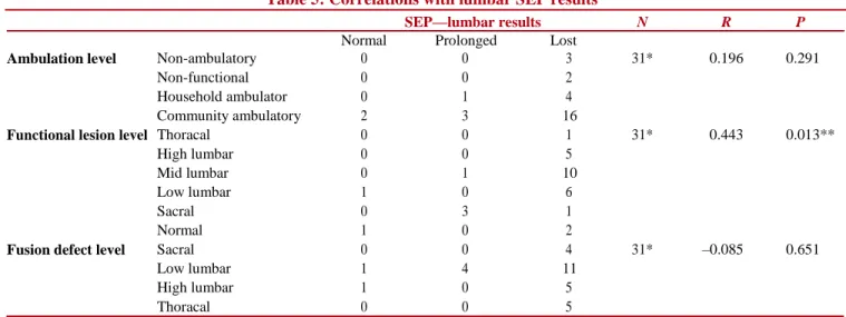
Lomber results were named as “normal,” “lost,” “artefact,” and “prolonged” according to reference values [Figure 2].
Ambulation levels
We evaluated the ambulation levels according to the study of Hoffer et al.[6] as community ambulators,
household ambulators, non-functional ambulators, and non-ambulators.
1. Community ambulatories: Patients who were able to walk indoors and outdoors most of the time. They may use a wheelchair for long commutes.
2. Household ambulatories: Patients who walk only indoors and use the wheelchair for any other activities in the community.
3. Non-functional ambulatories: Patients who use the wheelchair for all activities indoors and outdoors. They can only walk in therapy sessions.
4. Non-ambulatories: Wheelchair dependent patients but most of the time they can transfer themselves between the bed and the chair.
Clinical functional lesion levels
Clinical functional lesion levels were classified according to neurologic deficits level with numbers from 0 to 5. 0. Normal, no deficit.
1. Sacral: Foot plantar flexion.
2. Lower lumbar: foot dorsiflexion. 3. Mid lumbar: Knee extension. 4. Upper lumbar: Hip flexion. 5. Thoracic: Flask lower extremities.
Fusion defect levels
Fusion defect levels were assigned according to operation notes, MRI results, and direct radiographs. They were evaluated in four groups, according to the beginning of the posterior fusion defects from cranial to caudally. 1. Thoracic: Thoracic levels.
2. Upper Lumbar: L1 and L2 levels. 3. Lower Lumbar: L3, L4 and L5 levels. 4. Sacral: S1 and distal levels.
Statistical evaluations
Statistical evaluations were made by using IBM SPSS Statistic version 22 (USA) program. Correlations between SEP results and ambulation levels, clinical functional lesion levels and fusion defects levels were evaluated with Spearman test. Comparison of MR findings with ambulation levels and SEP results were made using the Fisher test.
R
esultsAmbulation levels—SEP results
The correlation between ambulation levels and SEP results of patients were significant according to Spearman analyse (r: 0.460, P < 0.01) [Table 2].
Canaz, et al.: Neurophysiological assessment of myelomeningocele
Figure 2: (A) 6-year-old male patient. Latencies of cortical potentials are longer than normal values on left (right: 37.6ms, left: 55.4 ms) (Group 2). (B) 7-year-old male patient. Latencies of cortical potentials are bilaterally prolonged (right: 60.1ms, left: 58.6ms) (Group 3). (C) 14-year-old female patient. The cortical potentials are lost on left, and lumbar potentials are bilaterally lost. On the right, the amplitude of the cortical potentials are low (1 µV) and latency is slightly prolonged (43 ms) (Group 4)
Clinical functional lesion levels—SEP results The correlation between clinical functional lesion levels and SEP results of patients were highly significant according to Spearman analyse (r: 0.477, P < 0.01) [Table 3].
Fusion defect levels—SEP results
The correlation between fusion defect levels and SEP results of patients were highly significant according to Spearman analyse (r: -0.528 P < 0.05) [Table 4].
Canaz, et al.: Neurophysiological assessment of myelomeningocele
Table 2: The correlation between ambulation levels and SEP results
SEP result
Ambulation Normal Unilat. Bilat. Unilat. lost Bilat. lost N R P
prolonged prolonged level 4 36 0.46 0.005* Non-ambulatory Non-functional 1 1 Household ambulator 2 3 Community ambulatory 9 2 2 5 6
*Correlation is significant at the 0.05 level according to Spearman test (2-tailed)
Table 3: The correlation between clinical functional lesion levels and SEP results
SEP result
Functional lesion Normal Unilat. Bilat. Unilat. lost Bilat. lost N R P
prolonged prolonged level 2 36 0.477 0.003* Thoracal High lumbar 1 2 3 Mid lumbar 2 1 1 8 Low lumbar 2 2 1 1 Sacral 2 1 1 Normal 2 1 1
*Correlation is significant at the 0.01 level according to Spearman test (2-tailed)
Table 4: The correlation between fusion defect levels and SEP results
SEP result
Fusion defect Normal Unilat. Bilat. Unilat. lost Bilat. lost N R P
prolonged prolonged level 2 1 36 –0.528 0.002* Sacral Low lumbar 6 1 2 3 4 High lumbar 1 2 3 Thoracal 1 6
*Correlation is significant at the 0.01 level according to Spearman test (2-tailed) Lumbar SEP results
There was a significant correlation between only lumbar SEP results and functional lesion levels, and it was in medium level (r: 0.443 P < 0.05) [Table 5].
MRI findings
In the MRI scans of the patients, which were performed in a close time with SEP tests, hydrocephalus (HC), Chiari anomaly (CH), tethered cord (TC), syringomyelia (SM), and diastomyelia (DM) were investigated [Table 6]. TC was spotted in 20/36 patients (%55.5). Among the 20 patients, 13 were community ambulatory. Only four patients were non-ambulatory.
Comparisons between TC presence with ambulation levels, functional lesion levels, and SEP scores revealed no significant relationship [Table 7].
D
iscussionOur study revealed that the cortical responses of posterior tibial nerve SEP are significantly correlated with ambulation levels, functional lesion levels and fusion defect levels. On the other hand, we found that
MRI findings of the patients have no relation with patients’ neurologic status.
In spina bifida patients, one of our major goals is to preserve ambulation. Life quality of MMC patients is highly related to their ambulation status.[7-9] In addition to
psychological effects, ambulation takes a big role in conditions with bone metabolism, such as osteoporosis, and in terms of cardiopulmonary capacity. In pediatric patients, we should consider mobility in regards to development. For a baby, the “community” concept is the house. As the child grows, first the outside and then the school become a part of this concept. Being “community ambulatory” and joining public activities is important for both, child’s growth and personal development. An instrument used to assess the neurological condition of a spina bifida patient is expected to be highly correlated with ambulatory levels. Hoffer et al.’s ambulation classification for patients with myelomeningocele, which we used in our study, had been widely accepted and used in many studies including researches about spinal injury.[6,10,11] One of the importance of this classification
Canaz, et al.: Neurophysiological assessment of myelomeningocele
Table 5: Correlations with lumbar SEP results
SEP—lumbar results N R P
Normal Prolonged Lost
Ambulation level Non-ambulatory 0 0 3 31* 0.196 0.291
Non-functional 0 0 2
Household ambulator 0 1 4
Community ambulatory 2 3 16
Functional lesion level Thoracal 0 0 1 31* 0.443 0.013**
High lumbar 0 0 5
Mid lumbar 0 1 10
Low lumbar 1 0 6
Sacral 0 3 1
Normal 1 0 2
Fusion defect level Sacral 0 0 4 31* –0.085 0.651
Low lumbar 1 4 11
High lumbar 1 0 5
Thoracal 0 0 5
*In 5 patient, lumbar responses could not be received due to artefacts **Correlation is significant at the 0.05 level according to Spearman test (2-tailed)
is that patients are not classified according to their orthoses or any other helping devices. For example, a patient with a wheelchair who can join public activities is in the same group with a patient who does not use any orthosis. The purpose is to evaluate the ability to join the community, independent from the method.
The evaluation of SEP results in the pediatric patient population has been a topic of debate for many years. The height of the patient is an important variable in SEP interpretation and increase in children’s height with the maturation of neural tissues are making it difficult to determine age-related normal values. In some studies, researchers had tried to formulate this proportion, but the general opinion is that the increase in the height and neural maturation balances each other’s effects.[5] Some clinics use
the values they obtained during their own practice to determine normal values. In our study, we used Boor et al.’s[5,12] recent study, including 89 children and 19 adults
SEP results after posterior tibial nerve stimulation. In the recent study, they updated their reference values, which they had reported in 1998, and classified normal values more detailed according to age. Patient cooperation is important in performing SEP test and this is another difficulty with the pediatric population in terms of the application and assessment of the test. During the application, between stimulus and measurement episodes, maintaining the minimal muscle activity in children are up to the skill of the technician or doctor. Although it is known that sedation effects the test results, there are still studies reporting the use of sedation for SEP measurements in children.[13,14] In our
study, all SEP studies were performed by an expert neurologist with no sedation used. The difficulty of the application of SEP
in the pediatric population is a factor that prevents the widespread use of the SEP tests.
MR imaging is a gold standard examination in spina bifida, just as in all spinal pathologies. MRI provides us with important information about the position of conus, anatomic integrity of posterior components of the vertebra, sac ingredients and roots’ relationship with neighbouring tissues. These make MRI vital especially for operation planning.[15] However, in operated patients,
due to fibrosis and granulation in operation space, it is hard to evaluate anatomical structures and functional integrity of neural tissues.[16,17] One of three patients who
was operated for MMC needs a reoperation for TC release in the following period.[3,18,19] This ratio is
changing between 17 and 60% in the literature. The main reason for this difference is the indecision about the indications. As we mentioned earlier, MRI is not a reliable method to follow these patients and does not provide sufficient findings to decide the operation. TC presents or not, in case of clinical deterioration, it is hard to decide indications or plan the possible new operation with the help of only MRI. In a study by Just et al.,[20] 114
patients, who had been operated for MMC, were evaluated and it is reported that 89% of the patients had MRI findings indicating TC syndrome. Tamaki et al.[21]
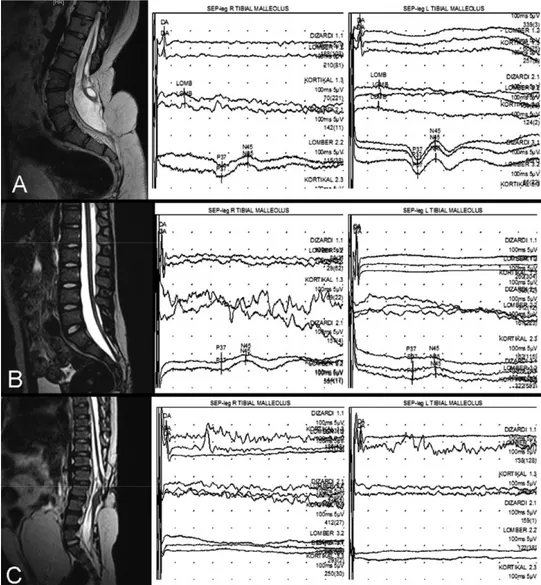
examined 29 patients’ MRI results, who had been previously operated for MMC. All 29 patients’ MRI results were reported to be compatible with TC, but only in 8 patients (27.5%), clinical symptoms were reported. In our study group too there were patients with normal examinations and SEP results despite pathologic MRI findings [Figure 3]. On the other hand, only four patients who were non-ambulatory and had bilaterally lost SEP responses have TC findings in their MRIs [Figure 3].
Canaz, et al.: Neurophysiological assessment of myelomeningocele
Table 6: Patients’ MRI findings, SEP results and ambulation levels
Patients Anomalies detected on MRI SEP results Ambulation levels
1 HC 4 1 2 SM 5 4 3 SM+TC 4 1 4 CH 5 1 5 SM+TC 5 2 6 None 5 1 7 CH+SM+TC 5 4 8 HC 5 2 9 HC 5 3 10 None 1 1 11 SM 1 2 12 SM+TC+DM 5 4 13 SM+DM+TC 1 1 14 SM+TC+CH+HC 3 1 15 SM+TC 1 1 16 SM+DM 5 1 17 CH+SM+DM+TC 4 3 18 TC+SM+HC 5 4 19 CH+SM+TC 2 1 20 SM+TC 1 1 21 SM+TC 1 1 22 None 1 1 23 SM+DM+TC 4 1 24 SM+TC+HC 5 4 25 HC 3 1 26 SM+LM+TC+HC 4 1 27 None 5 2 28 TC 1 1 29 SM+TC 4 1 30 None 1 1 31 None 1 1 32 TC 5 1 33 HC 5 1 34 SM+TC+LM 2 1 35 None 1 2 36 SM+TC 5 1
HC = hydrocephalus, CH = Chiari anomaly, TC = tethered cord, SM = syringomyelia, DM = diastomyelia, LM = lipoma
Table 7: Comparison of tethered cord presence with SEP results, ambulation levels and functional lesion levels, using Fisher’s test
SEP results N P
Tethered Normal Unilat. prolonged Bilat. prolonged Unilat. lost Bilat. lost
+ 5 2 1 5 7 36 0.710
cord
– 6 0 1 1 8
Ambulation levels
Community ambulatory Household Non-functional Non-ambulatory
ambulatory ambulatory
+ 14 1 1 4 36 0.213
– 10 4 1 1
Functional lesion levels
Normal Sacral Low lumbar Mid lumbar High lumbar Thoracal
+ 2 3 5 5 3 2 36 0.748
Canaz, et al.: Neurophysiological assessment of myelomeningocele
Figure 3: (A) 16-year-old female patient. She was operated for MMC closure in the first 48h of life. She has normal posterior tibial nerve SEP results and no lower extremity deficits. In MRI, TC findings and SM cyst are present. (B) 5-year-old female patient. She was operated for sacral MMC on the eighth day of life. She was community ambulatory and had no lower extremity deficit. Posterior tibial nerve SEP results are normal. In MRI, evident low conus and TC are present. (C) 7-year-old male patient. He was operated for high lumbar MMC on the 40th day of life. He is non-ambulatory and posterior tibial nerve SEP is bilaterally lost. In MRI, distinct tethering is visible on L1-2-3 levels. The patient has urinary incontinence, and increased bladder volume is visible
Neurophysiological tests take an important place in spina bifida and TC operations. The benefits of using intraoperative neurophysiologic monitoring (IONM) is widely accepted and were reported in plenty of studies. [22-24] In similar spinal pathologies and spina bifida patients,
there are studies suggesting the advantages of using neurophysiological tests in preop and postop follow-ups.[25-27] There are also important reports about
urodynamic studies which include electrophysiological methods. In separate studies of Alatas et al.[3] and Tarcan et al.,[28] preop and postop urodynamic study results of
newborns, operated for MMC, were evaluated and significant healing (75–66%) was reported in urodynamic parameters. In the light of these studies, it has been suggested that the urodynamic
parameters may help to detect the deterioration in autonomic and motor functions.
The main limitations of our study are the age range and number of patients, as it is in many similar studies. Different pathologic MRI findings like hydrocephalus, TC, SM, and lipomas may cause our patient population to be too heterogeneous, but these anomalies have a high concomitance with MMC. Because of this, we did not add them to criteria. The presence of TC on MRI was decided according to the reports of the neuroradiology department.[29] In the literature, there are studies which
are evaluating each anomaly’s relation to ambulation.[4]
The next step of this study should be the evaluation of the patients who were operated for MMC closure with regular SEPs.
Canaz, et al.: Neurophysiological assessment of myelomeningocele
C
onclusionsRadiological studies are insufficient when evaluating the functionality of the central nervous system. The full evaluation of functionality, especially in operated patients, is possible with electrophysiological studies. Electrophysiological studies should be accepted as the gold standard especially in the spina bifida patients group, so that neurological development as well as accompanying pathologies or treatment results can be followed closely. As our study supports, SEP is a reliable tool for the follow-ups of MMC patients. For results with higher levels of evidence, wider evaluations with multicenter studies and larger patient groups should be done.
Acknowledgements
We would like to thank Dr. Caglar CURA for illustrations.
Financial support and sponsorship Nil.
Conflicts of interest
There are no conflicts of interest.
R
eferences1. Copp AJ, Adzick NS, Chitty LS, Fletcher JM, Holmbeck GN,
Shaw GM. Spina bifida. Nat Rev Dis Primers 2015;1:15007.
2. Tunçbilek E, Boduroğlu K, Alikaşifoğlu M. Neural tube defects
in turkey: prevalence, distribution and risk factors. Turk J Pediatr 1999;41:299-305.
3. Alatas I, Demirci H, Canaz H, Akdemir O, Baydin S, Ozel K.
The role of urodynamic studies in the diagnosis and treatment of patients with spina bifida. Asian J Neurosurg 2015;10:83-6.
4. Dicianno BE, Karmarkar A, Houtrow A, Crytzer TM, Cushanick
KM, McCoy A, et al. Factors associated with mobility outcomes in a national spina bifida patient registry. Am J Phys Med Rehabil 2015;94:1015-25.
5. Boor R, Li L, Goebel B, Reitter B. Subcortical somatosensory
evoked potentials after posterior tibial nerve stimulation in children. Brain Dev 2008;30:493-8.
6. Hoffer MM, Feiwell E, Perry R, Perry J, Bonnett C. Functional
ambulation in patients with myelomeningocele. J Bone Joint Surg Am 1973;55:137-48.
7. Bartonek A, Saraste H, Danielsson A. Health-related quality of
life and ambulation in children with myelomeningocele in a swedish population. Acta Paediatr 2012;101:953-6.
8. Danielsson AJ, Bartonek A, Levey E, McHale K, Sponseller P,
Saraste H. Associations between orthopaedic findings, ambulation
and health-related quality of life in children with
myelomeningocele. J Child Orthop 2008;2:45-54.
9. Rocque BG, Bishop ER, Scogin MA, Hopson BD,
Arynchyna AA, Boddiford CJ, et al. Assessing health-related quality of life in children with spina bifida. J Neurosurg Pediatr 2015;15:144-9.
10. Vogel LC, Mendoza MM, Schottler JC, Chlan KM, Anderson CJ. Ambulation in children and youth with spinal cord injuries. J Spinal Cord Med 2007;3(Suppl 1):S158-64.
11. Lonstein JE, Akbarnia A. Operative treatment of spinal deformities in patients with cerebral palsy or mental retardation.
An analysis of one hundred and seven cases. J Bone Joint Surg Am 1983;65:43-55.
12. Boor R, Goebel B, Doepp M, Taylor MJ. Somatosensory evoked potentials after posterior tibial nerve stimulation—normative data in children. Eur J Paediatr Neurol 1998;2:145-52.
13. Fagan ER, Taylor MJ, Logan WJ. Somatosensory evoked potentials: part I. A review of neural generators and special considerations in pediatrics. Pediatr Neurol 1987;3:189-96. 14. Hashimoto T, Tayama M, Hiura K, Endo S, Fukuda K, Tamura Y,
et al. Short latency somatosensory evoked potential in children.
Brain Dev 1983;5:390-6.
15. Mehta DV. Magnetic resonance imaging in paediatric spinal dysraphism with comparative usefulness of various magnetic resonance sequences. J Clin Diagn Res 2017;11:TC17-22. 16. Halevi PD, Udayakumaran S, Ben-Sira L, Constantini S. The
value of postoperative MR in tethered cord: a review of 140 cases. Childs Nerv Syst 2011;27:2159-62.
17. Thakkar RS, Malloy JP IV, Thakkar SC, Carrino JA, Khanna AJ. Imaging the postoperative spine. Radiol Clin North Am 2012;50:731-47.
18. Kellogg R, Lee P, Deibert CP, Tempel Z, Zwagerman NT,
Bonfield CM, et al. Twenty years’ experience with
myelomeningocele management at a single institution: lessons learned. J Neurosurg Pediatr 2018:1-5.
19. Yamada S, Zinke DE, Sanders D. Pathophysiology of “tethered cord syndrome”. J Neurosurg 1981;54:494-503.
20. Just M, Schwarz M, Ludwig B, Ermert J, Thelen M. Cerebral and spinal MR-findings in patients with postrepair myelomeningocele. Pediatr Radiol 1990;20:262-6.
21. Tamaki N, Shirataki K, Kojima N, Shouse Y, Matsumoto S. Tethered cord syndrome of delayed onset following repair of myelomeningocele. J Neurosurg 1988;69:393-8.
22. Sala F, Bricolo A, Faccioli F, Lanteri P, Gerosa M. Surgery for intramedullary spinal cord tumors: the role of intraoperative (neurophysiological) monitoring. Eur Spine J 2007;16(Suppl 2):S130-9.
23. Cho SK, Lenke LG, Bolon SM, Kang MM, Zebala LP, Pahys JM,
et al. Progressive myelopathy patients who lack spinal cord
monitoring data have the highest rate of spinal cord deficits following posterior vertebral column resection surgery. Spine Deform 2015;3:352-9.
24. Costa P, Bruno A, Bonzanino M, Massaro F, Caruso L, Vincenzo I, et al. Somatosensory- and motor-evoked potential monitoring during spine and spinal cord surgery. Spinal Cord 2007;45:86-91. 25. Cheng JC, Guo X, Sher AH, Chan YL, Metreweli C. Correlation
between curve severity, somatosensory evoked potentials, and magnetic resonance imaging in adolescent idiopathic scoliosis. Spine (Phila Pa 1976) 1999;24:1679-84.
26. Kale SS, Mahapatra AK. The role of somatosensory evoked potentials in spinal dysraphism—do they have a prognostic significance? Childs Nerv Syst 1998;14:328-31; discussion 332. 27. Thomas DT, Yener S, Kalyoncu A, Uluc K, Bayri Y, Dagcinar A,
et al. Somatosensory evoked potentials as a screening tool for
diagnosis of spinal pathologies in children with treatment refractory overactive bladder. Childs Nerv Syst 2017;33:1327-33. 28. Tarcan T, Onol FF, Ilker Y, Simsek F, Simek F, Ozek M. Does
surgical release of secondary spinal cord tethering improve the
prognosis of neurogenic bladder in children with
myelomeningocele? J Urol 2006;176:1601-6; discussion 1606. 29. Raghavan N, Barkovich AJ, Edwards M, Norman D. MR imaging
in the tethered spinal cord syndrome. AJR Am J Roentgenol 1989;152:843-52.