The Treatment Method and Results of Percutaneous Pinning
and Dynamic External Fixator Application for Unstable Distal
Radius Fractures
Address for correspondence: Dr. Adnan Kara. Department of Orthopedics and Traumatology, Medipol University Faculty of Medicine, Istanbul, Turkey Phone: +90 505 395 14 75 E-mail: [email protected]
Submitted Date: April 11, 2018 Accepted Date: April 20, 2018 Available Online Date: September 27, 2018 ©Copyright 2018 by The Medical Bulletin of Sisli Etfal Hospital - Available online at www.sislietfaltip.org This is an open access article under the CC BY-NC-ND license (http://creativecommons.org/licenses/by-nc/4.0/).
D
istal radius fracture is the most common type offrac-ture among all bone fracfrac-tures in the whole body.[1] A standard treatment method has not been established al-though various surgical intervention methods and detec-tion materials in the treatment of unstable fractures have been described. Surgical alternatives include grafting, arthroscopy-assisted reduction and stabilization in addi-tion to percutaneous pinning or external fixator
applica-tion after closed reducapplica-tion, pinning after limited open or open reduction, internal fixation, and combinations of all these interventions.[2, 3]
Radius distal tip fracture is the most common type of frac-ture in whole body bone fracfrac-tures.[1] Though various sur-gical intervention methods and fixation materials in the treatment of unstable fractures have been described, a standard treatment method has not been established yet. Objective: The aim of the present study was to present the results of patients with unstable distal radius fracture treated with
closed reduction and percutaneous fixation followed by application of the Pennig dynamic wrist fixator to allow early wrist motion.
Methods: Twenty-five patients diagnosed with distal radius fracture and treated with closed reduction and percutaneous fixation
followed by application of a dynamic wrist fixator were included in the study. There were 15 (60%) male and 10 (40%) female patients. The mean age of the patients was 47.32 (20–76) years. The mean period between initial trauma and operation was 8.52 (1–23) days. All patients were allowed active shoulder, elbow, and finger exercises immediately after surgery.
Results: Radiological evaluation was performed according to the criteria described by Sarmiento and modified by Lidström.
Re-sults were excellent in 12 (46.15%), good in 11 (42.30%), and fair in 3 (11.55%) patients. No patient had poor result. Functional scores were assessed according to the Gartland–Werley classification and modified by Sarmiento. Results were excellent in 14 (56%), good in 8 (32%), and moderate in 3 (12%) patients.
Conclusion: Use of the Pennig dynamic wrist fixator in the treatment of unstable distal radius fractures has advantages, such
as ease of use, minimal surgical trauma, allowing early rehabilitation, and early return to daily activities as well as increased anatomical and functional results.
Keywords: Distal radius fracture; dynamic fixator; unstable fracture.
Please cite this article as ”Kara A, Ertürer E, Seçkin F, Akman Ş, Öztürk İ. The Treatment Method and Results of Percutaneous Pinning and Dynamic External Fixator Application for Unstable Distal Radius Fractures. Med Bull Sisli Etfal Hosp 2018;52(3):173–178”.
Abstract
DOI: 10.14744/SEMB.2018.07078
Med Bull Sisli Etfal Hosp 2018;52(3):173–178
Research Article
Adnan Kara,
1Erden Ertürer,
2Faik Seçkin,
3Şenol Akman,
3İrfan Öztürk
41Department of Orthopedics and Traumatology, Medipol University Faculty of Medicine, Istanbul, Turkey 2Department of Orthopedics and Traumatology, Istinye University Faculty of Medicine, Istanbul, Turkey 3Department of Orthopedics and Traumatology, Bilim University Faculty of Medicine, Istanbul, Turkey 4Department of Orthopedics and Traumatology, Istanbul University Faculty of Medicine, Istanbul, Turkey
Surgical treatment alternatives include percutaneous pin-ning or external fixator application after closed reduction, pinning after limited open or open reduction, and internal fixation. In addition to these interventions, combinations of all these interventions and arthroscopy-assisted reduc-tion and stabilizareduc-tion may be enumerated.[2, 3]
Whatever method is chosen, the basic prerequisite is to bring the radial length, radial inclination, and palmar incli-nation to the most appropriate level so as to allow anatom-ical repair of the distal radial articular surface.[4]
In the present study, patients with distal radius fractures treated with the Pennig-type dynamic external fixator (Orthofix Srl, Italy) were followed up, and the results of treatment were evaluated radiologically, clinically, and functionally.
Methods
In the present study, 26 unstable distal radius fractures were evaluated in 25 patients surgically treated with a dynamic wrist external fixator. Fractures were evaluated according to the AO,[5] Frykman,[6] and Gustilo–Anderson open frac-ture classification.[7] The radiological and anatomical results of the cases were evaluated according to the radiological criteria developed by Lidström and modified by Sarmiento. Statistical analysis was performed between pre- and post-reduction radial angulation, loss in radial height and pal-mar angulation measurements, and these measurements after removal of a fixator following achievement of dy-namization. The measurements were compared with those of the robust side.
The functional results were evaluated by the scoring sys-tem developed by Gartland–Werley[9] and modified by Sarmiento. Complications were recorded. Trauma etiolo-gies of the patients were evaluated. Pediatric patients and open wrist fractures with vascular lesions were excluded from the study.
Ethics approval was obtained (decision no. 10840098/159). Radial angulation, radial height, and palmar angulation were evaluated using the Student's t-test. Based on the two-way p-value, etiologies for trauma and their distribu-tion according to age groups were expressed as percent-ages (%).
Surgical Technique
Patients were brought to the operating room and placed in the supine position. The fracture was reduced after ap-plication of general or axillary block anesthesia. After the desired reduction was achieved, two K-wires crossing each other were sent proximally from the radial styloid and dor-somedial aspect of the radius under fluoroscopic control. In
addition, to prevent radial shortening, a parallel K-wire was sent to the radioulnar joint.
During application of a fixator, the distal Schanz pins were placed first. A 2 cm long dorsoradial incision site near the second metacarpal base was determined for the placement of the distal Schanz pins. Then, a 2 cm incision site at the dorsolateral aspect of the 10 cm proximal part of the radial styloid was determined for the placement of the proximal Schanz pins.
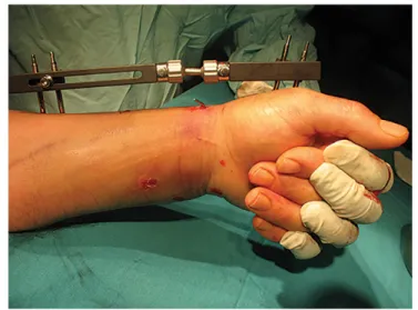
After placement of the Schanz pins, the mobile joint of the external fixators was adapted statically at 10°–20° flexion and 15° ulnar deviation so as to place them on the proxi-mal pole of the head of the capitolunate joint, which is the center of the rotational movement of the wrist. Distraction was then performed with the aid of the traction unit distal to the fixator. When appropriate reduction and distraction were achieved, the incision sites were sutured and closed with a dressing (Fig. 1).
Results
The median follow-up period of the patients was 20.3 (8–60) months. The median age of the patients was 47.32 (20–76) years. The median ages of the female (n=10, 40%) and male (n=15, 60%) patients were 41.26 (20–75) and 56.4 (44–76) years, respectively. Of the patients, 11 (44%) had right, 13 (52%) had left, and 1 (4%) had fractures on both wrists.
In the etiological evaluation, fractures occurred due to fall-ing on the floor at home (n=5, 20%), on flat ground outside the home (n=9, 36%), and from height (n=6, 24%); traffic accidents (n=4, 16%); and direct trauma (n=1, 4%) (Table 1). Five (19.23%) patients had open fractures. According to the Gustilo–Anderson classification, fractures were
fied as type I (n=1, 3.85%), II (n=3, 11.53%), and IIIb (n=1, 3.85%) open fractures. According to the Frykman classifi-cation, type I–II (n=3.11, 53%), III–IV (n=4, 15.38), V–VI (n=9, 34.61%), and VII–VIII (n=10, 38.46%) fractures were seen in respective number of patients. According to the AO/ASIF classification, type C (n=15, 57.69%), A2–3 (n=9, 34.61%), B1 (n=1, 3.84%), and B3 (n=1, 3.84) displaced fractures were detected. Nine (34.61%) patients had A2–3, 1 (3.84%) pa-tient had type B1, and 1 (3.84%) papa-tient had type B3 dis-placed fractures.
Patients were operated within an average of 8.52 (1–23) days after hospitalization. Prior to the application of an external fixator, fractures were fixed using closed reduc-tion and percutaneous fixareduc-tion with K-wires (n=24, 92.3%) and limited open reduction and fixation with K-wires (n=2, 7.7%) in an indicated number of patients. None of the frac-tures underwent grephonage.
A double articulated dynamic wrist fixator (Pennig external fixator; Orthofix Srl, Italy) was used for 24 out of 25 patients,
and a single articulated dynamic wrist fixator (Stableloc external fixator; Acumed, USA) was used for the remaining patient. Patients were discharged on an average of 3.48 (1–12) days postoperatively. An external fixator was dy-namized by loosening its articulated part at the center of the capitolunate joint.
After a median period of 6.8 (5–8) weeks, a radiologically adequate union was observed, and the fixators were re-moved with their fixation wires (Fig. 2). Statistically signif-icant improvements in radial length (p<0.0001), radial an-gulation (p<0.001), and palmar inclination (p<0.001) of our patients were detected during treatment and post-fixation examinations when compared with pretreatment measurements (Table 2).
According to Lidström's radiological–anatomical scoring system, 12 out of 26 (46.15%) distal radius fractures of 25 patients were excellent, 11 (42.30%) were good, and 3 (11.55%) were moderately satisfactory results. Poor out-comes were not obtained in any patient. One patient died Table 1. Distribution of etiologies of trauma according to age groups
Age groups Fall incident Fall on a flat Fall from Traffic accident Direct trauma Total
(years) at home surface outside height
the home 20-29 2 2 30-39 3 2 1 6 40-49 2 1 1 2 1 7 50-59 2 2 1 1 6 60-69 1 1 70≥ 3 3 Total (%) 5 9 6 4 1 25 20 36 24 16 4 100
of cardiopulmonary arrest on postoperative day 14 and was excluded from the functional follow-up group. Accord-ing to the clinical scorAccord-ing system of Gartland–Werley and modified by Sarmiento in cases with 25 distal radius frac-tures, excellent (n=24, 56%), good (n=8, 32%), and moder-ate (n=3, 12%) results were achieved in an indicmoder-ated num-ber of patients. The grip strength of the patients was found to be 87.5%.
Complications
During the early postoperative period, 3 (11.53%) patients developed superficial pin root infection responding to dressings and antibiotics. Reflex sympathetic dystrophy was seen in 2 (7.69%) patients. One (3.84%) patient had hypoes-thesia on the area innervated by the sensory branch of the radial nerve. Two (7.69%) patients developed finger stiffness. K-wires applied with a fixator after dynamization were seen to be loosened that required their removal in 2 (7.69%) pa-tients with advanced osteoporosis. The fixator joint com-pletely loosened, and distraction disappeared in week 3 of the control visit in 1 (3.84%) patient with poor cooperation.
Discussion
The basic prerequisite that must be met in the treatment of unstable radius fractures is to bring the radial length, radial inclination, and palmar inclination to the optimal level to pro-vide anatomical repair of the distal radial joint surface.[10, 11] Altessimi et al.[12] reported long-term results of their 217 patients who had undergone conservative treatment and observed abnormal radial angulation, dorsal angulation, and radial height; loss of grip strength; pain; and neu-ropathies. In conclusion, they indicated that in the man-agement of distal radius fractures, conservative treatment should not be the only option.
In his 10-year follow-up study, Warwick[13] reported that cast application cannot provide adequate radial length,
and that an external fixator should be preferred. We have first tried closed reduction and casting in all our patients. However, we did not insist on conservative treatment in patients without the appropriate reduction criteria. Brad-way[14] reported that he had achieved 80% satisfactory re-sults with internal fixation in the long term in cases with comminuted intraarticular fractures, suggesting open duction and, if necessary, grafting if articular surface re-pair cannot be achieved by closed methods.
In their study of 90 patients, Kapoor et al.[15] stated that the best treatment alternative for unstable intraarticular distal radius fractures is fixation with an external fixator. In our study, we could not achieve joint reduction with closed methods in 2 (7.70%) patients. K-wires and Pennig external fixators were used together with limited open re-duction of joints. No grafting was needed in any of our patients.
Klein et al.[16] treated 103 patients with distal radius frac-tures using the Pennig-type external fixator, and in 61% of them, they used additional methods, such as K-wires, bone grafting, and radioulnar stabilization. They indi-cated that with additional treatments, the anatomical in-tegrity of the joints is achieved and maintained more fa-vorably, which exerted a positive effect on the functional outcomes. In our study, all of our patients were managed with an external fixator and K-wires as an additional fixa-tion method.
It has been observed that combining the external fixa-tor with percutaneous K-wire aids in accomplishing both anatomical reduction and also increased stability, allow-ing for earlier intervention, even in comminuted fractures, by reducing the need for traction, thereby achieving suc-cessful results.
When the radiological results of our study were exam-ined, in 23 (88.47%) of our patients, loss of radial length was found to be <5 mm. Palmar inclination and neutral or volar angulation could be obtained in 17 (65.38%) pa-tients. In our study, the average value of radial angulation was 11.15° before reduction and increased to 20.76° after reduction, whereas it increased to 20.07° after dynamiza-tion and removal of the fixator.
In the present study, the easiest and best corrected pa-rameter in accordance with the literature was radial an-gulation, and the least corrected parameter was dorsal angulation. In cases with excessive dorsal angulation, it has been reported that there were loss of flexion and wrist deformity with a cosmetic appearance of dinner fork de-formity due to loss of function.[17]
Various angulations and shortenings that occur in the wrist can also lead to negative results in hand grip Table 2. Comparison of radiological values of the fractured side
measured before reduction and after both reduction and removal of the fixator with parameters of the intact side
Period Radial Radial Palmar
length angulation inclination
(mm) (°) (°)
Median Median Median
Robust 11.65 21.92 11.23 Preoperative 0.84 11.15 -17.84 Postoperative 9.88 20.76 2.04 After removal 9.11 20.07 1.15 of the fixator
Dorsal angulation was demonstrated as (−) value; p<0.001 (statistically and extremely significant).
strength. In their study, Jenkis et al.[18] reported lost in grip strength when fractures healed with radial shift >2 mm, radial angulation <10°, and dorsal angulation >20°. Cooney[19] reported that 50% restoration of grip strength is achieved. In our study, this rate was 87.5%. In accor-dance with the literature findings, we have thought that our high rate of grip strength can be attributed to the ability to restore fractures to the appropriate angle and length with a fixator, initiation of active shoulder elbow and finger movements immediately after surgery, and pa-tients’ use of their hands in daily activities and after dy-namization process.
Loss of reduction seen after removal of the external fix-ator is a major problem. Cooney et al.[14] and Szabo and Weber[15] reported reduction loss of 4% and 7.6%, respec-tively. There was no significant loss of reduction in any of the patients in our series. In our study, this is thought to be due to the use of an external fixator in combination with percutaneous K-wires in all of our patients.
Distal radius fractures have higher complication rates. Fac-tors including patients’ personal characteristics, presence of osteoporosis, and compliance with treatment influence complication rates. Szabo and Weber,[20] Solgaard,[21] and Vaughan[22] reported the complication rates as 61%, 45%, and 14%, respectively. In our series, complications were seen in 11 (42.3%) patients. None of our complications re-quired termination of therapy and resulted in poor func-tional outcome. We have not encountered complications, such as tendon rupture, compartment syndrome, and nonunion reported at different rates in the literature as a result of our external fixator application in distal radius fractures.
In conclusion, we consider the use of the Pennig-type dynamic wrist external fixator as a preferable treatment modality for the management of unstable distal radius fractures in that it can be applied easily with minimal sur-gical trauma; provides earlier rehabilitation, allowing pa-tients to return to their normal daily activities in a short period; and has successful anatomical and functional out-comes.
Disclosures
Ethics Committee Approval: Ethics approval was obtained (decision no. 10840098/159).
Peer-review: Externally peer-reviewed. Conflict of Interest: None declared.
Authorship contributions: Concept – İ.Ö., E.E.; Design – A.K.;
Su-pervision – E.E.; Materials – A.K.; Data collection &/or processing – A.K., F.S.; Analysis and/or interpretation – A.K., Ş.A.; Literature search – F.S.; Writing – A.K.; Critical review – İ.Ö., Ş.A.
References
1. Ekin A, Yaldiz K, Boya H, Turkyilmaz M: Distal radius kırıklarında açık redüksiyon, plak ve / veya eksternal fiksatör uygulamaları. XV Milli Türk Ortopedi ve Travmatoloji Kongre Kitabı. Ankara: Türk Hava Kurumu Basımevi; 1997. p.117-21.
2. Seitz WH, Froimson AI, Brooks DB, Postak P, Polando G,Greenwald AS. External fixator pin insertion techniques: biomechanical anal-ysis and clinical relevance. J Hand Surg Am 1991; 16: 560-3. 3. Werber KD, Raeder F, Brauer RB, Weiss S. External fixation of
dis-tal radial fractures: four compared with five pins: a randomized prospective study. J Bone Joint Surg Am 2003; 85: 660-6.
4. Markiewitz AD, Gellman H. Five-pin external fixation and early range of motion for distal radius fractures. Orthop Clin North Am 2001; 32: 329-35.
5. Kreder HJ, Hanel DP, McKee M, Jupiter J, McGillivary G, Swion-tkowski MF. Consistency of AO fracture classification for the distal radius. J Bone Joint Surg Br 1996; 78: 726-31.
6. Illarramendi A, González Della Valle A, Segal E, De Carli P, Maignon G, Gallucci G. Evaluation of simplified Frykman and AO classifica-tions of fractures of the distal radius. Assessment of interobserver and intraobserver agreement. Int Orthop 1998; 22: 111-5. 7. Horn BD, Rettig ME. Interobserver reliability in the Gustilo and
Anderson classification of open fractures. J Orthop Trauma 1993; 7: 357-60.
8. Sarmiento GW, Zagorski JB, Sınclair WF: Functional bracing of Colles fractures: A prospective study of immobilization in supina-tion, pronation. Clin Orthop 1980; 146: 175-183.
9. Gartland Jr JJ, Wer]ey CW. Evaluation of healed Colles’ fractures. J Bone Joint Surg 1951; 33: 895-907.
10. Ring D. Intra articular fractures of the distal radius. J Hand Surg Am 2002; 2: 60-77.
11. Rogachefsky RA, Lipson SR, Applegate B, Ouellette EA, Savenor AM, McAuliffe JA. Treatment of severely comminuted intra-artic-ular fractures of the distal end of the radius by open reduction and combined internal and external fixation. J Bone Joint Surg Am 2001; 83: 509-19.
12. Altissimi M, Antenucci R, Fiacca C, Mancini GB. Long-term results of conservative treatment of fractures of the distal radius. Clin Orthop Relat Res 1986; 206: 202-10.
13. Warwick D, Prothero D, Field J, Bannister G. Radiological measure-ment of radial shortening in Colles' fracture. J Hand Surg Br 1993; 18: 50-2.
14. Bradway JK, Amadio PC, Cooney WP. Open reduction and internal fixation of displaced, comminuted intra-articular fractures of the distal end of the radius. J Bone Joint Surg Am 1989; 71: 839-47. 15. Kapoor H, Agarwal A, Dhaon BK. Displaced intra-articular
frac-tures of distal radius: a comparative evaluation of results follow-ing closed reduction, external fixation and open reduction with internal fixation. Injury 2000; 31: 75-9.
transarticular fixator application in distal radius fractures. Injury 2000; 31: 71-7.
17. Older TM, Stabler EV, Cassebaum WH. Colles Fracture: Evaluation and Selection of Therapy. J Trauma 1965; 5: 469-76.
18. Jenkins NH, Jones DG, Johnson SR, Mintowt-Czyz WJ. External fix-ation of Colles' fractures. An anatomical study. J Bone Joint Surg Br 1987; 69: 207-11.
19. Cooney WP. Distal radius fractures: external fixation proves best. J
Hand Surg Am 1998; 23: 1119-21.
20. Szabo RM, Weber SC. Comminuted intraarticular fractures of the distal radius. Clin Orthop 1988; 230: 39-47.
21. Solgaard S. External fixation or a cast for Colles' fracture. Acta Orthop Scand 1989; 60: 387-91.
22. Vaughan PA, Lui SM, Harrington IJ, Maistrelli GL. Treatment of un-stable fractures of the distal radius by external fixation. J Bone Joint Surg Br 1985; 67: 385-9.