Published: 2020.08.12
1664
—
3
24
Pancreatic Teratoma in Infancy: A Case Report
BCDEF 1
Ayse Pinar Cemeroglu
BCDE 2
Faik Sarialioglu
BCDF 2
Fatma Burcu Belen-Apak
BDEF 3
Yunus Kasim Terzi
Corresponding Author: Ayse Pinar Cemeroglu, e-mail: [email protected]
Conflict of interest: None declared
Patient: Female, 6-month-old
Final Diagnosis: Hyperinsulinemic hypoglycemia with abdominal teratoma
Symptoms: Hypoglycemia
Medication: —
Clinical Procedure: Surgery removal
Specialty: Endocrinology and Metabolic
Objective: Unusual clinical course
Background: Pediatric intraabdominal pancreatic teratomas have been rarely reported. This is the first case of severe hyper-insulinemic hypoglycemia in a 6-month-old infant secondary to an intraabdominal teratoma. The hypoglyce-mia resolved after surgical removal.
Case Rreport: A 6-month-old infant was seen in a pediatric emergency department with complaints of lethargy and abnor-mal eye movements. She was diagnosed with hyperinsulinemic hypoglycemia and started on diazoxide. A CT and MRI of the abdomen revealed a 165×77×72 mm cyst with a 51×45×30 mm solid structure connecting to the wall of the cyst by a stalk, raising suspicion of a fetus in fetu. The mass had no connection to her pancreas. Following total excision of the intraabdominal mass, her hypoglycemia resolved. Histopathological examina-tion showed immature fetal pancreatic tissue consistent with a mature teratoma. Whole exon sequencing of the infant’s peripheral blood showed a negative mutation of ABCC8 and presence of heterozygous variations of HNF1b and IRS1 genes.
Conclusions: This is the first case report of an infant with severe hyperinsulinemic hypoglycemia secondary to a pancreatic teratoma. The heterozygous variations of HNF1b and IRS1 genes likely played a role in the embryogenesis, causing a pancreatic teratoma and hyperinsulinemic hypoglycemia.
MeSH Keywords: Hyperinsulinism • Hypoglycemia • Infant, Newborn • Teratoma
Abbreviations: MRI – magnetic resonance imaging; US – ultrasound; CT – computed tomography; FIF – fetus in fetu; MCT – mature cystic teratoma; HNF1b – hepatocyte nuclear factor-1 beta; IRS1 – insulin receptor sub-strate 1 Full-text PDF: https://www.amjcaserep.com/abstract/index/idArt/925273 Authors’ Contribution: Study Design A Data Collection B Statistical Analysis C Data Interpretation D Manuscript Preparation E Literature Search F Funds Collection G
1 Deparment of Pediatric Endocrinology, Faculty of Medicine, Baskent University, Ankara, Turkey
2 Deparment of Pediatric Hematology Oncology, Faculty of Medicine, Baskent University, Ankara, Turkey
3 Deparment of Clinical Genetics, Faculty of Medicine, Baskent University, Ankara, Turkey
Background
Persistent hyperinsulinemic hypoglycemia of infancy (PHHI) is a rare genetic disorder in newborns and infants. It is com-monly due to mutations of genes that regulate insulin secre-tion from the beta cells of the pancreas, resulting in focal or diffuse pancreatic hyperplasia [1–5]. The incidence of PHHI is about 1 in 35 000 live births, but it is more prevalent in coun-tries with a high consanguinity rate [1,2]. Severe cases usually present in the newborn period, whereas diagnosis of less se-vere forms may be delayed until 3 to 6 months of age. PHHI presents with an inappropriately high insulin level in the face of hypoglycemia and an absence of ketosis. Hyperinsulinism suppresses ketone production and gluconeogenesis, mak-ing infant brains even more vulnerable to hypoglycemic dam-age [1–4]. Prompt diagnosis and treatment is therefore cru-cial to prevent irreversible brain damage. We are presenting a case of severe hyperinsulinemic hypoglycemia with an ab-dominal mass in a 6-month-old infant.
Case Report
This full-term, 3370 gm infant was delivered by Cesarean sec-tion. The pregnancy was uneventful except for right hydro-nephrosis and a 30 mm amniotic surface cystic lesion of the placenta on prenatal ultrasound (US) at 25 weeks’ gestation. Physical exam was normal at 4 weeks of age. At the age of 2 months she presented with abdominal distention. An US and magnetic resonance imaging (MRI) of the abdomen revealed a retroperitoneal 15-cm pure cystic mass (Figure 1A) displacing the left kidney inferiorly and a right grade III hydronephrosis. Laboratory work-up to rule out possible malignancy included alpha-fetoprotein, human chorionic gonadotropin, and neuron sensitive enolase, which were normal. The blood glucose con-centration was 91 mg/dL. Since she was in significant respi-ratory distress, 700 mL of cystic fluid was aspirated under US guidance. Biochemical analysis was not consistent with urine, and no malignant cells were detected. Within a 3-day period, 400 mL of fluid reaccumulated and was again aspirated, leav-ing a smaller cystic mass on US. At age 3 months a repeat US of the abdomen revealed the presence of a small, solid com-ponent within one of the bibulated cysts (25×25×9 mm and 28×27×13 mm in diameter) (Figure 1B). A voiding cystoure-throgram revealed a right ureteropelvic junction stenosis and grade III hydronephrosis. At age 5.5 months she was seen by a pediatric neurologist for concerns of staring spells. An MRI of the brain and EEG were normal. A week later she presented to the pediatric emergency department with lethargy, sleepiness, and abnormal eye movements. Laboratory studies revealed a blood glucose concentration of 23 mg/dL and a simultane-ous insulin level of 18.8 μU/mL (which should be undetect-able with hypoglycemia). Blood ketones were negative, and
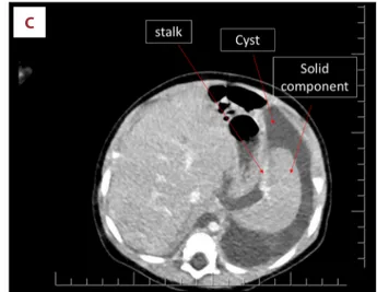
cortisol concentration was 25.7 μg/dL (5–10 μg/dL). She was given an intravenous bolus of 10% dextrose and was admitted to the pediatric intensive care unit with the diagnosis of hyper-insulinemic hypoglycemia. She required a 15% dextrose infu-sion via central line to maintain a blood glucose concentration above 70 mg/dL. Concomitant oral diazoxide was started and increased to 15 mg/kg/day. She demonstrated an abnormally elevated response to glucagon administration during a hypo-glycemic episode with a rise in blood glucose concentration from 37 to 96 mg/dL. This confirmed the diagnosis of hyperin-sulinemic hypoglycemia. Despite a good oral intake (breastmilk and formula) and the diazoxide, she required 6.25 mg/kg/min glucose infusion to maintain a blood glucose concentration above 70 mg/dL. An MRI and CT of the abdomen showed a cys-tic mass of 104×61×48 mm in diameter with a 50×40×36 mm heterogeneous solid component (Figure 1C). At 6 months of age a through-cut biopsy was performed. Histopathological examination showed ductal structures within a myxoid stro-ma with no atypical cells and a low level of mitosis. A deci-sion was made to surgically excise the intra-abdominal mass. Surgical findings are depicted in Figure 2.
Histopathological findings
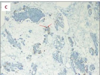
The entire solid mass consisted of fetal pancreatic tissue simi-lar to that at 3 months’ gestation, based on embryologic stag-ing of pancreatic ductal and acinar structures. Almost 95% of the mass consisted of islets of Langerhans. The immune-histo-chemical testing revealed an abundance of insulin-secreting beta cells with scattered areas of glucagon and few soma-tostatin-secreting cells, which are seen in the early embryo-logic stage of development (Figure 3).
Genetic evaluation
Her karyotype was 46 XX with no deletion, duplication, or ab-errant methylation profile of one or more sequences of the KvDMR and H19DMR domains in the 11p15 BWS/Russell-Silver syndrome region. Chimerism testing of the surgically removed material and the blood sample of the infant was performed by using an AmpFlSTR Identifiler PCR Amplification kit (Applied Biosystems, Warrington, UK). Briefly, we used 17 different STR markers located at 2p23-2per, 2q35-37.1, 3p21.31, 4q28, 5q21-31, 5q33.3-34, 7q11.21-22, 8q24.3, 11p15.5, 12p12-pter, 13q22-31, 16q24-qter, 18q21.3, 19q12-13.1, 21q11.2-q21, Xp22.1-22.3, and Yp11.2. The analysis showed that the surgi-cal material and the baby were genetisurgi-cally identisurgi-cal. The zy-gocity testing of the surgical material demonstrated homo-zygocity at the centromeric region. Whole exon sequencing (WES) of the infant’s peripheral blood showed negative mu-tation of the ABCC8 gene and heterozygous variation of hepa-tocyte nuclear factor-1 beta (HNF1b), (exon 1 (rs747555052),
Cemeroglu A.P. et al.: Hyperinsulinemic hypoglycemia and abdominal mass © Am J Case Rep, 2020; 21: e925273
c.79G>C, p.V27L) and insulin receptor substrate 1 (IRS1) (exon 1 (rs779584301), c.1860 C>T, p.P454S) genes.
Postoperatively she had marked hyperglycemia, but after 24 h her blood glucose normalized with complete resolution of her hyperinsulinemic hypoglycemia. At age 8.5 months she was di-agnosed with a congenital hiatal hernia and underwent correc-tive surgery at 11 months. Histologic examination of the sur-gical material showed mature squamous epithelium and was thought to be residue of the previously removed mature tera-toma. The patient is currently 2.5 years of age and has had no subsequent episodes of clinical or biochemical hypoglycemia.
Discussion
The mechanism of action was initially unclear in this case of hyperinsulinemic hypoglycemia with teratoma in this 6-month-old infant. Insulinoma is a very rare cause of hyperinsulinemic
hypoglycemia in childhood [6] and therefore had not been con-sidered due to her age and the presence of an intra-abdom-inal mass without connection to her normal appearing pan-creas. The solid component of the mass connected to the wall of the cyst by a 2.5-cm stalk raised the suspicion of a case of fetus in fetu (FIF). The differential diagnoses included pan-creatic heterotopias, mature cystic teratoma (MCT), and FIF. Pancreatic heterotopia is a rare congenital anomaly in which there is pancreatic tissue with no anatomic connection to the pancreas itself [7]. Pancreatic heterotopia is commonly an in-cidental finding in adults and very rarely reported to cause hyperinsulinemic hypoglycemia [8]. In MCT the presence of various tissue types in a cystic mass is characteristic. The pres-ence of mature pancreatic tissue in mediastinal teratomas has been reported; however, none of the cases resulted in hyper-insulinemic hypoglycemia [8–11]. Although diagnostic criteria have been proposed for FIF [12], the differentiation between MCT and FIF is still controversial [13–16]. All cases of FIF occur in identical twins and are always genetically identical to the host. It has been reported that MCT and FIF could be differen-tiated by zygosity [16]. Teratomas are always homozygous at or near the centromeric regions [17]. Although this mass ful-filled the diagnostic criteria of FIF [12], it was homozygous at the centromeric region, confirming the diagnosis of an MCT. The presence of a squamous epithelium in a small region was also consistent with a teratoid maturation.
A
C
B
Figure 1. MRI and US findings of the intraabdominal lesion. (A) MRI at 2 months of age, a pure cystic lesion. (B) US of the abdomen at 3 months of age showing a vague small solid component within 1 of the bilobulated cysts of 25×25×9 mm and 28×27×13 mm in diameter. (C) CT at 6 months of age showing a 50×40×36 mm solid component within a 104×61×48 mm cystic lesion connecting with a 2-cm stalk. Please note that this image has a striking similarity to a prenatal US image of a fetus.
The most interesting point of this case is that the mature ter-atoma with immature fetal pancreatic tissue lacked the nor-mal control mechanisms of insulin secretion. Having extra beta cells does not cause hyperinsulinemic hypoglycemia if the beta cell function is properly regulated. During the first trimester, beta cells have immature glucose sensing mecha-nisms [18]. The insulin secretion from the fetal pancreas dur-ing early stages of maturation responds to amino acid rather than to glucose concentrations, and at 12 to 22 weeks’ gesta-tion, basal insulin secretion from the fetal pancreas is 4-fold higher [19]. Therefore, immaturity of the fetal pancreatic tis-sue may be the reason for the uncontrolled insulin secretion and partial response to diazoxide treatment. Unfortunately, WES on the tumor tissue could not be performed due to the quality and quantity of the available DNA, and we cannot to-tally exclude the possibility of a mutation in the teratoma that could have caused uncontrolled insulin secretion. The WES of the peripheral blood, however, detected a heterozygous vari-ation of HNF1b and IRS1 genes but no mutvari-ation of the ABCC8 gene. The heterozygous IRS1 variation detected in this case is known to be associated with type 2 diabetes and susceptibility to insulin resistance [20] and therefore could be a coinciden-tal finding. However, the combination of heterozygous varia-tions of the HNF1b and IRS1 genes may be a new finding for the pathogenesis of persistent hyperinsulinemic hypoglycemia
in infants. HNF1b plays an important role in organogenesis, especially of the liver, kidney, and pancreas. Mutations in the HNF1b gene cause monogenic diabetes in youth type 5 (MODY5), renal cysts, genital malformations, and pancreas at-rophy [21]. Mutations in the HNF1b gene have not been re-ported to cause hyperinsulinemic hypoglycemia. This infant had a right ureteropelvic junction stenosis and grade III hy-dronephrosis which could have been due to the detected vari-ation of the HNF1b gene. About one-third of patients with an HNF1b mutation have pancreatic structural anomalies, but a pancreatic teratoma has never been reported with variations or mutations of the HNF1b gene [22]. The specific variation in the HNF1b gene detected in this infant has been reported previously and is considered to be a benign variant or varia-tion of unknown significance [23,24]. This variavaria-tion was locat-ed at the dimerization domain of the HNF1b protein, which is important for protein function and therefore might have im-pacted the embryogenesis of the pancreas, causing a pancre-atic teratoma with uncontrolled insulin secretion and severe hyperinsulinemic hypoglycemia in this infant.
A
B
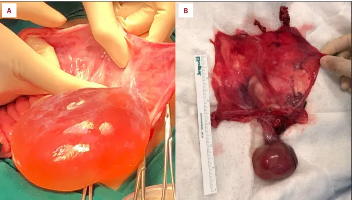
Figure 2. During laparotomy, a clear serous fluid-filled intact sac with a solid component was detected (A). The spleen and pancreas appeared normal and were easily detached from the cyst sac. The left adrenal gland was removed as it could not be detached from the wall of the cyst. The wall of the cyst was cut open exposing the solid component connected to the wall of the cyst with a stalk. A homogenous, smooth, ovoid shaped rubbery solid component connected to the cyst wall with a 2.5-cm stalk was removed (B).
Cemeroglu A.P. et al.: Hyperinsulinemic hypoglycemia and abdominal mass © Am J Case Rep, 2020; 21: e925273
Conclusions
The combination of heterozygous variations of HNF1b and IRS1 genes in this infant likely played a role in the embryogenesis in utero, causing a pancreatic teratoma and severe hyperin-sulinemic hypoglycemia. Therefore, this should be considered
References:
1. Stanley CA: Perspective on the genetics and diagnosis of congenital hyper-insulinism disorders. J Clin Endocrinol Metab, 2016; 101: 815–26 2. Nessa A, Rahman SA, Hussain K: Hyperinsulinemic hypoglycemia – the
mo-lecular mechanisms. Front Endocrinol (Lausanne), 2016; 7: 29
3. Kaczirek K, Niederle B: Nesidioblastosis: An old term and a new under-standing. World J Surg, 2004; 28: 1227–30
4. Sweet CB, Grayson S, Polak M: Management strategies for neonatal hypo-glycemia. J Pediatr Pharmacol Ther, 2013; 18: 199–208
5. De Leon D, Stanley CA: Congenital hypoglycemia disorders: New aspects of etiology, diagnosis, treatment and outcomes. Pediatr Diabetes, 2017; 18: 3–9
6. Escartín R, Brun N, García Monforte MN et al: Insulinoma: A rare cause of hypoglycemia in childhood. Am J Case Rep, 2018; 19: 1121–25
7. Seymore N, Zoghbi B, Sotelo-Avila C, Farmakis SG: Pancreatic heterotopia in a neonatal abdominopelvic cyst. Pediatr Radiol, 2019; 49: 415–18 8. Hussain K, Seppânen M, Nântö-Salonen K et al: The diagnosis of ectopic
focal hyperinsulinism of infancy with [18F]-dopa positron emission tomog-raphy. J Clin Endocrinol Metab, 2006; 91: 2839–42
Figure 3. Histopathologic examination: Macroscopically, the surgical material consisted of a cystic mass with a diameter of 175×80×40 mm and a solid component with a diameter of 66×44×40 mm with a total diameter of 205×80×40 mm. (A) Pattern is that of a pancreatic tissue in early embryonic development with islets much higher in number than expected (Hematoxylene and Eosine X100 original magnification, shown in arrows). (B) Neuroendocrine islets showing extensive positivity for insulin (Anti-insulin antibody immunohistochemistry staining, ×100 original magnification, shown in arrows). (C) Neuroendocrine islets showing sparse glucagon positive cells (Anti-glucagon antibody immunohistochemistry staining, ×100 original magnification, shown in arrows).
A
B
C
in the pathogenesis of persistent hyperinsulinemic hypogly-cemia in infants.
Conflict of interest
None.
9. Dunn PJ: Pancreatic endocrine tissue in benign mediastinal teratoma. J Clin Pathol, 1984; 37: 1105–9
10. Agrawal T, Blau AJ, Chwals WJ, Tischler AS: A Unique case of mediastinal teratoma with mature pancreatic tissue, nesidioblastosis, and aberrant is-let differentiation: A case report and literature review. Endocr Pathol, 2016; 27: 21–44
11. Weichert W, Koch M, Schmidt B et al: Mature mediastinal teratoma with subtotal unidirectional pancreatic differentiation. Pathol Res Pract, 2010; 206: 346–48
12. Gupta SK, Singhal P, Arya N: Fetus – in-fetu: A rare congenital anomaly. J Surg Tech Case Rep, 2010; 2: 77–80
13. Spencer R: Parasitic conjoined twins: External, internal (fetuses in fetu and teratomas), and detached (acardiacs). Clin Anat, 2001; 14: 428–44 14. Satgé D, Jaubert F, Sasco AJ, Vekemans MJ: Are fetus-in-fetu highly
differ-entiated teratomas? Practical implications. Pediatr Intern, 2003; 45: 368 15. Weiss JR, Burgess JR, Kaplan KJ: Fetiform teratoma (homonculus). Arch
Pathol Lab Med, 2006; 130: 1552–56
16. Greenberg JA, Clancy TE: Fetiform teratoma (homunculus). Rev Obstet Gynecol, 2008; 1: 95–76
17. Linder D, McGaw BK, Hecht F: Pathogenic origin of benign ovarian terato-mas. N Eng J Med, 1975; 292: 63–66
18. Hoffman L, Mandel TE, Carter WM et al: Insulin secretion by fetal human pancreas in organ culture. Diabetologia, 1982; 23: 426–30
19. Fowden AL, Hill DJ: Intra-uterine programming of the endocrine pancreas. Br Med Bull, 2001; 60: 123–42
20. Kido Y, Burks DJ, Withers D et al: Tissue-specific insulin resistance in mice with mutations in the insulin receptor, IRS-1, and IRS-2. J Clin Invest, 2000; 105: 199–205
21. Heidet L, Decramer S, Pawtowski A et al: Spectrum of HNF1B mutations in a large cohort of patients who harbor renal diseases. Clin J Am Soc Nephrol, 2010; 5: 1079–90
22. Madariaga L, García-Castaño A, Ariceta G et al., Spanish group for the study of HNF1B mutation. Variable phenotype in HNF1B mutations: Extrarenal manifestations distinguish affected individuals from the population with congenital anomalies of the kidney and urinary tract. Clin Kidney J, 2018; 12: 373–79
23. Edghill EL, Bingham C, Ellard S, Hattersley AT: Mutations in hepatocyte nu-clear factor-1-beta and their related phenotypes. J Med Genet, 2006; 43: 84–90
24. Urakami T: Maturity-onset Diabetes of the Young (MODY): Current perspec-tives on diagnosis and treatment. Diabetes Metab Syndr Obes, 2019; 12: 1047–56
Cemeroglu A.P. et al.: Hyperinsulinemic hypoglycemia and abdominal mass © Am J Case Rep, 2020; 21: e925273