Address for Correspondence/Yazışma Adresi
Oxidant and Antioxidant Balance in Patients with
Childhood Non-Cystic Fibrosis-Related Bronchiectasis
Kistik Fibrozis Dışı Bronşiektazili Çocuk Hastalarda Oksidan ve
Antioksidan Denge
Sevgi PEKCAN1, Seda KÖSE KARATAŞ1, Bahar GÖKTÜRK2, Sevil KURBAN3, Şükrü Nail GÜNER4
1 Department of Pediatric Pulmonology, Necmettin Erbakan University, Meram School of Medicine, Konya, Turkey
Necmettin Erbakan Üniversitesi, Meram Tıp Fakültesi, Çocuk Göğüs Hastalıkları Bilim Dalı, Konya, Türkiye
2 Department of Pediatric Allergy and Immunology, Baskent University, Faculty of Medicine, Ankara, Turkey
Başkent Üniversitesi, Tıp Fakültesi, Çocuk Alerji ve İmmünoloji Bilim Dalı, Ankara, Türkiye
3 Department of Biochemistry, Necmettin Erbakan University, Meram School of Medicine, Konya, Turkey
Necmettin Erbakan Üniversitesi, Meram Tıp Fakültesi, Biyokimya Ana Bilim Dalı, Konya, Türkiye
4 Department of Pediatric Allergy and Immunology, Necmettin Erbakan University, Meram School of Medicine, Konya, Turkey
Necmettin Erbakan Üniversitesi, Meram Tıp Fakültesi, Çocuk İmmünoloji ve Alerji Bilim Dalı, Konya, Türkiye
ÖZ
Amaç: Çocuklarda, oksidan ve antioksidan dengenin kistik fibrosis dışı bronşiekatazi (non-CF BE) patogenezi ve prognozundaki etkisinin değerlendirilmesi.
Gereç ve Yöntem: Haziran 2009 ve Ekim 2010 tarihleri arasında non-CF BE tanılı 29 çocuk çalışmaya alındı. Otuz sağlıklı çocuk kontrol grubunu oluşturdu. Sağlıklı ve hasta çocuklarda paraoksonaz1 (PON1), total oksidan durum (TOS) ve total antioksidan durum (TAS) serum düzeyleri stabil ve alevlenme dönemlerinde ölçüldü. Bulgular: Hasta grubunda PON1 ve TAS düzeyleri alevlenme döneminde kontrol gurbuna göre daha düşüktü (p= 0.05 ve p= 0.01, sırasıyla). İmmün yetmezlikli hastalarda oksidatif stresi gösteren TOS düzeyleri ve TAS/TOS oranı kontrol grubuna göre daha düşüktü (p= 0.008 ve p= 0.01, sırasıyla). TAS düzeyleri ve PON1/ TOS oranı orta-şiddetli bronşiektazili hastalarda hafif bronşektazili hastalardan daha düşüktü (p= 0.04 ve p= 0.03, sırasıyla).
Sonuç: Alevlenme döneminde non-CF BE çocuklarda oksidatif stres artmış ve anti-oksidan kapasite azalmıştır. Antioksidan tedavi özellikle immün yetmezliği ve/veya orta-şiddetli bronşiaktazisi olan non-CF BE hastalarda oksidatif stresin neden olduğu hasarın şiddetini azaltmaya yardımcı olacak, özellikle daha iyi prognoza katkıda bulunacaktır.
ABSTRACT
Objective: To evaluate the role of the oxidant and antioxidant balance in the pathogenesis and prognosis of non-cystic fibrosis bronchiectasis (non-CF BE) in children.
Materials and Methods: Twenty-nine children with non-CF BE were enrolled between June 2009 and October 2010. Thirty healthy children were enrolled as controls. Paraoxonase 1 (PON1), total oxidant status (TOS), and total antioxidant status (TAS) serum levels were measured in controls and in patients when stable and at acute exacerbation.
Results: PON1 and TAS levels were lower in patients at acute exacerbation than in controls (P= 0.05 and P= 0.01, respectively). TOS levels indicative of oxidative stress were higher, and TAS/ TOS levels were lower, in immune-deficient patients than control group (P= 0.008 and P= 0.01, respectively). TAS levels and PON1/ TOS ratio were significantly lower in patients with moderate–severe bronchiectasis than in patients with mild bronchiectasis (P= 0.043 and P= 0.03, respectively).
Conclusion: Oxidative stress was increased and antioxidant capacity decreased in patients with non-CF BE during the exacerbation period. Antioxidant treatment in patients with non-CF BE, especially in patients with immunodeficiency and/or with moderate–severe bronchiectasis, could be helpful to reduce the frequency and severity of the attacks by reducing oxidative stress-induced damage, ultimately contributing to a better prognosis.
INTRODUCTION
Bronchiectasis is a chronic inflammatory disease of the bronchial wall characterized by congenital or acquired irreversible dilation (1). In developed countries, the most common cause is cystic fibrosis with other types of bronchiectasis being defined as non-cystic fibrosis bronchiectasis (non-CF BE). At tissue level, bronchiectasis is considered to arise from inflammatory destruction of muscle, elastic tissue, and bronchial wall cartilage resulting from long-term contact of inflamed mucoid fluid with the bronchial wall (2,3).
Free radical production and elimination rates are well balanced under physiological conditions. This balance is known as oxidative balance. However, an increase in the production rate or a decrease in the elimination rate leads to imbalance between free radical production and antioxidant defense mechanisms, thereby causing tissue injury (4-6). This imbalance is known as oxidative stress. If not neutralized, free radicals degrade cellular membrane proteins, inhibit cellular functions, and make DNA susceptible to breaks and mutations by affecting nuclear genetic material. Chronic diseases and cell death occur as the result of oxidative stress and subsequent tissue injury.
Paraoxonase (PON) is an enzyme associated with high-density lipoproteins that exerts antioxidant effects by inhibiting fatty acid oxidation in low-density lipoproteins. Increased lipid and protein oxidation products and oxidative stress caused by a reduction in antioxidant enzymes and vitamins reduce PON level. An organism’s oxidant– antioxidant balance can be determined by measuring the antioxidant enzyme activity and the oxidant/antioxidant level, but an easier way is to measure the total oxidant status (TOS) and the total antioxidant status (TAS). Oxidants and antioxidants are involved in the pathogenesis of many renal, pulmonary, cardiac, hematologic, and neurologic diseases. Studies based on the measurement of oxidants and antioxidants have been conducted for pulmonary diseases such as chronic obstructive pulmonary disease (COPD), tuberculosis, and cystic fibrosis (7-11). In this study, we evaluated the role of the oxidant and antioxidant balance
in the pathogenesis and prognosis of pediatric non-cystic fibrosis bronchiectasis (non-CF BE) by measuring serum PON1, TAS, and TOS levels.
MATERIALS and METHODS
A total of 29 patients who were being followed up at the Division of Pediatric Pulmonology of Necmettin Erbakan School of Medicine were included in this prospective case-control study between June 2009 and October 2010. Twenty-nine patients were evaluated at acute exacerbation and 28 cases were evaluated during the stable period following exacerbation (one patient died during the exacerbation period). Thirty healthy children whose age and sex were matched to the study group and who were admitted to a general pediatrics clinic for regular check-up and did not have any diseases were included as controls.
Ethics committee approval was obtained from Necmettin Erbakan University Meram School of Medicine. Support was provided by Scientific Research Projects of Necmettin Erbakan University. All parents were informed about the content and aim of the study, and informed consent was obtained. Patients whose parents did not agree to participate and those who had cystic fibrosis-related bronchiectasis were excluded from the study.
Date of participation, age, sex, height, weight, percentiles, body mass index (BMI), body temperature,
respiratory rate, oxygen (O2) saturation, physical
examination findings, smoking status at home, number of family members, status of chest physiotherapy, yearly influenza vaccination status, duration of follow-up, and duration of therapy during the exacerbation period were recorded at acute exacerbation. Height, weight, percentiles, BMI, body temperature, respiratory rate, O2 saturation, and physical examination findings were recorded again during the stable period.
Increase in sputum production, change in the quality of sputum, increase in cough, dyspnea, fever, elevated C-reactive protein (CRP), procalcitonin, and novel infiltration on chest radiography were accepted as exacerbation criteria. Postero-anterior chest radiography, Anahtar kelimeler: Çocuk, kistik fibrosis dışı bronşiektazi, oksidatif denge, paraoksonaz
Key words: Children, non-cystic fibrosis-related bronchiectasis, oxidative balance, paraoxonase
Geliş Tarihi: 30/11/2016 • Kabul Tarihi: 10/02/2017 Received: 30/11/2016 • Accepted: 10/02/2017
complete blood count, erythrocyte sedimentation rate (ESR), CRP, procalcitonin, sputum culture, and blood gas analysis were obtained from the patients in order to determine the exacerbation criteria. Respiratory function test was performed in patients compliant to the test. During this period, patients received antibiotic therapy for 14 days either in outpatient or in inpatient setting.
Previously or newly obtained tomographic scans of all patients were evaluated. Anatomic spread of bronchiectasis was evaluated by a radiologist blinded to the clinical condition of the patients and scored individually for each lobe: grade 0, no bronchiectasis findings; grade 1, mild bronchiectasis (<25% of the lobe); grade 2, moderate bronchiectasis (25–50% of the lobe); grade 3, severe bronchiectasis (> 50% of the lobe). Scores of all lobes were summed up and an overall score was obtained according to the severity of bronchiectasis (11). A total score of 0–6 indicated mild bronchiectasis, 7–12 indicated moderate bronchiectasis, and 13–18 indicated widespread bronchiectasis.
Auscultation findings were recorded separately as crackles and rhonchi according to zones. Crackles were not classified according to being inspiratory or expiratory or to type.
Blood (5 cc) was withdrawn from controls and from patients with bronchiectasis at acute exacerbation and 1 month following antibiotherapy, during the stable period. Blood was centrifuged at 2500 rpm for 10 min and sera were separated. Serum samples were stored at -80°C until use. PON1, TOS, and TAS levels were determined at the Central Laboratory of Biochemistry, Necmettin Erbakan University Meram School of Medicine.
Paraoxone was used as substrate for measurement of PON1 activity and absorbance of the color formed upon hydrolysis of paraoxone was recorded at 412 nm and 37°C. Paraoxonase activity was measured as basal activity and results were given in U/L. TOS was determined by using an automatized measurement method (12,13). Oxidants in the sample convert ferrous ion-o-dianisidine to ferric ion. Ferric ion forms a color complex with xylenol orange in an acidic environment. Density of the color measured spectrophotometrically is related with the total amount of oxidant molecules. Measurement was calibrated with hydrogen peroxide (H2O2) and the results were given in µmol H2O2 equiv/L (12,13). TAS was determined with an automatized measurement method based on the formation
of a characteristic pale color upon addition of antioxidants to a solution containing 2,2’-azino-bis (3-ethylbenz-thiazoline-6-sulfonic acid) (ABST). Results were given in mmol Trolox equivalent/L (12,13).
The Statistical Package for the Social Sciences (SPSS) for Windows version 15.0 was used as the statistical analysis program to assess the data. Tests of normality were conducted to compare the general characteristics between patients and controls. The chi-square and Fisher’s exact test was used to compare the differences between the frequency rates of the categorical data. Student’s t test and the Mann–Whitney U test were used to compare the difference between the averages of the data that did and did not comply with normal distribution, respectively. For repetitive measurements, one-way analysis of variance was used. Statistical significance was set at P<0.05, and the confidence interval (CI) was determined at a 95% level.
RESULTS
Of 29 patients with non-CF BE, 14 (48.3%) were boys and 15 (51.7%) were girls. Their mean age was 10.55 ± 4.31 years (16–202 months). The control group consisted of 14 (46.7%) boys and 16 (53.3%) girls. There was no statistically significant difference between groups in terms of age and sex (P > 0.05).
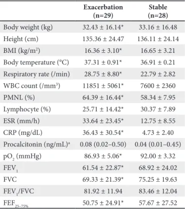
A respiratory function test could not be done in five patients due to non-compliance to the test. Decreased FEV1 (<80%) was detected in 18 patients (75%), decreased FVC (<80%) was detected in 19 patients (79.1%), and decreased FEF25–75% (<80%) was detected in 23 patients (95.8%). Body weight, BMI, FEV1, FVC, FEF25–75%, pO2 values of non-CF BE patients were significantly higher during the stable period than during the exacerbation period (P<0.05). Fever, respiratory rate, white blood cell count, neutrophil and lymphocyte ratios, ESR, and CRP values were significantly lower during the stable period than during the exacerbation period (P<0.05). Weight gain was significantly higher during the stable period (P= 0.001).
There was no statistically significant difference between the exacerbation and stable periods in terms of height, procalcitonin, and FEV1/FVC ratio (P > 0.05) (Table I).
Complaints reported during the exacerbation period were cough (100%), increased sputum (93.1%), change in the color of sputum (79.3%), dyspnea (44.8%), fever (31%),
fatigue (31%), hemoptysis (6.9%), and chest pain (3.4%). Physical examination findings during the exacerbation period were as follows: crepitant rale (24 patients [82.7%]), rhonchus (17 patients [58.6%]), dyspnea (13 patients [44.8%]), decreased O2 saturation (≤ 92%) (11 patients [37.9%]), fever (9 patients [31.0%]), tachypnea (9 patients [31%]), and decreased respiratory sounds (3 patients [10.3%]). Chest deformity and clubbing were detected in 9 (31%) and 5 patients (17.2%), respectively. Six patients (20.6%) had undergone surgery for bronchiectasis.
Distribution of cases according to etiology was as follows: previous lung infection (14 patients [48.3%]), asthma (8 patients [27.6%]), immune deficiency (4 patients [13.8%]), tuberculosis (1 patient [3.4%]), Swyer– James syndrome (1 patient [3.4%]), and primary ciliary dyskinesis (1 patient [3.4%]).
Anatomic spread of bronchiectasis was as follows: mild (24 patients [82.7%]), moderate (4 patients [13.8%]), and severely widespread (1 patient [3.4%]).
PON1 and TAS levels indicative of antioxidant activity were decreased during the exacerbation period in non-CF BE patients compared to controls. Although not statistically significant, TOS levels indicative of oxidant activity were elevated slightly during the exacerbation period in non-CF BE patients compared to controls. Although not statistically significant, PON1 and TAS levels were decreased slightly, and TOS levels did not change during the exacerbation period compared to the stable period. Therefore, antioxidant activity appears to be decreased slightly in non-CF BE patients at acute exacerbation compared to the stable period (Table II). PON1, TOS, and TAS levels were similar in non-CF BE patients during the stable period and in controls (P= 0.33, P= 0.89, and P= 0.33, respectively). PON1/TOS level was higher in controls than in non-CF BE patients; among non-CF BE patients, PON1/TOS level was higher during the stable period than at acute exacerbation. PON1/TOS, and TAS/TOS levels were similar between groups (Table II).
When patients were classified according to the etiology of bronchiectasis, TOS levels indicative of oxidative stress were higher (P= 0.008), and TAS/TOS level was lower (P= 0.01), in immune-deficient patients. PON1 levels were lower in immune-deficient patients compared to other groups, but not significantly so (Table II).
Oxidative balance was evaluated according to severity of bronchiectasis based on thoracic computed tomography scores. Serum TAS and PON1/TOS levels were significantly lower in patients with moderate–severe bronchiectasis compared to patients with mild bronchiectasis (P= 0.043 and P= 0.03, respectively). Although TAS/TOS and PON1 levels were lower and TOS levels were higher in patients with moderate–severe bronchiectasis, the differences were not statistically significant (Table II). It is possible that oxidative stress increases as the anatomic spread of bronchiectatic areas increases, but antioxidant capacity is insufficient.
There were no correlations between the FEV1 and PON1(r=-0.03; p=0,69), FEV1 and TAS (r=-0.05; p=0.81), FEV1 and TOS (r=-0.10; p=0,66), FVC and PON1 (r=0,23; p=0.33), FVC and TAS (r=0,08; p=0,71), FVC and TOS (r=-0.11; p=0.61).
Table I. Clinical and laboratory characteristics of patients with non-CF BE during both exacerbation and stable periods Exacerbation (n=29) (n=28)Stable Body weight (kg) 32.43 ± 16.14* 33.16 ± 16.48 Height (cm) 135.36 ± 24.47 136.11 ± 24.14 BMI (kg/m2) 16.36 ± 3.10* 16.65 ± 3.21 Body temperature (°C) 37.31 ± 0.91* 36.91 ± 0.21 Respiratory rate (/min) 28.75 ± 8.80* 22.79 ± 2.82 WBC count (/mm3) 11851 ± 5061* 7600 ± 2360 PMNL (%) 64.39 ± 16.44* 58.34 ± 7.95 Lymphocyte (%) 25.71 ± 14.42* 30.37 ± 7.89 ESR (mm/h) 33.64 ± 23.45* 12.75 ± 8.55 CRP (mg/dL) 36.43 ± 30.54* 4.73 ± 2.40 Procalcitonin (ng/mL)a 0.08 (0.02–0.50) 0.04 (0.01–0.45) pO2 (mmHg) 86.93 ± 5.06* 92.00 ± 3.32 FEV1 61.54 ± 22.87* 68.92 ± 24.02 FVC 69.33 ± 21.39* 75.25 ± 19.63 FEV1/FVC 81.92 ± 11.94 83.46 ± 12.04 FEF25–75% 50.75 ± 24.91* 57.67 ± 27.52
Data are the mean ± standard deviation.
a Median (minimum–maximum), * P<0.05
BE: Bronchiectasis, BMI: Body mass index, WBC: White blood cell, PMNL: Polymorphonuclear leukocyte, ESR: Erythrocyte sedimentation rate, CRP: C-reactive protein, FEV1: Forced expiratory volume in one second, FVC: Forced vital capacity, FEF25–75%: Forced expiratory flow 25–75%.
DISCUSSION
The incidence of bronchiectasis is approximately 5/1.000.000 in the pediatric population, 1/25000 in patients aged 18–34 years, and 272/100.000 in patients aged 75 years and older. Its prevalence is estimated to be higher in developing countries (14-19).
Proteases and toxic oxygen radicals released from neutrophils that have migrated to the inflammation site have been shown to result in the inflammatory destruction of smooth muscle, elastic tissue, and the cartilage structure of the bronchial wall, injury to the ciliary structures, and gland hypersecretion (2,20). If oxygen radical formation exceeds the capacity of the antioxidant defense system under oxidative stress, increased free oxygen radicals interact with various cellular and extracellular components, resulting in structural and functional impairment. This imbalance may occur in favor of either oxidants or antioxidants (6,20). TAS reflects the total effect of antioxidants in plasma and body fluids, whereas TOS reflects the total effect of oxidants. Although oxidant–antioxidant/oxidant status can be determined by measuring oxidant and antioxidant molecules separately, these measurements are time consuming, costly, and complex. Therefore, we chose to determine the oxidant–antioxidant/oxidant status by measuring TOS and TAS.
Infections lead to oxidative damage by facilitating accumulation and activation of phagocytic cells in the lungs. However, increased O2 production during the exacerbation period declines to normal levels during the stable period in COPD patients (10). In our study, PON1 and TAS levels were found to be lower during the exacerbation period than during the stable period in patients with non-CF BE.
Increased oxidative stress and decreased antioxidant system were studied in children with asthma, allergic rhinitis, and cystic fibrosis; and in adults with COPD, bronchiectasis, emphysema, and asthma (21). However, the number of studies in children is limited. In a study with 25 patients with pulmonary tuberculosis and 33 healthy controls, Selek et al. (22) found that PON, arylesterase, and total free sulfydryl levels were lower, and TOS and lipid hydroperoxide levels were higher in patients than in controls. Rai et al. (23) reported that oxidant and antioxidant status varied across different respiratory diseases. Malondialdehyde (MDA)—an indicator of plasma oxidant status—is significantly increased in patients with COPD, emphysema, bronchiectasis, and bronchial asthma. Antioxidant vitamins, namely vitamin E and vitamin C, are lower in those patients than in controls. Similarly, in our study, PON1 and TAS levels were lower in non-CF BE patients at acute exacerbation than in controls (decreased antioxidant activity), but TOS values were higher Table II. Comparison of PON1, TOS, TAS, PON1/TOS (antioxidant status), and TAS/TOS (oxidant status) levels of non-CF BE patients according to periods, etiology, and severity of bronchiectasis
PON1 TAS TOS PON1/TOS TAS/TOS
Type Exacerbation 47.67 ± 31.48* 1.96 ± 0.35* 40.22 ± 26.67 2.10 ± 2.02 0.08 ± 0.05 Stable 52.01 ± 34.96* 2.14 ± 0.37* 40.78 ± 26.20 1.81 ± 1.51 0.07 ± 0.03 Control 66.55 ± 45.06* 2.16 ± 0.27* 37.30 ± 19.21 2.56 ± 2.82 0.08 ± 0.05 Etiology PPE 39.90 ± 22.91 2.04 ± 0.44 31.47 ± 18.78** 1.89 ± 1.68 0.09 ± 0.05** ID 26.57 ± 14.65 1.98 ± 0.44 84.86 ± 14.22** 0.31 ± 0.15 0.02 ± 0.01** Asthma 62.67 ± 41.62 1.93 ± 0.45 35.12 ± 22.32** 2.90 ± 2.57 0.07 ± 0.04** Tx CT score 0–6 49.57 ± 30.10 2.02 ± 0.35*** 37.63 ± 26.79 1.31 ± 1.07*** 0.05 ± 0.03 7–18 29.76 ± 35.70 1.67 ± 0.21*** 52.99 ± 24.24 0.56 ± 0.45*** 0.03 ± 0.02
PPE: Previous pulmonary infection, ID: Immunodeficiency, Tx CT: Thoracic computed tomography. * PON1 and TAS levels were lower at exacerbation than in controls (P= 0.05 and P= 0.01, respectively)
** TOS level was higher, and TAS/TOS level was lower, in the ID group than in other groups (P= 0.008 and P= 0.01, respectively) *** TAS and PON1/TOS levels were lower in the moderate–severe BE group than in the mild BE group (P= 0.04 and P= 0.03, respectively).
(increased oxidant activity). Impairment of oxidant– antioxidant balance is common in cystic fibrosis patients, as inflammation and infection are constantly present (24). However, there is no constant inflammation in non-CF BE. Elevated oxidative stress was shown not only in lung tissue but also in circulation of children with asthma and cystic fibrosis-related bronchiectasis (25). Horvath et al. (26) suggested carbon monoxide as an oxidative stress marker in bronchiectasis patients; indeed, its levels were shown to be high in exhaled air despite anti-inflammatory therapy. Rahman et al. (27) reported lower plasma TAS levels in asthma patients than in controls. Stanojkovic et al. (28) found that TOS levels were elevated but returned to normal during the activation period in COPD patients; on the other hand, TAS levels were lower than normal both in activation and stable periods, and PON1 enzymatic activity was similar to that of controls. In a study by Gumral et al. (29), for the patients with bronchial asthma and chronic obstructive pulmonary disease, oxidative stress was found to increase both in activation and stable periods, while antioxidant enzymes were decreased. Similar results were obtained in non-CF BE patients both in activation and stable periods in our study. Two possible explanations are the induction of oxidative stress in the lung due to inflammation and the impairment of oxidant–antioxidant balance.
In a study by Olveira et al. (20), oxidative stress was increased, and antioxidant capacity was decreased, in 90 adult patients with bronchiectasis. No difference was found between cystic fibrosis-related bronchiectasis and non-CF BE. In our study, TOS level and oxidative stress were significantly higher (P= 0.008) in patients with immune deficiency compared to patients who had recurrent pulmonary infections and asthma with causes other than immune deficiency. Immune-deficient patients are more commonly exposed infections and thus to constant oxidative stress. It is possible that the antioxidant system is unable to cope with constant oxidative stress.
In this study, another factor affecting oxidative stress was the extent of the bronchiectatic area. Oxidative stress increased, and antioxidant capacity decreased, as the bronchiectatic area increased. To our knowledge, no other studies have reported similar findings. On the contrary, Palop-Cervera et al. (30) failed to identify a relationship between inflammation markers in the exhaled air and the severity of clinical and radiologic findings, respiratory function tests, quality of life; however, the authors found a relationship between inflammation markers and bacterial colonization.
In a study with asthma patients, Guo et al. (31) showed that nutritional support improves oxidant–antioxidant imbalance, inflammation, immune response, and pulmonary functions. We also consider that antioxidant or nutritional supplement administration would positively affect the prognosis of pediatric non-CF BE patients during the exacerbation period, with positive consequences to the quality of life.
The results of our study suggested that changes in PON1 activity could play a role in the pathogenesis of bronchiectasis, and that elevated TOS and decreased TAS levels could be the pathologic result of oxidative stress in bronchiectasis. Therefore, we suggest that plasma TOS and TAS levels, especially PON1/TOS levels, may be used as inflammation markers depending on the presence and the degree of inflammation in bronchiectasis patients. These markers’ being elevated in widespread bronchiectasis may reflect prognosis during follow-up and even radiation exposure of children if oxidative stress markers stay stable during stable periods. Studies conducted with more patients are needed to understand progression of bronchiectasis through measurement of total oxidative stress and total antioxidant capacity measurement. Understanding the mechanism of action of oxidative stress in COPD, bronchiectasis, and other lung diseases would enable future potent antioxidant therapies.
In conclusion, oxidative stress increased, and antioxidant capacity decreased, in patients with non-CF BE at acute exacerbation. Antioxidant treatment in patients with non-CF BE, especially in patients with immunodeficiency and/ or with moderate–severe bronchiectasis, could be helpful to reduce the frequency and severity of the attacks.
AUTHORSHIP and CONTRIBUTORSHIP
Sevgi Pekcan and Seda Kose Karatas conceived the study. Sevgi Pekcan and Bahar Gokturk participated in its design and drafted the manuscript. Sevil Kurban performed the laboratory studies. Sevgi Pekcan and Bahar Gokturk revised the manuscript. Sukru Nail Guner performed the statistical analysis and revised the manuscript. All authors read and approved the final manuscript.
ETHICS
This study has been approved by the ethics committee at the Necmettin Erbakan University Meram Medical Faculty, Konya, Turkey.
CONFLICT of INTEREST
The authors have stated explicitly that there are no conflicts of interest in connection with this article.
REFERENCES
1. Dagli E. Non cystic fibrosis bronchiectasis. Paediatr Respir Rev 2000;1:64-70.
2. Lichter JP. Bronchiectasis. In: Bardow RA, Ries AL, Morris TA (eds). Manual of Clinical Problems in Pulmonary Medicine. 6th ed. Philadelphia, PA: Lippincott Williams and Wilkins, 2005. 306-13.
3. King PT. The pathophysiology of bronchiectasis. Int J Chron Obstruct Pulmon Dis 2009;4:411-9.
4. Atlan N, Dinçel AS, Koca C. Diabetes mellitus and oxidative stress. Turk J Biochem 2006;31:51-6.
5. Serafini M, Del Rio D. Understanding the association between dietary antioxidants, redox status and disease: Is the total antioxidant capacity the right tool? Redox Report 2004;9:145-52. 6. Del Maestro RF. An approach to free radicals in medicine and
biology. Acta Physiol 1980;492:153-68.
7. Kusano C, Ferrari B. Total antioxidant capacity: A biomarker in biomedical and nutritional studies. J Cell Mol Biol 2008;7:1-15. 8. Cross CE, Vander Vliet A, O’Neill CA, Eiserich JP. Reactive
oxygen species and the lung. Lancet 1994;343:930-3.
9. Pryor WA, Godber SS. Non-invasive measures of oxidative stress status in humans. Free Radic Biol Med 1991;10:177-84.
10. Vliet AV, Cross CE. Oxidants, nitrosants, and the lung. Am J Med 2000;109:398-421.
11. Ooi GC, Khong PL, Chan-Yeung M, Ho JC, Chan PK, Lee JC, Lam WK, Tsang KW. High-resolution CT quantification of bronchiectasis: Clinical and functional correlation. Radiology 2002;225:663-72.
12. Erel O. A novel automated direct measurement method for total antioxidant capacity using a new generation, more stable ABTS radical cation. Clin Biochem 2004;37:277-85.
13. Erel O. A new automated colorimetric method for measuring total oxidant status. Clin Biochem 2005;38:1103-11.
14. Säynäjäkangas O, Keistinen T, Tuuponen T, Kivelä SL. Evaluation of the incidence and age distribution of bronchiectasis from the Finnish hospital discharge register. Cent Eur J Public Health 1998;6:235-7.
15. Pifferi M, Caramella D, Bulleri A, Baldi S, Peroni D, Pietrobelli A, Boner AL. Pediatric bronchiectasis: Correlation of HRCT, ventilation and perfusion 72 scintigraphy, and pulmonary function testing. Pediatr Pulmonol 2004;38:298-303.
16. Weycker D, Edelsberg J, Oster G, Longini IM Jr, Nizam A, Ciuryla V, Oster G. Prevalence and economic burden of bronchiectasis. Clin Pulm Med 2005;12:205-9.
17. Karadag B, Karakoc F, Ersu R, Kut A, Bakac S, Dagli E. Non-cystic-fibrosis bronchiectasis in children: A persisting problem in developing countries. Respiration 2005;72:233-8.
18. Barker AF. Bronchiectasis. N Engl J Med 2002;346:1383-93. 19. Bilton D. Update on non-cystic fibrosis bronchiectasis. Curr
Opin Pulm Med 2008;14:595-9.
20. Olveira G, Olveira C, Dorado A, García-Fuentes E, Rubio E, Tinahones F, Soriguer F, Murri M. Cellular and plasma oxidative stress biomarkers are raised in adults with bronchiectasis. Clin Nutr 2013;32:112-7.
21. MacNee W. Pulmonary and systemic oxidant/antioxidant imbalance in chronic obstructive pulmonary disease. Proc Am Thorac Soc 2005;2:50-60.
22. Selek S, Cosar N, Kocyigit A, Erel O, Aksoy N, Gencer M, Gunak F, Aslan M. PON1 activity and total oxidant status in patients with active pulmonary tuberculosis. Clin Biochem 2008;41:140-4.
23. Rai RR, Phadke MS. Plasma oxidant-antioxidant status in different respiratory disorders. Indian J Clin Biochem 2006;21:161-164.
24. Cobanoğlu N, Ozçelik U, Göçmen A, Kiper N, Doğru D. Antioxidant effect of beta- carotene in cystic fibrosis and bronchiectasis: Clinical and laboratory parameters of a pilot study. Acta Paediatr 2002;91:793-8.
25. Palop-Cervera M, de Diego Damiá A, Martínez-Moragón E, Cortijo J, Fullana J, León M. Inflammation markers in the exhaled air of patients with bronchiectasis unassociated with cystic fibrosis. Arch Bronconeumol 2009;45:597-602.
26. Horvath I, Loukides S, Wodehouse T, Kharitonov SA, Cole PJ, Barnes PJ. Increased levels of exhaled carbon monoxide in bronchiectasis: A new marker of oxidative stress. Thorax 1998;53:867-70.
27. Rahman I, Morrison D, Donaldson K, MacNee W. Systemic oxidative stress in asthma, COPD, and smokers. Am J Respir Crit Care Med 1996;154:1055-60.
28. Stanojkovic I, Kotur-Stevuljevic J, Milenkovic B, Spasic S, Vujic T, Stefanovic A, Llic A, Ivanisevic J. Pulmonary function, oxidative stress and inflammatory markers in severe COPD exacerbation. Respir Med 2011;105:31-7.
29. Gumral N, Naziroglu M, Ongel K, Beydilli ED, Ozguner F, Sutcu R, Caliskan S, Akkaya A. Antioxidant enzymes and melatonin levels in patients with bronchial asthma and chronic obstructive pulmonary disease during stable and exacerbation periods. Cell Biochem Funct 2009;27:276-823.
30. Palop-Cervera M, de Diego Damiá A, Martínez-Moragón E, Cortijo J, Fullana J, León M. Inflammation markers in the exhaled air of patients with bronchiectasis unassociated with cystic fibrosis. Arch Bronconeumol 2009;45:597-602.
31. Guo CH, Liu PJ, Lin KP, Chen PC. Nutritional supplement therapy improves oxidative stress, immune response, pulmonary function, and quality of life in allergic asthma patients: An open-label pilot study. Altern Med Rev 2012;17:42-56.