https://doi.org/10.31925/farmacia.2019.5.24 ORIGINAL ARTICLE
THE ANTIOXIDANT AND ANTIGENOTOXIC POTENTIAL OF
PELTIGERA CANINA AND UMBILICARIA NYLANDERIANA BASED
ON THEIR PHENOLIC PROFILE
BUGRAHAN EMSEN *
Department of Biology, Kamil Özdağ Faculty of Science, Karamanoğlu Mehmetbey University, Karaman, Turkey *corresponding author: [email protected]
Manuscript received: October 2018
Abstract
The present study aimed to assess the biological effects of methanol and water extracts obtained from Peltigera canina and Umbilicaria nylanderiana on human peripheral lymphocytes. Chromosome aberration and micronucleus tests evaluated genotoxicity levels of the extracts on the cells. In both tests, it was determined that the treatments at concentrations of 1 - 50 mg/L did not significantly increase genotoxicity. When investigating the total oxidative stress (TOS) levels of the cells exposed to the tested extracts, it was revealed that water extracts decreased TOS in the cells. In order to determine the antioxidant capacities of the extracts, DPPH radical scavenging, metal chelating, reducing power activities and besides, total antioxidant capacity in the cells were measured. Concentration-dependent increases were observed for each antioxidant activity. In addition, there were detected the main phenolic compounds in the extracts by an HPLC method. Consequently, it was revealed that lichen-derived extracts are worth investigating as possible antioxidative agents.
Rezumat
Studiul prezintă efectele biologice ale extractelor apoase și metanolice ale speciilor Peltigera canina și Umbilicaria nylanderiana asupra limfocitelor umane periferice. Mutațiile cromozomiale și testele realizate pe micronuclei au demonstrat nivelul de genotoxicitate celulară. În ambele teste, concentrații cuprinse între 1 și 50 mg/L nu au prezentat efecte semnificative asupra genotoxicității. În ceea ce privește nivelul stresului oxidativ total (TOS), extractele apoase au scăzut TOS în celule. Au fost determinate activitatea de chelatare a radicalilor liberi și a metalelor, activitatea reducătoare și capacitatea totală antioxidantă. De asemenea, a fost determinat conținutul fenolic printr-o metodă HPLC. În consecință, extractele pe bază de licheni prezintă un interes deosebit pentru potențialul lor antioxidant remarcabil.
Keywords: antioxidant, cytotoxic, genotoxic, lichen, Peltigera canina, Umbilicaria nylanderiana Introduction
All multicellular organisms try to protect themselves against harmful microorganisms that can cause disease. Immunity describes the reaction and response of an organism to an agent that can cause all kinds of diseases such as bacteria, viruses, fungi [23]. Lymphocytes are essential cells of the acquired immune system. For this reason, protection of lymphocytes is of great importance for human health [14].
Free radicals that cause oxidative stress in the human body are important elements that negatively affect the immune system. Reactive oxygen and nitrogen-induced oxidative stress have been demonstrated to play a role in a variety of inflammatory and degenerative diseases such as cancer, cataracts, ageing, rheumatoid arthritis and diabetes [4]. The living metabolism that has an established balance between endogenous and exogenous antioxidants undergoes oxidative stress in some conditions. In this context, when occasionally endogenous antioxidants such as catalase, superoxide dismutase, melatonin, uric acid and bilirubin are inadequate, supplementation of exogenous antioxidants
may prevent the formation of oxidative stress [18]. Exogenous antioxidants are mostly molecules that can be taken from food or some preparations and usually support the antioxidant system directly or indirectly [25]. Phenolic compounds are the most important among the antioxidant substances found in plant extracts identified as biological materials. Plant-derived antioxidants especially phenols function as free radical scavengers, peroxide scavengers, enzyme inhibitors and synergists [12]. They have positive effects on health by preventing the harmful effects of free radicals, low-density lipoproteins and lipoprotein oxidation [1].
Lichens are among the sources of natural antioxidant substances [6]. They are morphological and physiological associations of fungi brought together by algae. In the lichens, fungi use algae as a carbon source for survival, growth and reproduction. Algae take the mineral, water for photosynthesis from the fungi and are protected by the fungi against adverse conditions such as high temperature, harmful rays, and high humidity [2]. The lichens have long been used as traditional medicinal
913
products in many countries. There is much evidence that they are used as a decoction or infusion in the treatment of various diseases [7]. Scientists have also carried out numerous studies in order to demonstrate the protective properties of the lichens by the anti-oxidant components found in their structures [19]. However, to the best of our knowledge, no adequate information is available regarding the biological activities of Peltigera canina (L.) Willd. and Umbilicaria
nylanderiana (Zahlbr.) H. Magn. on human peripheral
lymphocytes (HPLs). Therefore, this study aimed to investigate the antioxidant, oxidative, genotoxic and cytotoxic capacities of methanol and water extracts obtained from P. canina and U. nylanderiana in HPLs and detect the main phenolic compounds of the tested extracts by high-performance liquid chromatography (HPLC) method.
Materials and Methods
Materials
Collection and identification of the lichen samples
The samples were collected from Eastern Anatolian region of Turkey (40°35′N - 41°49′E). Necessary morphological and ecological characteristics of the samples were recorded and they were photographed in their natural habitats. After collecting the materials, they were exposed to a dry atmosphere in room conditions. Lichen herbaria were obtained and kept for the records. Comparing the obtained macroscopic and microscopic data with literature [20, 28], they were identified as P. canina (KKEF-803) and U.
nylanderiana (KKEF-804). Voucher specimens are
kept in the herbarium of Faculty of Pharmacy, Van Yüzüncü Yıl University, Van, Turkey.
Preparation of the extracts
20 g of the lichen samples were dried for 7 days under room conditions and powdered under liquid nitrogen. Then, bioactive ingredients were extracted by 250 mL of methanol and water solvent systems using a Soxhlet extraction apparatus throughout 2 days. After extraction, solvents were evaporated with a rotary evaporator (IKA, Staufen, Germany) under
vacuum to dryness and lyophilized to get ultra-dry
powders that were solubilized with the minimum amount of sterile distilled water. While methanol (PME) and water (PWE) extracts isolated from P.
canina yielded 11.87% and 13.64% (w/w) of lichens
substances, respectively, methanol (UME) and water (UWE) extracts of U. nylanderiana were obtained with yields of 11.94% and 12.45% (w/w), respectively. The stock solutions were prepared with distilled water.
Identification and quantification of main phenolic compounds by HPLC
The method described by Rodríguez-Delgado et al. [22] was used to separate the phenolic compounds by HPLC. The samples taken after the extraction were centrifuged at 15000 rpm for 15 min. The
supernatant was filtered through a 0.45 µm Millipore filter and collected in the vials. The vials were placed in HPLC autosamplers. Chromatographic separation was carried out on an Agilent 1100 HPLC system using a diode array detector and a 250 × 4.6 mm, 4 µm octadecyl-silica column. Methanol:acetic acid:water (10:2:88) and methanol:acetic acid:water (90:2:8) solvents were used as mobile phase. HPLC conditions for the separation of phenolic compounds were set at 254 and 280 nm wavelength. The flow rate and injection volume were determined: 1 mL/min and 10 µL, respectively. Finally, eleven phenolic compounds (chlorogenic acid, p-coumaric acid, q-coumaric acid, ferulic acid, gallic acid, phlorodizin, protocatechuic acid, resveratrol, rutin, syringic acid and valinic) in the extracts were detected.
Blood samples
Heparinised blood samples were collected from healthy individuals without any disease or active infection. The present study was performed with the approval of the Ethics Committee of the Faculty of Health Sciences of Karamanoğlu Mehmetbey University.
Total antioxidant capacity (TAC) assay
TAC analysis was performed in plasma samples obtained from heparinised blood cultures for 2 h and a commercially available kit (Rel Assay Diagnostics, Gaziantep, Turkey) was used for this purpose, in order to determine the antioxidant levels of samples by inhibiting the formation of a free radical, 2,2′-azino-bis (3-ethylbenzothiazoline-6-sulfonic acid) (ABTS) compound.
The cells were incubated for 24 h. The applications of standard, reagent 1 and reagent 2 solutions were completed in the wells of the trial plate. Spectro-photometric readings were performed at 660 nm. The positive control (control+) was ascorbic acid
(Sigma-Aldrich, Darmstadt, Germany at 4 × 10⁻⁷ M).
Total oxidative stress (TOS) assay
TOS analysis was performed in plasma samples obtained from heparinised blood cultures for 2 h and a commercially available kit (Rel Assay Diagnostics, Gaziantep, Turkey) was used. The assay implies that complexes with ferric ion are oxidized to ferrous ion by oxidants presented in the sample. The oxidation reaction is carried out with strengthening molecules in the reaction medium. Ferrous ions form a coloured structure with the chromogen in the acidic environment. The colour intensity measured spectrophotometrically is related to the total amount of oxidant molecules in the sample.
The cells were incubated for 24 h. The applications of standard, reagent 1 and reagent 2 solutions were completed in the wells of the trial plate. Spectro-photometric readings were performed at 530 nm.
Control+ was hydrogen peroxide (H
2O2)
2,2-diphenyl-1-picrylhydrazyl (DPPH) radical scavenging activity
Free radical scavenging activities of the extracts were determined by monitoring DPPH reduction. Gallic acid (0.01 - 0.5 mM) was used as standard antioxidant molecule and in order to determine the radical scavenging activities, 20 µL of various concentrations of standard or extracts were mixed with 180 µL of DPPH solution (0.06 mM in methanol) and incubated for 1 h at dark in microtiter plates. Blank measurements without standard or extracts were also performed. After reading the absorbance at 517 nm, the reduction of the DPPH radical was determined as percent discoloration of DPPH which was calculated according to the following formula:
Radical Scavenging Activity (RSA) (%) = (DPPH absorbance-DPPH and extract absorbance)/(DPPH
absorbance) × 100
The extract concentrations providing 50% inhibition (IC50) were calculated from extract amount that was
used for the comparison of different extracts of tested lichens.
Metal chelating activity
The chelating abilities of the extracts were determined using ethylenediaminetetraacetic acid (EDTA) as a standard chelating agent. Different concentrations of extracts (1, 5, 10, 25, 50 and 100 mg/L) and standard (0.1 - 5 mM) were added to microplate wells (50 µL) and mixed with 10 µL ferrozine (5 mM), 5 µL iron(II)
chloride (2 mM) and 185 µL absolute methanol.
After incubation for 10 min at room temperature, absorbances were read at 562 nm and IC50 values
were calculated.
Reducing power activity
The reducing powers of the extracts were determined with an adaptation to microplate measurement. Gallic acid (0.01 - 0.1 mM) was used as standard anti-oxidant. In this method, various concentrations of 50 µL lichen extracts (1, 5, 10, 25, 50 and 100 mg/L) were mixed with 75 µL phosphate buffer (0.2 M pH: 6.6) and 75 µL potassium ferricyanide (1% w/v) in a total volume of 200 µL and incubated at 50°C for 20 min. After adding 75 µL trichloroacetic acid (10% w/v), samples were centrifuged for 10 min at 1000 g. The supernatants (75 µL) were transferred to another microtiter plate and mixed with 75 µL distilled water and 15 µL iron (III) chloride (0.1% w/v). After reading the absorbance at 700 nm, effective concentrations (EC50) were calculated.
Chromosome aberration (CA) assay
The samples were added to chromosome medium (Chromosome Medium B, Biochrom, Berlin) and final concentrations of 1, 5, 10, 25, 50 and 100 mg/L of the lichen extracts were added to the medium after 24 h. Cultures were incubated at 37°C for 72 h. A negative control (control-) and a control+ (Mitomycin-C
(C15H18N4O5, Sigma, St, Louis/MO, USA, at 10-7 M))
were also used. The cells were exposed to colchicine (Sigma, St, Louis/MO, USA) 2 h before harvesting. At the end of the incubation, cells were harvested by centrifugation at 900 rpm for 10 min. Then, the cells were treated with KCl as hypotonic solution for 15 min and fixation was performed (methanol:glacial acetic acid 3:1). The cell suspension was centrifuged at 900 rpm for 10 min after each fixative treatment. After the last fixation process, drops of the fixed cell suspension were dropped on a clean slide and air-dried. The slides were stained in 3% Giemsa solution in phosphate buffer (pH 6.8) for 15 min. For each treatment, 30 well-spreaded metaphases were analysed to detect the presence of chromosomal aberrations (CA). In order to classify the different types of aberrations, chromatid/chromosome gaps, chromatid/ chromosome breaks and fragments were determined. In addition, the effect of the experiments on cell proliferation was supported by mitotic index (MI) analysis. This analysis was detected by the following equation:
MI = (number of dividing cells/total cell count) × 100.
Micronucleus (MN) assay
In this experiment, similar applications to chromosome protocol were performed. Briefly, the heparinized blood was mixed with chromosome medium (Chromosome Medium B, Merck, Berlin). Five samples of the extracts at different concentrations (1, 5, 10, 25, 50 and 100 mg/L) were added to cultures 24 h after the beginning of incubation. In addition, a positive control (+) (Mitomycin-C, 10-7 M) and negative control (-) group with no extract were included to the treatments. 44 h after the beginning of incubation cytochalasin B (Sigma, St, Louis/MO, USA) was added to the culture tubes. After the fixation process, the cell suspensions were dropped onto clean slides, air-dried and stained with 3% Giemsa solution. 1000 binuclear cells per concentration were examined for micronucleus (MN) scoring and to determine the number of cells with 1, 2, 3 and 4 nuclei. Nuclear division index (NDI) was calculated using the formula:
NDI = [(1 × N1) + (2 × N2) + (3 × N3) + (4 × N4)]/ n (total cell count).
In this formula, N1-N4 represents the number of cells with 1 to 4 nuclei.
Statistical analyses
Different results of the treatments were interpreted using the analysis of variance followed by appropriate
post-hoc test (Duncan test) and differences were accepted
as statistically significant at p < 0.05. Probit regression analysis was used for calculating EC50 andIC50 values.
Statistical Package for Social Sciences (SPSS, version 21.0, IBM Corporation, Armonk, NY) software was used for all analyses.
915 Results and Discussion
Identification and quantification of main phenolic compounds by HPLC
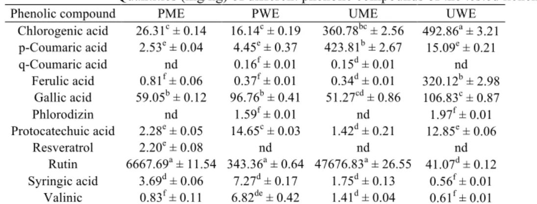
HPLC analysis revealed the presence of eleven phenolic compounds (chlorogenic acid, p-coumaric acid, q-coumaric acid, ferulic acid, gallic acid, phlorodizin, protocatechuic acid, resveratrol, rutin, syringic acid and valinic) in PME, PWE, UME and UWE. When the amounts of the phenolic compounds detected in methanol and water extracts were compared, PWE outweighed than PME. UME was in a position equivalent to UWE. When all the compounds were examined, the high amount of rutin in PME, PWE and UME was noteworthy. It is known that rutin is used in many pharmacological experiments because of its potent antioxidant property. Therefore, the high content of this compound in the extracts is of great importance for
drug development [24]. Rutin was also used in many lichen-antioxidant studies as standard compound [26]. Another remarkable point was that resveratrol was only detected in PME. As for UWE, it was determined that it was especially rich in chlorogenic and ferulic acid. While q-coumaric acid was not detected in PME and UWE, it was detected in the lowest quantity in PWE and UME. Ferulic and syringic acid were the antioxidants that were present in the lowest amounts in PME and UWE, respectively (Table I). Ristic et al. [21] revealed various antioxidant activities of depsides, dibenzofurane and depsidones identified in Ramalina fraxinea and R.
fastigiata by HPLC. Similarly, Oran et al. [17]
proposed that Usnea fulvoreagens, U. intermedia and
U. filipendula could be used as antioxidant agents
due to their total phenolic components.
Table I
Quantities (mg/kg) of different phenolic compounds of the tested lichen extracts
Phenolic compound PME PWE UME UWE
Chlorogenic acid 26.31c ± 0.14 16.14c ± 0.19 360.78bc ± 2.56 492.86a ± 3.21 p-Coumaric acid 2.53e ± 0.04 4.45e ± 0.37 423.81b ± 2.67 15.09e ± 0.21 q-Coumaric acid nd 0.16f ± 0.01 0.15d ± 0.01 nd Ferulic acid 0.81f ± 0.06 0.37f ± 0.01 0.34d ± 0.01 320.12b ± 2.98 Gallic acid 59.05b ± 0.12 96.76b ± 0.41 51.27cd ± 0.86 106.83c ± 0.87 Phlorodizin nd 1.59f ± 0.01 nd 1.97f ± 0.01 Protocatechuic acid 2.28e ± 0.05 14.65c ± 0.03 1.42d ± 0.21 12.85e ± 0.06 Resveratrol 2.20e ± 0.08 nd nd nd Rutin 6667.69a ± 11.54 343.36a ± 0.64 47676.83a ± 26.55 41.07d ± 0.12 Syringic acid 3.69d ± 0.06 7.27d ± 0.17 1.75d ± 0.13 0.56f ± 0.01 Valinic 0.83f ± 0.11 6.82de ± 0.42 1.41d ± 0.04 0.61f ± 0.01
Each value is expressed as mean ± standard deviation (n = 3); Values followed by different superscript letters in the same column differ significantly at p < 0.05; nd: not determined
TAC activity in the cells
When the effects of the tested extracts on TAC change in cells were examined, a concentration-dependent increase was observed. While the treatments at the lowest concentration (1 mg/L) of the extracts did not significantly (p > 0.05) increase TAC in HPLs compared with control-, TAC levels of the cells exposed to high concentrations (≥ 50 mg/L) were close to control+. The concentration of 100 mg/L of PWE
attracted the attention as this experiment resulted in the highest TAC level. Furthermore, the concentration of 50 mg/L of PWE was not statistically significant (p > 0.05) from the previous treatment. Although TAC levels of PME and UME were lower compared to PSW, there was significantly (p > 0.05) no difference between concentrations of 50 and 100 mg/L of them. The lowest results among the extract tests belonged to UWE (Figure 1a).
TOS activity in the cells
Oxidative stress levels in extract and control groups in HPLs were assessed with TOS. In this assay, control+ was separated from other experiments by high TOS value (37.49 µmol H2O2 equivalent/L). In
general, it was determined that the extracts did not raise the oxidative stress level in HPLs at a dangerous rate. Furthermore, while both methanol and water extracts did not significantly (p > 0.05) increase TOS level in the cells compared with control- (12.57 µmol
H2O2 equivalent/L), the maximum concentration of
PWE (9.86 µmol H2O2 equivalent/L) and UWE
(11.23 µmol H2O2 equivalent/L) statistically (p < 0.05)
reduced TOS rate. Besides, it was a remarkable result that the TOS activity caused by the treatment with 50 mg/L of PWE was not statistically significant (p > 0.05) from the maximum concentration of UWE (Figure 1b). Other studies concluded that the reducing effect of α-glucan obtained from Cladina rangiferina against Pb2+-induced oxidative is of interest and it
was revealed that the tested lichen decreased reactive oxygen species (ROS) generation in alveolar epithelium cells [11]. Another study assessed the inhibition of oxidative stress on neurons and astrocytes cells. In this research, it was reported that a secondary metabolite of Cetraria islandica, fumarprotocetraric acid showed a protective role against H2O2-induced oxidative
Figure 1.
TAC (a) and TOS (b) levels in the HPLs in the presence of different lichen extracts
Each value represents the mean standard deviation of three experiments. Different small letters indicate significant differences among treatments at p < 0.05
Scavenging ability on DPPH radicals
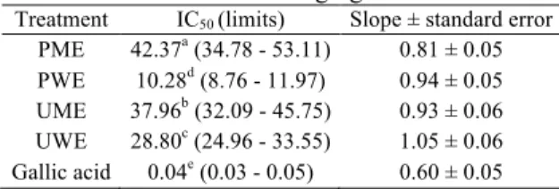
When investigating the DPPH scavenging activities, increases were registered with increasing concentrations 50 and 100 mg/L of PWE were noted with activities over 70%. There was also a no statistically (p > 0.05) significant difference between these treatments. Another critical extract that contributed to the capture of DPPH radicals was UWE. High concentrations (25 - 100 mg/L) of UWE had 60 - 65% DPPH scavenging ability. Based on the highest concentrations, the scavenging activities of the extracts on DPPH radicals were in the descending order PWE > UWE > PME > UME (Figure 2a). Based on the IC50 values that helped
to bring out the effects on the DPPH radical of the extracts, PWE was found to be effective with the lowest IC50 value (10.28 mg/L). The highest value
belonged to PME with 42.37 mg/L. In addition, it was seen that IC50 values of all extract treatments and
gallic acid used as standard antioxidant molecule were statistically (p < 0.05) different from each other (Table II).
Table II
IC50 values (mg/L) of extracts and standard for
scavenging on DPPH radicals
Treatment IC50 (limits) Slope ± standard error
PME 42.37a (34.78 - 53.11) 0.81 ± 0.05
PWE 10.28d (8.76 - 11.97) 0.94 ± 0.05
UME 37.96b (32.09 - 45.75) 0.93 ± 0.06
UWE 28.80c (24.96 - 33.55) 1.05 ± 0.06
Gallic acid 0.04e (0.03 - 0.05) 0.60 ± 0.05
Values followed by different superscript letters in the same column differ significantly at p < 0.05
Chelating ability of metal ions
When focused on metal chelating activities of the tested lichen extracts, it was revealed that there was a correlation between the chelating capacities of the extracts and their concentrations. There were observed close metal chelating activities between PWE and UWE; PME and UME treatments. While concentrations of 25 - 100 mg/L of PWE and UWE caused high results (60 - 70%), in PME and UME, concentrations of 50 and 100 mg/L (55 - 60%) were remarkable (Figure 2b). Moreover, the IC50 values calculated
from the results of the chelating activity showed that the potent inhibitor was PWE (13.24 mg/L). The standard chelating agent, EDTA possessed the lowest
IC50 value among the treatments (0.16 mg/L).
According to IC50 values, the extracts were in the
ascending order of PWE < UWE < UME < PME and they were statistically (p < 0.05) different from each other (Table III).
Table III
IC50 values (mg/L) of extracts and standard for
chelating on ferrous ions
Treatment IC50 (limits) Slope ± standard error
PME 50.33a (40.88 - 64.14) 0.81 ± 0.05 PWE 13.24d (10.86 - 16.10) 0.71 ± 0.05 UME 43.46b (35.89 - 54.09) 0.84 ± 0.05 UWE 25.23c (21.95 - 29.19) 1.06 ± 0.06 EDTA 0.16e (0.14 - 0.18) 1.21 ± 0.06 0 2 4 6 8 10 12 14 16 18 20 P M E P W E U M E U W E Co n tr ol + Co n tr ol -TAC (mmol Trolox equivalent/L) T re at m en t 1 mg/L 5 mg/L 10 mg/L 25 mg/L 50 mg/L 100 mg/L a l l l l l l l kl jk jk j i i i h gh fg f e de cd cd cdbc b a 0 5 10 15 20 25 30 35 40 P M E P W E U M E U W E Co n tr ol + Co n tr ol -TOS (µmol H2O2 equivalent/L) T re at m en t 1 mg/L 5 mg/L 10 mg/L 25 mg/L 50 mg/L 100 mg/L a g f ef de de de bc cd cd bcd bcd bcd bcd bcd bcdbcd bcd bcd bcd bcd bcd bcd cd bc b b
917
Values followed by different superscript letters in the same column differ significantly at p < 0.05
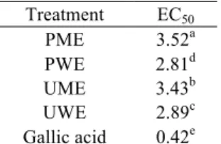
Reducing power activity
In the experiments testing the reducing power activities of lichen extracts at different concentrations, concentrations of 50 and 100 mg/L of PWE that were not statistically significant (p > 0.05) showing towering activity. The reducing power activity emerging by the trials at high concentration (50 and 100 mg/L) of UWE was close to PWE. Additionally, there was no difference (p > 0.05) between concentrations of 50 and 100 mg/L of PME and UME. Considering the lowest concentrations, it was defined that there was no statistically significant difference between PWE, PME and UME (Figure 2c). EC50 values also supported the
results in Figure 2c. As shown in Table IV, EC50
values of PME-UME and PWE-UWE were very close. However, EC50 values of the extracts and standard for
reducing power were as follows: gallic acid < PWE < UWE < UME < PME.
Table IV
EC50 values (mg/L) of extracts and standard for
reducing power Treatment EC50 PME 3.52a PWE 2.81d UME 3.43b UWE 2.89c Gallic acid 0.42e
Values followed by different superscript letters in the same column differ significantly at p < 0.05
Figure 2.
DPPH (a) scavenging, metal chelating (b) and reducing power activities (c) of the different lichen extracts
Each value represents the mean standard deviation of three experiments. Different small letters indicate significant differences among treatments at p < 0.05
As in many herbal product trials, antioxidant substances are at the forefront in the anticancer or therapeutic effect studies [16, 29]. The results of the experiments showed that anticytotoxic and antigenotoxic potentials of P. canina and U. nylanderiana on HPLs were attributed to their antioxidative capacities. Leandro et
al. [15] contributed to the literature by proving that
the chemical agent-induced genotoxicity on hamster lung fibroblast was reduced by (+)-usnic acid, a lichen secondary metabolite. They reported that the anti-oxidant activity of (+)-usnic acid was effective in this regard. Some researchers have determined the total antioxidant capacity-enhancing effects of different lichen species such as Dermatocarpon intestiniforme,
Aspicilia calcerea, Cetraria chlorophylla [3], Hypogymnia
physodes, Usnea florida and Ramalina polymorpha [27]
on lymphocytes due to their antioxidant properties without genotoxic activity.
Genotoxic/CA and MN activities
In order to assess the genotoxic effects of PME, PWE, UME and UWE against HPLs, CA and MN analyses were performed. In CA assays, chromatid/chromosome breaks, gaps and fragments appeared in HPLs were determined (Figure 3a). In these treatments, the highest CA frequency belonged to control+ with 0.67. As
determined in Figure 4a, the results of CA in the cells exposed to all of the other lichen extracts except for the maximum concentration of UME did not differ from the control-. Moreover, a concentration-dependent effect was only observed for UME treatment.
0 10 20 30 40 50 60 70 80 90 P M E P W E U M E U W E DPPH scavenging activity (%) T re a tm e n t 1 mg/L 5 mg/L 10 mg/L 25 mg/L 50 mg/L 100 mg/L a o o n n mn mn lml k j i h g f e e de dc cc b a a 0 10 20 30 40 50 60 70 80 90 P M E P W E U M E U W E Metal chelating activity (%) T re a tm e n t 1 mg/L 5 mg/L 10 mg/L 25 mg/L 50 mg/L 100 mg/L a p o o no n n m l kl k j j i h g gf ef de cde bcd bc ab b 0.0 0.1 0.2 0.3 0.4 0.5 0.6 0.7 0.8 0.9 1.0 P M E P W E U M E U W E Reducing power activity (absorbance-700 nm) T re a tm e n t 1 mg/L 5 mg/L 10 mg/L 25 mg/L 50 mg/L 100 mg/L a a m m lm kl k k j j i h h h g f e e e d d d cb c
Figure 3.
Images of (a) chromatid/chromosome breaks, gaps, fragments and (b) micronucleated cell that emerge in the experiments (10 ×100 magnification)
In the MN assay, micronucleated cells appearing in HPLs were calculated (Figure 3b). Control+ markedly had a high MN ratio (5.81) and this value was statistically (p < 0.05) different from all other experiments. It was revealed that UME, UWE and PME statistically (p < 0.05) induced the formation of MN at a concentration of 100 mg/L. Concentrations of 1 - 50 mg/L of the tested extracts did not significantly (p > 0.05) increase MN frequency compared to control- (1.87) (Figure 4b). Based on the genotoxic effects of P. canina and U. nylanderiana, CA and MN tests provided insights about potential effects for genetic damage formation of the tested extracts on
HPLs. To date, the genotoxic effects of many lichen species were discovered by scientists using different methods. Similar to the antigenotoxic part of the present study, Kosanić et al. [13] examined the genotoxic effects of Lasallia pustulata on peripheral blood lymphocytes through MN test and revealed that the frequency of MN did not significantly change compared with control-. In another research, Guterres et al.
[10] pronounced that salazinic and psoromic acid isolated from Parmotrema cetratum and Usnea
jamaicensis, respectively, significantly decreased by
doxorubicin-induced genetic damage against somatic cells of Drosophila melanogaster.
Figure 4.
Frequencies of (a) CA and (b) MN in the HPLs exposed to the different lichen extracts
Each value represents the mean standard deviation of three experiments. Different small letters indicate significant differences among treatments at p < 0.05
0.0 0.1 0.2 0.3 0.4 0.5 0.6 0.7 PM E PW E U M E U W E Co nt ro l + Co nt ro l -CA/Cell Tr ea tm en t 1 mg/L 5 mg/L 10 mg/L 25 mg/L 50 mg/L 100 mg/L a c b bc bc bc bc bc bc bc bc bc bc bc bc bc bc bc bc bc bc bc bc bc bc bc b 0 1 2 3 4 5 6 PM E PW E U M E U W E Co nt ro l + Co nt ro l -MN/1000 cells Tr ea tm en t 1 mg/L 5 mg/L 10 mg/L 25 mg/L 50 mg/L 100 mg/L a c c b d d d d d d d d d d d d d d d d d d d d d d a
919
Cytotoxic/MI and NDI activities
MI and NDI determined the effects of PME, PWE, UME and UWE on the proliferation of HPLs. When investigating MI percentage in the HPLs exposed to the lichen extracts, close results were observed. However, it was found that the treatments at the concentration of 100 mg/L of PME and UME lowered MI. Besides, there was no significant difference between the concentrations of 50 and 100 mg/L of UME. Other treatments of lichen extracts were not statistically
(p > 0.05) different from control- (MI% = 5.18)
(Figure 5a).
NDI was calculated based on the nucleus numbers in HPLs (Figure 6). In NDI analyses, the value of control+ that negatively affected the nuclear division was 1.27 and data of other trials was significantly (p < 0.05) different compared with the previously mentioned value. None of the treatments of the tested extract was statistically (p > 0.05) different from NDI (1.51) of control- (Figure 5b).
Figure 5.
MI percentage (a) and NDI (b) in the HPLs exposed to different lichen extracts
Each value represents the mean standard deviation of three experiments. Different small letters indicate significant differences among treatments at p < 0.05
Figure 6.
Images of mononucleated (a), binucleated (b), trinucleated (c) and tetranucleated (d) cells that emerge in the experiments (10 ×100 magnification) 0 1 2 3 4 5 6 P M E P W E U M E U W E Co n tr ol + Co n tr ol -MI (%) T re at m en t 1 mg/L 5 mg/L 10 mg/L 25 mg/L 50 mg/L 100 mg/L e d d d c bc abc abc abc abc abc abc abc abc abc abc ab ab ab ab ab ab ab a a a a 0.0 0.2 0.4 0.6 0.8 1.0 1.2 1.4 1.6 1.8 2.0 P M E P W E U M E U W E Co n tr ol + Co n tr ol -NDI T re at m en t 1 mg/L 5 mg/L 10 mg/L 25 mg/L 50 mg/L 100 mg/L e e d cd bcd bcd bcd bcd bcd abcd abcd abcd abcd abcd abcd abcd abcd abcd abcd abcd abcd abc abcabc abc ab a b
Information on whether the lichens show cytotoxic properties on different cells is available in the literature. Emsen et al. [8] proved that the viability of human amnion fibroblasts exposed to olivetoric and physodic acid of lichen secondary metabolites, was affected depending on the dose of metabolite. However, they suggested that low concentrations of these metabolites did not significantly indicate cytotoxicity on the fibroblasts. In a different experiment carried out on lymphocytes, it was detected that Cetrelia olivetorum,
Cetraria aculeata and Cladonia chlorophaea did
not have a significant anti-mutagenic effect [5].
Conclusions
In conclusion, in the present study, it was revealed that PME, PWE, UME and UWE except for their high concentrations did not show significant cytotoxic, genotoxic and pro-oxidative effects on HPLs. There were correlations between different antioxidant activities of the extracts and TAC levels in HPLs exposed to them. In addition, eleven phenolic compounds identified in the chemical composition of P. canina and U.
nylanderiana by HPLC analysis scientifically reinforce
their antioxidant capacities. Taking into account all of the study findings, we can conclude that PME, PWE, UME and UWE applications in specific concentrations (1 - 25 mg/L) may increase TAC in HPLs without cytotoxic and genotoxic effects. PME, UME, UWE and especially PWE from lichen-derived extracts are worth investigating as possible anti-oxidative agents.
References
1. Anand David A, Arulmoli R, Parasuraman S, Overviews of biological importance of quercetin: a bioactive flavonoid. Pharmacogn Rev., 2016; 10(20): 84-89.
2. Aschenbrenner IA, Cernava T, Berg G, Grube M, Understanding microbial multi-species symbioses. Front Microbiol., 2016; 7: 1-9.
3. Aydin E, Turkez H, Antioxidant and genotoxicity screening of aqueous extracts of four lichens collected from North East Anatolia. Fresenius Environ Bull., 2011; 20(8A): 2085-2091.
4. Bala A, Mondal C, Haldar PK, Khandelwal B, Oxidative stress in inflammatory cells of patient with rheumatoid arthritis: clinical efficacy of dietary antioxidants. Inflammopharmacology, 2017; 25(6): 595-607.
5. Ceker S, Orhan F, Sezen S, Gulluce M, Ozkan H, Aslan A, Agar G, Anti-mutagenic and anti-oxidant potencies of Cetraria aculeata (Schreb.) Fr., Cladonia chlorophaea (Flörke ex sommerf.) spreng. and Cetrelia olivetorum (Nyl.) W.L. Culb. & C.F. Culb.). Iran J Pharm Res., 2018; 17(1): 326-335.
6. Demir L, Togar B, Turkez H, Sozio P, Aslan A, Stefano ADi, The investigation of cytogenetic and oxidative effects of diffractaic acid on human
lymphocyte cultures. Brazilian Arch Biol Technol., 2015; 58(1): 75-81.
7. Emsen B, Aslan A, Use of lichens as natural insecticide. Anatol J Bot., 2018; 2(1): 22-27. 8. Emsen B, Turkez H, Togar B, Aslan A, Evaluation of
antioxidant and cytotoxic effects of olivetoric and physodic acid in cultured human amnion fibroblasts. Hum Exp Toxicol., 2017; 36(4): 376–385.
9. Fernández-Moriano C, Divakar PK, Crespo A, Gómez-Serranillos MP, In vitro neuroprotective potential of lichen metabolite fumarprotocetraric acid via intracellular redox modulation. Toxicol Appl Pharmacol., 2017; 316: 83-94.
10. Guterres ZDR, Honda NK, Coelho RG, Alcantara GB, Micheletti AC, Antigenotoxicity of depsidones isolated from Brazilian lichens. Orbital., 2017; 9(1): 50-54.
11. Huang X, Ma J, Wei L, Song J, Li C, Yang H, Du Y, Gao T, Bi H, An antioxidant α-glucan from Cladina rangiferina (L.) Nyl. and its protective effect on alveolar epithelial cells from Pb 2+-induced oxidative
damage. Int J Biol Macromol., 2018; 112: 101-109. 12. Karatas M, Dogan M, Emsen B, Aasim M, Determination
of in vitro free radical scavenging activities of various extracts from in vitro propagated Ceratophyllum demersum L. Fresenius Environ Bull., 2015; 24(9a): 2946-2952.
13. Kosanić M, Ranković B, Stanojković T, Stošić I, Grujičić D, Milošević-Djordjević O, Lasallia pustulata lichen as possible natural antigenotoxic, antioxidant, antimicrobial and anticancer agent. Cytotechnology, 2016; 68(4): 999-1008.
14. Kumar A, Suryadevara N, Hill TM, Bezbradica JS, Van Kaer L, Joyce S, Natural killer T cells: an ecological evolutionary developmental biology perspective. Front Immunol., 2017; 8: 1-19. 15. Leandro LF, Munari CC, Sato VLFL, Alves JM, de
Oliveira PF, Mastrocola DFP, Martins S de PL, Moraes T da S, de Oliveira AI, Tozatti MG, Cunha WR, Tavares DC, Assessment of the genotoxicity and antigenotoxicity of (+)-usnic acid in V79 cells and Swiss mice by the micronucleus and comet assays. Mutat Res., 2013; 753(2): 101-106. 16. Legouin B, Lohézic-Le Dévéhat F, Ferron S, Rouaud
I, Le Pogam P, Cornevin L, Bertrand M, Boustie J, Specialized metabolites of the lichen Vulpicida pinastri act as photoprotective agents. Molecules, 2017; 22(7): 1-17.
17. Oran S, Sahin S, Sahinturk P, Ozturk S, Demir C, Antioxidant and antimicrobial potential, and HPLC analysis of stictic and usnic acids of three Usnea species from Uludag mountain (Bursa, Turkey). Iran J Pharm Res., 2016; 15(2): 527-535.
18. Pisoschi AM, Pop A, The role of antioxidants in the chemistry of oxidative stress: a review. Eur J Med Chem., 2015; 97: 55-74.
19. Pol CS, Savale SA, Khare R, Verma N, Behera BC, Antioxidative, cardioprotective, and anticancer potential of two lichenized fungi, Everniastrum cirrhatum and Parmotrema reticulatum, from Western Ghats of India. J Herbs Spices Med Plants., 2017; 23(2): 142-156.
20. Purvis OW, Coppins BJ, Hawksworth DL, James PW, Moore DM, The lichen flora of Great Britain
921 and Ireland. Natural History Museum Publications in Association with the British Lichen Society, 2007, London.
21. Ristic S, Rankovic B, Kosanić M, Stamenkovic S, Stanojković T, Sovrlić M, Biopharmaceutical potential of two Ramalina lichens and their metabolites. Curr Pharm Biotechnol., 2016; 17(7): 651-658. 22. Rodríguez-Delgado MA, Malovaná S, Pérez JP,
Borges T, García Montelongo FJ, Separation of phenolic compounds by high-performance liquid chromatography with absorbance and fluorimetric detection. J Chromatogr A., 2001; 912(2): 249-257. 23. Santos PC, Teixeira MM, Souza DG, Opportunities for the development of novel therapies based on host-microbial interactions. Pharmacol Res., 2016; 112: 68-83.
24. Sharma S, Ali A, Ali J, Sahni JK, Baboota S, Rutin: therapeutic potential and recent advances in drug delivery. Exp Opin Investig Drugs., 2013; 22(8): 1063-1079.
25. Srinivasan K, Antioxidant potential of spices and their active constituents. Crit Rev Food Sci Nutr., 2014; 54(3): 352-372.
26. Thadhani VM, Choudhary MI, Ali S, Omar I, Siddique H, Karunaratne V, Antioxidant activity of some lichen metabolites. Nat Prod Res., 2011; 25(19): 1827-1837.
27. Turkez H, Aydin E, Aslan A, Effects of lichenic extracts (Hypogymnia physodes, Ramalina polymorpha and Usnea florida) on human blood cells: cytogenetic and biochemical study. Iran J Pharm Res., 2012; 11(3): 889-896.
28. Wirth V, Die flechten baden württembergs. Ulmer, 2008, Stuttgart.
29. Yeash EA, Letwin L, Malek L, Suntres Z, Knudsen K, Christopher LP, Biological activities of undescribed North American lichen species. J Sci Food Agric., 2017; 97(14): 4721-4726.