Development of Hodgkin lymphoma
in a patient with sarcoidosis
F. Gediz¹, A.F. Yilmaz¹, B. Payzin¹, T. Yuksel¹, A.O. Calli², S. Kobak³
¹Department of Hematology, Katip Celebi University, Ataturk Education and Research Hospital, Izmir, Turkey; ²Department of Pathology, Katip Celebi University, Ataturk Education and Research Hospital, Izmir, Turkey; ³Department of Rheumatology, Istinye University, Faculty of Medicine, LIV Hospital, Istanbul, Turkey
SUMMARY
Sarcoidosis is a chronic granulomatous disease of unknown etiology characterized by non-caseified granulo-mas in many different organs and systems. The disease most frequently manifests with bilateral hilar lymph-adenopathy and infiltrations in the lungs and skin, as well as with eye lesions. It may mimic a number of systemic diseases and/or accompany them. The development of lymphoma in patients with sarcoidosis or the co-occurrence of both diseases is rarely reported in the literature. In this paper we report a female patient fol-lowed up with sarcoidosis for three years who developed Hodgkin lymphoma, according to the results of the investigations and biopsy results.
Key words: Sarcoidosis; Hodgkin lymphoma; Development.
Reumatismo, 2017; 69 (2): 84-87
n INTRODUCTION
S
arcoidosis is a systemic disease of un-known etiology that is characterized by non-caseified granuloma reaction that can involve multiple organs (1). Although its pathogenesis is not clear, there appears to be a cellular immune system activation and a non-specific inflammatory response in the presence of some genetic and envi-ronmental factors. Th1 cells and macro-phages stimulated by pro-inflammatory cytokines induce the inflammatory cascade and the formation of granulomas occur as a result of tissue permeability, cellular influx and local cell proliferation (2). The crucial pathological evidence of sarcoid-osis is non-caseified epithelioid cellular granulomas. Epithelioid cells are trans-formed bone marrow monocytes that have prominent seretory activity. An increased number of active CD4 T lymphocytes are shown in sarcoidosis tissues. While these lymphocytes diffusely increase in the granulomatous lesion, smaller quantities of CD-8 T-Ly, B-cells, plasma and mast cells are found in the periphery of the granuloma. The fact that the disease showsdiffering prevalence, clinical findings and disease courses in different race and ethni-cal groups is an indicator that sarcoidosis is a heterogeneous disease (3). It is gener-ally seen in women and it appears at the 4th decade. While the incidence of sarcoid-osis in the white race of the USA is 10.9 in 100,000, this rate increases to 35.5 in Afro-Americans in whom the progress of the disease is more severe (4). Sarcoidosis is a chronic inflammatory disease that can manifest with various clinical findings. Chronic inflammation may be a risk fac-tor for the development of a malignancy and malignancy rates in sarcoidosis pa-tients were reported as 1.2-2.5% (5). Lung cancer, lymphoma and uterus cancers are frequently seen (6). This paper reports the co-occurrence of sarcoidosis and Hodgkin lymphoma, which is very rare.
n CASE REPORT
A 39-year-old female patient presented to our Hematology clinic with swellings on her neck and groin, fever, weight loss and night sweating. Her medical history revealed that she had been investigated
Corresponding author
Senol Kobak Department of Rheumatology Istinye University Faculty of Medicine, LIV Hospital, 34340 Istanbul, Turkey E-mail: [email protected]
Reumatismo 2/2017 85
Development of Hodgkin lymphoma in a patient with sarcoidosis CASE REPORT
evaluated by FDG positron emission mography associated with computed to-mography (PET/CT) for staging. Bilateral cervical, hilar, mediastinal, para tracheal, paracaval, iliac, inguinal lymphadenopa-thies and sclerotic changes in the bone were found on PET-CT and the disease was staged as 3B mixed cellular Hodgkin lym-phoma. The patient was started with ABVD (doxorubicin, bleomycin, vinblastine, and dacarbazine) chemotherapy regimen treat-ment and scheduled for follow up.
The PET/CT that was performed after the fourth course of the treatment revealed the
Figure 1 - Lymph node biopsy revealed non-caseified granuloma for-mation.
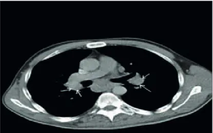
Figure 2 - Thorax CT showing bilateral hilar and mediastinal lymph-adenopaties.
and followed up in the Rheumatology out-patient clinic from the year 2012. On the basis of recurrent uveitis attacks and bilat-eral hilar lymphadenopathies detected on torax computed tomography (CT), she was suspected for sarcoidosis and the lymph node biopsy taken by endobronchial ul-trasonography (EBUS) method revealed non-caseified granuloma formation (Figure 1). Conditions that might cause granuloma-tous disease (such as bacterial and fungal infections, and lymphoma) had been ruled out and the patient had been diagnosed as sarcoidosis stage 1 according to the Scad-ding criteria. Because she had recurrent uveitis attacks, she had occasionally been treated with prednisone 20 mg/day. Three years later, the patient suffered from new complaints such as fever, weight loss and night sweating and was referred to our Hematology clinic. At physical examina-tion, bilateral cervical lymph nodes (7.5 x 6.5 mm) and three lymph nodes of 2 cm in here left groin were palpated. There were no organomegalies. Her laboratory exami-nation results were: Hgb: 12.4 g/L, MCV: 81, WBC: 27.100, Neu: 7710, Neu%28.5, Lym%62, Plt: 396000, Vitamin B12: 469 ng/L, Ferritin: 106 ng/mL.
Liver functions and kidney functions were normal. The acute phase reactants were: C-reactive protein (CRP): 5.38 mg/dl, eryth-rocyte sedimentation rate (ESR): 29 mm/h. The level of serum angiotensin converting enzyme (ACE) was 86 mg/dL (normal <45 mg/dL). There was polyclonal gammopa-thy in her protein electrophoresis. Bilateral hilar and mediastinal lymphadenopathies were present in her thorax CT (Figure 2). Even if she had been followed up because of sarcoidosis, besides cervical and ingui-nal lymphadenopathies, she had complaints such as weight loss and night sweating as well, which is why we performed a lymph node biopsy.
The lymph node was taken from the left cervical region and led to diagnosis of mixed cellular type Hodgkin lymphoma with markers; CD15 (+), CD30 (-), CD20 (-), CD45 (-) (Figure 3). A bone marrow biopsy was performed for staging which revealed no infiltration. The patient was
regression of nearly all the lymphadenopa-thies. The follow-up and the treatment of the patient, who remains clinically stable, is still in progress.
n DISCUSSION
AND CONCLUSIONS
In this paper, we report a patient who has been followed up for sarcoidosis and was subsequently diagnosed with Hodgkin lymphoma as a result of examinations per-formed because of increasing complaints. The initial diagnosis of sarcoidosis had been made by the rheumatologists with reference to clinical indications and symp-toms and histopathological confirmation of non-caseified granulomas on lymph node biopsy. The risk of malignancy develop-ment (especially lymphomas and lung cancer) is increased in patients with sar-coidosis. Chronic inflammation is an im-portant risk for the carcinogenesis process. Sarcoidosis-lymphoma syndrome was first described by Brinker (7). The etiopatho-genesis of this co-occurrence may be ex-plained by immunological abnormalities seen in several malignancies and lympho-proliferative disorders.
Sarcoidosis and lymphoma co-occurrence
may be detected simultaneously, but gener-ally lymphoma develops one or two years later. In our case, the development of lym-phoma from a sarcoidosis base took nearly three years. Patients with sarcoidosis have a high risk of development of malignant lymphoma, and this association was de-fined with the term sarcoidosis-lymphoma
syndrome. Sarcoidal granulomas can be
ob-served in patients with malignant tumors, including malignant lymphoma and carci-noma without history of systemic sarcoid-osis. Ishida et al. reported a 60-year-old Japanese female patient with extranodal marginal zone lymphoma of mucosa-asso-ciated lymphoid tissue (MALT lymphoma) who developed sarcoidosis two years after treatment (8).
Goswami et al. reported 4 patients diag-nosed with sarcoidosis either during or after the treatment of lymphoma (9). Al-though rare, there are previous case reports about the co-occurrence of sarcoidosis and Hodgkin disease. Silva et al. report-ed sarcoidosis diagnosreport-ed with biopsy in a 37-year-old female patient appearing with erythema nodosum and bilateral hilar lymphadenopathies (10). Three years later, this patient manifested widespread lymph-adenopathies and was diagnosed with mixed type Hodgkin lymphoma. However, the development of sarcoidosis in patients with lymphoma was reported too. London et al. reported sarcoidosis in 39 lymphoma (19 Hodgkin) patients (11). Mean devel-opment duration was 18 months and only 16 of the patients were treated. Complete remission was provided in 98% of the treated patients. Sarcoidosis is a chronic inflammatory disease characterized by non-caseified granulomas and lymphoid hyperplasia resulting in the dysfunction of the immunoregulatory pathways. There are many immunological similarities be-tween sarcoidosis and lymphoma; both have cutaneous anergy, peripheral lymph-adenopathies and over infiltration of T-helper cells in tissues. According to one theory, systemic sarcoidosis is initiated by the cell-mediated immune response that develops against the tumor antigens that were secreted by the non-caseified granu-Figure 3 - A) granu-Figure showing mixed cellular population in Hodgkin
lymphoma with various inflammatory cells, Reed-Stenberg cells (H&Ex220). B) Reed-Stenberg and Hodgkin cells selectively immu-nostained for CD-30 marker (x110).
Reumatismo 2/2017 87
Development of Hodgkin lymphoma in a patient with sarcoidosis CASE REPORT
loma inside the tumor (12). It is important to recognize that systemic sarcoidosis and sarcoidal reaction without evidence of systemic disease can occur after develop-ment of malignant lymphoma. Therefore, sarcoidal reaction must be included in the differential diagnostic consideration of re-current malignant lymphoma.
In conclusion, we reported the develop-ment of Hodgkin lymphoma in a patient followed up for a diagnosis of sarcoidosis. Sarcoidosis and lymphoma co-occurrence mainly consists of the development of lymphoma after sarcoidosis. Even rare sarcoidosis may develop in patients with lymphoma. Since clinical manifestations of both diseases are similar, especially if new findings such as fever, weight loss, splenomegaly and new lymphadenopathies appear, histolopathological confirmation is needed. New prospective studies into the pathogenesis of the sarcoidosis-lymphoma syndrome are necessary.
n REFERENCES
1. Newman LS, Rose CS, Maier LA. Sarcoid-osis. N Engl J Med. 1997; 336: 1224-34. 2. Hofmann S, Franke A, Fischer A, et al.
Genome-wide association study identifies
ANXA11 as a new susceptibility locus for sar-coidosis. Nat Genet. 2008; 40: 1103-6. 3. Chen ES, Moller DR. Etiology of sarcoidosis.
Clin Chest Med. 2008; 29: 365-77.
4. Smith G, Brownell I, Sanchez M, Prystowsky S. Advances in the genetics of sarcoidosis. Clin Genet. 2008; 73: 401-12.
5. Askling J, Grunewald J, Eklund A, et al. In-creased risk for cancer following sarcoidosis. Am J Respir Crit Care Med. 1999; 160: 1668-72.
6. Cohen PR, Kurzrock R. Sarcoidosis and ma-lignancy. Clin Dermatol. 2007; 25: 326-33. 7. Brincker H. The sarcoidosis-lymphoma
syn-drome. Br J Cancer. 1986; 54: 467-73. 8. Ishida M, Hodohara K, Furuya A, et al.
Sar-coidal granulomas in the mediastinal lymph nodes after treatment for marginal zone lym-phoma of the esophagus: report of a case with review of the concept of the sarcoidosis-lym-phoma syndrome. Int J Clin Exp Pathol. 2014; 7: 4428-32.
9. Goswami T, Siddique S, Cohen P, Cheson BD. The sarcoid-lymphoma syndrome. Clin Lym-phoma Myeloma Leuk. 2010; 10: 241-7. 10. Silva GN, Morais MF, Raposo J, et al.
Hodg-kin’s disease in a patient with sarcoidosis. Acta Med Portuguesa. 1996; 9: 287-93. 11. London J, Grados A, Fermé C, et al.
Sarcoid-osis occurring after lymphoma: report of 14 patients and review of the literature. Medicine (Baltimore). 2014; 93: e121.
12. Maayan H, Ashkenazi Y, Nagler A, et al. Sar-coidosis and lymphoma: case series and lit-erature review. Sarcoidosis Vasc Diffuse Lung Dis. 2011; 28: 146-52.