Embolization of a complex pulmonary arteriovenous fistula
and coarctation treatment with covered stent at the same
session
Elif Erolu-Günay1, Yalım Yalçın2, Nilüfer Çetiner1, Muhsin Bekir Arpaözü1, Berna Çevik1, Figen Akalın1
Divisions of Pediatric Cardiology, Department of Pediatrics1 Marmara University Faculty of Medicine and 2Bilim University Faculty of Medicine İstanbul, Turkey. E-mail: [email protected]
Received: 10 October 2014, Revised: 4 March 2015, Accepted: 18 November 2015
SUMMARY: Erolu-Günay E, Yalçın Y, Çetiner N, Arpaözü MB, Çevik B, Akalın F. Embolization of a complex pulmonary arteriovenous fistula and coarctation treatment with covered stent at the same session. Turk J Pediatr 2015; 57: 413-417.
Pulmonary arteriovenous fistula (PAVF) are rare malformations that may cause serious complications such as paradoxical embolism, stroke, pulmonary hemorrhage and hemoptysis. Accompanying cardiac malformations such as aortic coarctation were not reported previously. Here we present a case of complex PAVF associated with aortic coarctation. The patient was treated successfully by transcatheter embolization of PAVF with amplatzer vascular plug I and II and implantation of a covered CP stent for coarctation at the same session.
Key words: pulmonary arteriovenous fistula, aortic coarctation, transcatheter intervention, vascular plug, covered stent.
Pulmonary arteriovenous fistulas (PAVF) are abnormal connections of pulmonary arteries with pulmonary veins and they do not have capillary system. The pulmonary segments perfused by anomalous vessels do not participate in gas exchange. The arteriovenous connection causes right-to-left shunt, cyanosis and volume overload to the left ventricle. Serious complications like paradoxical embolization, stroke, transient ischemic attack and cerebral abscess or rupture of the malformation may occur in patients with PAVF, if left untreated. The diagnosis is based on clinical suspicion in patients with cyanosis, continuous murmur and persistent opacity on chest X-ray; and confirmed by contrast echocardiography and pulmonary angiography. Actual choice of treatment is transcatheter embolotherapy of the pulmonary arteriovenous malformation by coils or vascular plugs with satisfactory outcomes.
Aortic coarctation is not previously reported as an accompanying congenital heart disease in patients with PAVF.
We herein present a case of pulmonary arteriovenous malformation (PAVM) associated
with aortic coarctation in whom stent implantation for coarctation of aorta and transcatheter embolization of PAVM was performed at the same session.
Case Report
A 16-year-old Syrian boy was referred to our hospital because of persistent cough and hemoptysis. His symptoms have aggravated for the recent three months. However he had recurrent respiratory tract infections and pneumonia starting from one year of age. He had a surgical operation of the chest wall which was told to be due to a hamartoma. He had growth retardation, height and weight was below 3rd percentile according to the age. Heart rate was 97/min. blood pressure was 140/90 femoral pulses were weak. Cyanosis and clubbing of the fingernails were observed during physical examination. An old incision scar was seen on the right chest. Oxygen saturation was measured as 88%. A continuous murmur was audible at the interscapular area. On the chest X-ray, heart shadow was enlarged, cardiothoracic ratio was 0.50, the
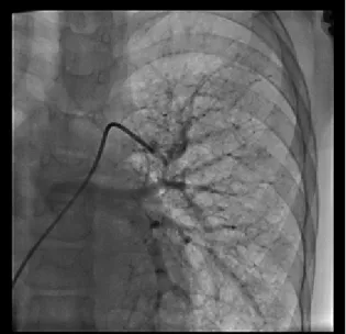
appearance of the lungs suggested chronic bronchopulmonary disease and an opacity was present on the lower lobe of the right lung which was found to be persisting on several of the chest roentgenograms. Electrocardiogram showed sinus rhythm, QRS axis and voltages were normal. High resolution computed tomography of the chest showed volume loss of the right lung which was occupied by nodular lesions. Echocardiographic examination revealed enlarged left atrium and left ventricle with normal systolic and diastolic functions, coarctation of the aorta at the classical site of the aortic arch and 98 mmHg systolic gradient, with a diastolic tail by Doppler echocardiography. Coarctation and chronic lung disease was the initial diagnosis, however cyanosis, radiologic appearance of opacity and presence of the continuous murmur suggested presence of pulmonary arteriovenous fistula. Contrast echocardiography confirmed the diagnosis which was performed using agitated saline given from a large vein, by visualization of the contrast bubbles within the left cardiac chambers. Cardiac catheterization and angiography demonstrated a long segment aortic coarctation with a length of 25.9 mm, diverticulum of the ductus arteriosus was arising from the coarcted segment, the gradient between the proximal and distal aorta was measured as 32 mmHg (Ascending aorta 130 mmHg, Descending aorta 98 mmHg). Massive collateral circulation was present between the proximal and distal parts of aorta. Selective pulmonary artery angiograms showed a very large arteriovenous fistula involving the middle and lower lobe of the right lung with multiple feeding arteries. No blood flow towards the upper lobe was observed. The patient had a history of excision of a hamartoma and the location of the incision scar suggested that the upper lobe of the right lung was the site of hamartoma. Almost all the pulmonary blood flow entering the right pulmonary artery turned to the left atrium without reaching the pulmonary capillaries (Fig. 1). In addition, a few other tiny arteriovenous malformations were visible on the contralateral lung (Fig. 2). A covered CP stent (NuMed, NY, USA) 28 mm in length, 10 mm in diameter mounted over Z-med balloon in balloon catheter was used for stenting the coarctation, post procedural gradient was 6 mmHg (Fig. 3).
In order to occlude the pulmonary arteriovenus fistula, the feeding arteries of the malformation were visualized selectively. Three main feeding arteries were visible and the right lung was entirely covered by the tangle of tortuous vessels, upper lobe was not perfused. At the beginning, 10 mm Amplatzer vascular plug I (St Jude Medical, Minnesota, USA) was deployed within the inferiorly located feeding artery in order to decrease the blood flow and then 12 mm Amplatzer vascular plug II (St Jude Medical, Minnesota, USA) was deployed beginning from the more superiorly located feeding artery and extending towards the right main pulmonary artery (Fig. 4). Pulmonary blood flow to right lung was entirely occluded. As no part of the right lung participated in gas exchange, after occlusion of the right pulmonary artery, peripheral oxygen saturation of the patient increased to 96%.
Nasal mucosa was examined and cranial magnetic resonance imaging (MRI) angiography and abdominal contrast MRI were performed in order to exclude hereditary hemorrhagic telangiectasia. Arteriovenous malformation was not found in nasal mucosa, cranial or liver tissue.
Discussion
Pulmonary arteriovenous malformations are abnormal connections of the arteries and veins without intervening capillary bed. This causes cyanosis, desaturation with resulting exercise intolerance, paradoxical embolism because of direct right to left shunt of the bloodstream without any filtration of its contents like thrombi or bacteria and hemoptysis. In order to prevent these complications, transcatheter embolotheraphy are recommended for PAVMs with a feeding artery larger than 3 mm in diameter1. Three different types of PAVMs
are defined; simple type PAVMs have one or more feeding arteries arising from the same segmental artery, complex PAVMs have multiple feeding arteries originating from different segmental arteries2 and the third type
of PAVMs are described as PAVMs involving at least one segment of the lung diffusely3.
In our patient, middle and lower lobes were involved by PAVM. PAVMs were also present on the contralateral lung but they were very small and clinically insignificant.
The diagnosis of the PAVF is based on clinical findings such as cyanosis, continuous murmur and persistent radiologic findings. Many of the patients have history of recurrent pneumonia which is usually a false diagnosis because of the radiologic appearance during febrile diseases. Our patient was also followed-up with the diagnosis of chronic lung disease. Contrast echocardiography is helpful for confirmation of the diagnosis by observation of the echo-contrast within the left heart chambers after infusion from a systemic vein.
Computed tomography and selective pulmonary angiography are needed for demonstration of detailed anatomy and lung segments involved. Hemodynamic study was not performed since there was a clear-cut indication for intervention. Since surger y is an invasive method, interventional methods with various devices have been developed. Recently, detachable coils or vascular plugs are available for occlusion of PAVM and being used with safety and low complication rates4,5. These devices may be Fig. 1. Right pulmonary artery angiogram shows PAVM
and early venous return. Fig. 2. Left pulmonary artery angiogram shows multiple tiny PAVMs.
Fig. 3. Aortagram showing fully expanded covered CP stent. Fig. 4. Angiogram of right pulmonary artery after occlusion of PAVM with amplatzer vascular plug I and II. Pulmonary blood flow to the right lung was blocked.v
used separately or together6,7. Balloon occlusion
is not preferred. We did not use detachable coils because there were so many tortuous vessels with high blood flow, we tried to avoid the risk of migration. Additionally, angioarchitecture was suitable for placing vascular plug 2 to the feeding artery nearest to the beginning of right pulmonary artery. After placing vascular plug 1 to the feeding artery of the inferiorly located PAVM, it became easier to occlude the superiorly located feeding artery by a vascular plug 2 and extend it towards right pulmonary artery since the amount of blood flow within the malformation decreased.
Choosing the modality of treatment in this case, surgery or intervention, was challenging. After occlusion of the main fistula the tiny malformations found on the contralateral lung could grow further and cause the same clinical findings, and lung transplantation would be the only option in this case. Both surgery and the interventional treatment could have the same consequences, therefore we chose the less invasive one and occluded the fistula by transcatheter route in the same session with interventional coarctation treatment. Total occlusion of a branch pulmonary artery did not cause any complication, since the lung tissue was not yet functional prior to intervention and necrosis did not occur since the lung tissue was perfused by bronchial arteries. Possibly the upper lobe had been resected during the surgical intervention for hamartoma; middle and lower lobes were invaded by arteriovenous malformation.
Since pulmonary arteriovenous malformation may be one of the manifestations of the hereditary hemorrhagic telangiectasia (HHT), we searched for telangiectasia in nasal mucosa and arteriovenous malformations in liver or brain by cranial MRI angiography and abdominal contrast MRI. No telangiectasia or arteriovenous malformations were detected by these investigations. Although some of the findings of HHT like epistaxis and telangiectasia may appear years or decades later, none of the relatives of our patient had known HHT diagnosis, therefore genetic test for HHT was not necessary.
Acquired PAVF may develop in patients with chronic liver disease and cause hepatopulmonary syndrome and in patients with Fontan
circulation which was not the cause in our patient. The surgery or pathology reports were not available, but the history of the previous surgery suggested that this arteriovenous fistula may be a part of a hamartomatosis syndrome. Association of coarctation of aorta with pulmonary arteriovenous fistula was not reported previously, presence of these two pathologies in the same patient was the other interesting feature of our patient. In patients with arteriovenous malformations of the central nervous system such as Galen vein aneurysm, coarctation or coarctation-like physiology causing persistent fetal circulation may be found8. However in these patients,
steal phenomenon because of increased cerebral blood flow and decreased flow in descending aorta may explain the association. In pulmonary arteriovenous fistula, aortic blood flow increases and there is no steal in systemic circulation. Therefore other unknown, probably genetic, mechanisms may be effective in development of the coarctation.
In conclusion, pulmonary arteriovenous malformations must be considered in differential diagnosis in chidren with cyanosis, persistent infiltration or opacity of a lung segment and a continuous murmur. Transcatheter occlusion of the feeding arteries is an effective treatment modality and less invasive comparing surgery. Aortic coarctation is a rare association which may also be treated by transcatheter stent implantation in the same session.
REFERENCES
1. White RI Jr, Lynch-Nylan A, Terry P, et al. Pulmonary arteriovenous malformations: techniques and long-term outcome of embolotherapy. Radiology 1988; 169: 663-669.
2. White RI Jr, Pollak JS, Wirth JA. Pulmonary arteriovenous malformations: diagnosis and transcatheter embolotherapy. J Vasc Interv Radiol 1996; 7: 787-804.
3. Pierucci P, Murphy J, Henderson KJ, Chyun DA, White RI Jr. New definition and natural history of patients with diffuse pulmonary arteriovenous malformations: twenty-seven-year experience. Chest 2008; 133: 653-661.
4. Ando K, Mochizuki A, Kurimoto N, et al. Coil embolization for pulmonary arteriovenous malformation as an organ-sparing therapy: outcome of long-term follow-up. Ann Thorac Cardiovasc Surg 2011; 17: 118-123.
5. Tapping CR, Ettles DF, Robinson GJ. Long-term follow-up of treatment of pulmonary arteriovenous malformations with AMPLATZER Vascular Plug and AMPLATZER Vascular Plug II devices. J Vasc Interv Radiol 2011; 22: 1740-1746.
6. Hundt W, Kalinowski M, Kiessling A, et al. Novel approach to complex pulmonary arteriovenous malformation embolization using detachable coils and Amplatzer vascular plugs. Eur J Radiol 2012; 81:e732-e738.
7. Trerotola SO & Pyeritz RE. Does use of coils in addition to amplatzer vascular plugs prevent recanalization. . AJR Am Roentgenol 2010; 195: 766-771.
8. Deverall PB, Taylor JF, Sturroc GS, Aberdeen E. Coarctation-like physiology with cerebral arteriovenous fistula. Pediatrics 1969; 44: 1024-1028.