The therapeutic results after laparoscopic hellers’ myotomy and
partial fundoplication for achalasia
Kamil Gülpınar1, Haydar Celasin2, Ulas Sözener3, Ahmet Türkcapar4 1Department of General Surgery, Ufuk University Faculty of Medicine, Ankara, Turkey 2Department of General Surgery, Ankara Kavaklıdere Umut Private Hospital, Ankara, Turkey 3Department of General Surgery, Ankara Medicana International Hospital, Ankara, Turkey 4Department of General Surgery, Ankara University Faculty of Medicine, Ankara, Turkey
ABSTRACT
Background/Aims: The aim of this prospectively designed study was to postoperatively assess the subjective com-plaints or relief of symptoms of achalasia patients’ who underwent Laparoscopic Hellers’ myotomy and partial fundoplication in our clinic.
Materials and Methods: 40 patients were enrolled in the study that underwent Laparoscopic Hellers’ myotomy and partial fundoplication (toupet or dor) for idiopathic achalasia in our clinic between years 2002 and 2012. Post-operative follow-up was conducted at 1st, 3rd. and 6th months in each patient for symptoms and a follow-up ques-tionnaire was completed which is classified according to Vantrappen and Hellemans’ modified classification. Results: 34 patients underwent Laparoscopic Heller myotomy with Toupet fundoplication whereas 6 patients un-derwent Laparoscopic Heller myotomy with Dor fundoplication. After 3 months, 2 patients’ complaints of dyspha-gia were detected (5%). In the 6 month follow-up, one of these two patient dysphadyspha-gia symptoms got worsened and a balloon dilatation was performed (2.5%). In the other one no clinical reason was found for dysphagia and medical therapy was initiated. In the 6 th month only three patients (7.5%) were presented with gastroesophageal reflux which successful medical treatment was initiated.
Conclusion: Laparoscopic myotomy with fundoplication seems to be the most effective surgical technique that provides both short and long term symptomatic relief with released hospitalization time and less complication rate.
Keywords: Laparoscopic Hellers’ myotomy and partial fundoplication, achalasia, surgical therapy
INTRODUCTION
Achalasia is an esophageal motor disorder that results from the absence of esophageal peristalsis combined with a defective relaxation of the lower esophageal sphincter (LES). Since the etiology of the disease is not known the therapies are based on reducing the out-flow resistance caused by the dysfunctional LES and prevent clinical symptoms like dysphagia, chest pain, regurgitation. Medical treatment, botulinum toxin in-jections, pneumatic dilatation (PD) and surgical my-otomy operations are currently available to achieve this goal. The treatment options are still revolving; more recently per oral endoscopic myotomy (POEM)
has been introduced as a new treatment for achalasia with promising short term results, but time will show its long term efficiency. Although controversial many surgeons agree upon the surgical treatment to be primary therapy in achalasia. In addition, minimally in-vasive techniques as laparoscopic hellers myomtomy and partial fundoplication in achalasia surgery have become the gold standard therapy in many clinics. The aim of our study was to postoperatively assess the subjective complaints or relief of symptoms of patients’ who underwent Laparoscopic Hellers’ myotomy and partial fundoplication in our clinic.
This study was presented at the European Surgery Congress 2012, İstanbul, Turkey.
Address for Correspondence: Kamil Gülpınar, Department of General Surgery, Ufuk University School of Medicine, Ankara, Turkey E-mail: [email protected]
Received: February 03, 2013 Accepted: March 16, 2013
© Copyright 2014 by The Turkish Society of Gastroenterology • Available online at www.turkjgastroenterol.org • DOI: 10.5152/tjg.2014.4944
Or
iginal Ar
MATERIALS AND METHODS
After patients written informed consent were sought and the study was approved by the Ankara University School of Medi-cine ethical committee, 40 patients were enrolled in the study that underwent Laparoscopic Hellers’ myotomy and partial fundoplication (toupet or dor) for idiopathic achalasia in our clinic. They all underwent operation by the same team of sur-geons (n:3) and using the same technique. The surgeries took place between 2002 and 2012. Medical records were reviewed for patient demographics, frequency, and duration of preop-erative dysphagia, reflux, regurgitation and chest pain. Preop-erative diagnostic tests including barium esophagography, esophageal pH monitoring, esophageal manometry and up-per gastrointestinal endoscopy were completed in all patients. Prior therapeutic interventions including endoscopic pneu-matic dilatation, botox injection or surgical myotomy were re-corded. Surgical technique in each patient was completed as described by Harold, 2004 and Morino, 1995 (1,2).
Briefly, under general anesthesia the patient placed in supine position with the surgeon being between legs. A standard 5-tro-char approach was used. The gastro hepatic ligament is initially divided to expose the right crus. The posterior attachments of the esophagus are bluntly divided, preserving the posterior va-gus nerve. The anterior phrenoesophagial ligament is incised, and the esophagus is circumferentially dissected and encircled with a Penrose drain. The esophagus is mobilized superiorly for at least 8cm into the mediastinum. The short gastric vessels are divided to allow for a tension free fundoplication. The anterior vagus nerve is also identified and bluntly dissected from the pro-posed myotomy site. The myotomy extends 4-5 cm above the gastroesophageal junction and at least 1-2 cm onto the stom-ach. Intraoperative endoscopy is routinely performed to ensure an adequate myotomy and identify mucosal injury in every patient. If an inadequate relaxation was encountered by using bougies, esophageal site myotomy was extended for another 2cm. After myotomy, a posterior partial fundoplication (Toupet) was performed. In cases with mucosal perforation, after muco-sal primary suturing is done, anterior fundoplication (Dor) was performed.
Postoperative follow-up was conducted at 1st, 3rd. and 6th months in each patient for symptoms of recurrent achalasia. The data was collected from routine office visits and our staff also contacted patients who were unable to come to visits by phone. A complete follow-up questionnaire was done for each patient and classified according to Vantrappen and Hellemans’ modified classification (3) as follows;
1. excellent: asymptomatic
2. good: occasional dysphagia, thoracic pain or heartburn re-quiring no therapy
3. fair: dysphagia, thoracic pain or heartburn more than once a week, requiring therapy although the patient was better than before the surgery.
4. poor: the patient worse than before surgery (dysphagia, thoracic pain, heartburn, regurgitation or weight loss) RESULTS
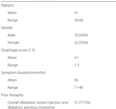
The 22 female (55%) and 18 male (45%) patients with a mean age of 41 (18- 60) was included in the study. All of the patients had more than 6 months of dysphagia, thoracic pain or heart-burn before surgery. The duration of symptoms, expressed as a median (range) was 36 months (6 months-20 years). 31 pa-tients (77,5%) have had previous ineffective medical therapies combined with balloon dilatation. 1 patient (2.5%) has had previous ineffective antireflux surgery (Nissen operation) a year ago and diagnosed as achalasia when he admitted to our hos-pital. Patient characteristics are shown in Table 1.
34 patients underwent Laparoscopic Heller myotomy with Toupet fundoplication whereas 6 patients underwent Laparo-scopic Heller myotomy with Dor fundoplication.
Intraoperative complications occurred in seven cases (17,5%) which were all esophageal mucosal perforations and primarily sutured during operation. All the esophageal mucosal perfora-tions occurred in patients who had previous balloon dilatation attempts (2 or 5 times) as a first line therapy for achalasia. There were no postoperative complications in patients except for one patient (2.5%) in whom esophageal perforation was detected on postoperative 1st day. He was taken to surgery and a primary repair of perforation was performed.
One month after the surgical treatment all patients had a significant reduction in symptoms of dysphagia and regur-gitation. After 3 months, 2 patients’ complaints of dysphagia were detected (5%). In the 6 month follow-up, one of these
Age(yrs) Mean 41 Range 18-60 Gender Male 18 (45%) Female 22 (55%) Dysphagia score (1-5) Mean 4.1 Range 1-5 Symptom duration(months) Mean 36 Range 7-140 Prior theraphy
Overall (dilatation, botox injection and 31 (77.5%) dilatation, previous myotomy)
Table 1. Patients undergoing surgery (n:46)
Or
iginal Ar
two patient dysphagia symptoms got worsened and a bal-loon dilatation was performed (2.5%). In the other one no clinical reason was found for dysphagia and medical therapy was initiated. In the 6th month only three patients (7.5%) were presented with gastroesophageal reflux which successful medical treatment was initiated. When the results interpreted in Vantrappen and Hellemans’ modified classification after 6months 92.5% of patients had satisfactory results whereas only 7.5% had unsatisfactory surgical results in terms of relief of symptoms.
When postoperative surgical results are compared in 1st ,3rd and 6th months the relief of symptoms due to Vantrappen and Helle-mans’ modified classification has shown no statistically different (p> 0.001) results during 6 months’ time where satisfactory rates are found to be 100%, 95% and 92,5% respectively (Table 2). Mean dysphagia score improved preoperatively from 4.1 to 1.6 (p<0.001) at six 6th months follow-up.
DISCUSSION
The optimum treatment of achalasia is agreed upon to be sur-gical with various sursur-gical options. In the last decade a radical change in the surgical treatment of achalasia has been per-formed with addition of minimally invasive techniques. After surgical myotomy was demonstrated to treat achalasia in 1913 (4), the technique was preferred and advanced rapidly due its excellent results of symptom relief of dysphagia. Minimally in-vasive surgery as called laparoscopic Heller myotomy has be-come the most commonly performed procedure and again laparoscopic myotomy with partial fundoplication reported to be the most effective surgical technique compared with all en-doscopic and other surgical approaches, with a low complica-tion rate and minimal development of gastroesophageal reflux (5). The minimally invasive approach has many advantages of better visualization of the gastroesophageal junction, magni-fied view of both layers of the esophageal muscularis propria as well as of the gastric oblique fibers. In addition laparoscopic technique has led to better recognition of gastroesophageal junction and to examine the completeness of the myotomy as well as to discover occult perforations by transillumination and air insufflation.
While there is settlement about the requirement for a fundo-plication in conjunction with the myotomy, it is still a debate which type of fundoplication should be performed. Total fun-doplication had good early results, but too much resistance at the level of the gastroesophageal junction obstructing the emptying of food from the esophagus into the stomach by gravity causing persistent or recurrent dysphagia after this procedure in long term, has required new surgery technique search. Total fundoplication was abandoned based on the long term results showing esophageal decompensation and recur-rence of symptoms ultimately occur in most patients (2).This problem was achieved by performing partial fundoplication. In a study; with a long term results of 125 months follow-up time, the recurrence rate of dysphagia was found to be signifi-cantly higher among the patients who underwent Nissen fun-doplication when compared with Dor funfun-doplication and Dor fundoplication was suggested to be the preferred method to re-establish GER control in patients undergoing laparoscopic Heller myotomy (6).
Partial fundoplication surgery is either done by anteriorly (Dor) or posteriorly (Toupet). There are no published data comparing both. Some surgeons prefer a posterior approach as it keeps the edges of the myotomy separated and it may be more ef-fective for antireflux operation while others prefer an anterior method and say it is easier to perform without the need of posterior dissection and it adds the advantage of covering the exposed mucosa (3, 7-10). In our study group we mostly pre-ferred to perform a Laparoscopic Heller myotomy with Toupet fundoplication (34 patients) as it is suggested in many reports (10). We believe if surgically possible a posterior approach with a retro esophageal dissection advance mediastinal esophageal mobilization for the long esophageal myotomy and additional-ly holds the myotomy open for a better antireflux valve. That is why, we believe, depending on our experience, Toupet proce-dure is a better technique in preventing reflux when compared with Dor especially in long term.
But in 6 cases we prefer to perform a Dor fundoplication where posterior dissection were not possible due their anatomic structures and in 4 of them mucosal perforation were en-countered during retro esophageal dissection and eventually switched to anterior approach.
Heller myotomy with fundoplication has few complications (11-13). In a review, the efficacy and safety of Heller myotomy were sought and published studies on treatment of achalasia with HM in PubMed from 1966 to 2008 were evaluated. The most common intraoperative complication of Heller myotomy in that analysis were reported to be gastrointestinal perfora-tion including gastric and esophageal perforaperfora-tion with an in-cidence of 1.5%- 20% (14). Consistent with these studies, our most common intraoperative complication was also esopha-geal mucosal perforation with a 17.5% incidence in 34 patients and primarily sutured during operation.
1st month 3rd month 6th month
Satisfactory 100% 95% 92,5% Excellent 32 (80%) 28 (70%) 22 (55%) Good 8 (20%) 10 (25%) 15 (37.5%) Unsatisfactory none 5% 7.5% Fair 2 (5%) 2 (5%) Poor 1 (2.5%)
Clinical results with Vantrappen and Hellemans’ modified classification
Table 2. Patient characteristics
Or
iginal Ar
Interestingly in our study, all perforations occurred in patients who have had previous balloon dilatation procedures; 5 of the patients have had previous 5 balloon dilatation attempts and the others had previous 2 balloon dilatation attempts before surgeries.
Pneumatic dilatation is the first attempt and an effective form of treatment of achalasia. Although it appears to be the most cost effective alternative, there is controversy whether pre-operative endoscopic treatment might affect subsequent surgical outcomes. It has been reported that previous endo-scopic therapy with pneumatic dilatation or Botox injections may lead to the development of inflammation between the surgical planes thus complicating the surgical procedure and increasing the likelihood of developing intraoperative perfo-ration(15-17). Some other studies showed no difference in outcomes regardless of whether prior endoscopic therapy was rendered (18, 19). A population based study reported that in terms of relief of symptoms both Heller myotomy and balloon dilatation provided good results however the cu-mulative risk of subsequent intervention for achalasia at 10 years was 63.5% for patients initially treated with dilatation compared to 37.5% for patients initially treated with surgical myotomy(20).
In our experience of patients, we believe that balloon dilatation as an initial treatment in achalasia should be debated well be-fore performed as it might interfere with the following surgical outcomes.
There is a remarkable increase in the number of patients re-ferred for surgery every year (21) and fortunately rere-ferred pa-tient incidence without previous treatment is increased as well (22). This fact simply amplifies surgery success with lower per-foration rates.
Comprehensive exploration for esophageal perforations and primary reparation during operations is extremely important in preventing postoperative leakage. Perforation recognition is relatively easy with laparoscopic technique with the use of intraoperative endoscopy that identifies occult perforations by transillumination and air insufflation. In our series, recognizing and repairing even very small perforations less than 1cm that only required 1 stitch for primary repair were enough for pre-venting leakage and serious post-operative complications. We did not encounter any complications afterwards.
In the review where the efficacy and safety of Heller myotomy were sought and published studies on treatment of achalasia with HM in PubMed from 1966 to 2008 were evaluated; during follow-up gastroesophageal reflux was found to be a frequent symptom presenting at a rate of 2.6%-20%. Our study group presented a slightly lower rate of 7.5% during a 6 th month fol-low-up that is why we believe the outcome of the surgery is fairly good with symptom relief.
When compared with other treatment options the best out-comes for achalasia disease seems to be Heller myotomy and fundoplication. In a meta-analysis of 105 articles reporting on 7855 patients; laparoscopic myotomy when combined with an antireflux procedure, provided better symptom relief (90%) than all endoscopic and other surgical approaches and a low complication rate (6.3%) (23). In our study group, constant with previous studies, 6 months follow-up showed a 95% of preop-erative dysphagia, reflux, regurgitation and chest pain symp-tom relief with a complication rate of 17.5% (24). Indubitably, we have to admit the weakness of our results depending on small patient group and relatively short follow-up time limits making assumptions. But our results will advance by time with increased surgical experience and higher number of patient records.
In this report, we believe it is important to emphasize the peroral endoscopic myotomy (POEM) in the achalasia treatment options which appears to be a promising, incisionless selective myoto-my, and a less invasive surgical treatment. In 2008, Haru Inoue, a surgical endoscopist in Yokohama presented 4 clinical cases of submucosal myotomy for achalasia at Digestive Diseases Week and subsequently published his early results (25). He coined the acronym POEM (per-oral endoscopic submucosal myotomy), which is widely used for this approach. Currently, more than 700 clinical cases of POEM have been performed worldwide and en-thusiasm for the procedure is growing. Despite this, only a few series have been conducted in peer-reviewed literature and these have concentrated on the technique and immediate out-comes. Future work comparing its long term results with Hellers myotomy and fundoplication should be conducted.
In conclusion, depending on our results and to date literature, we believe laparoscopic myotomy with fundoplication is the most effective minimally invasive surgical technique that pro-vides both short and long term symptomatic relief with de-creased hospitalization time and less complication rate. Disclosures and conflicts of interest:
Corresponding author (corresponding author) and all other co-authors (HC and AT) have no conflict of interest.
Conflict of Interest: No conflict of interest was declared by the authors.
REFERENCES
1. Harold KL, Matthews BD, Kercher KW, Sing RF, Heniford BT. Surgi-cal treatment of achalasia in the 21st century. South Med J 2004; 97: 7-10.
2. Morino M, Rebecchi F, Festa V, Garrone C. Laparoscopic Heller cardiomyotomy with intraoperative manometry in the manage-ment of oesophageal achalasia. Int Surg. 1995; 80: 332-5. 3. Vantrappen G, Hellemans J. Treatment of achalasia and related
motor disorders. Gastroenterology 1980; 79: 144-54.
Or
iginal Ar
4. H E. Extra mucous cardioplasty in chronic cardiospasm with dila-tation of the esophagus Mitt Grenzgels Med Chir 1913; 27: 141. 5. Burpee SE, Mamazza J, Schlachta CM, Bendavid Y, Klein L, Moloo
H, et al. Objective analysis of gastroesophageal reflux after laparo-scopic heller myotomy: an anti-reflux procedure is required. Surg Endosc 2005; 19: 9-14.
6. Ackroyd R, Watson DI, Devitt PG, Jamieson GG. Laparoscopic cardiomyotomy and anterior partial fundoplication for achalasia. Surg Endosc 2001; 15: 683-6.
7. Yamamura MS, Gilster JC, Myers BS, Deveney CW, Sheppard BC. Laparoscopic heller myotomy and anterior fundoplication for achalasia results in a high degree of patient satisfaction. Arch Surg 2000; 135: 902-6.
8. Perrone JM, Frisella MM, Desai KM, Soper NJ. Results of laparo-scopic Heller-Toupet operation for achalasia. Surg Endosc 2004; 18: 1565-71.
9. Khajanchee YS, Kanneganti S, Leatherwood AE, Hansen PD, Swanstrom LL. Laparoscopic Heller myotomy with Toupet fundo-plication: outcomes predictors in 121 consecutive patients. Arch Surg 2005; 140: 827-33.
10. Wright AS, Williams CW, Pellegrini CA, Oelschlager BK. Long-term outcomes confirm the superior efficacy of extended Heller myot-omy with Toupet fundoplication for achalasia. Surg Endosc 2007; 21: 713-8.
11. O’Connor JB, Singer ME, Imperiale TF, Vaezi MF, Richter JE. The cost-effectiveness of treatment strategies for achalasia. Dig Dis Sci 2002;47(7):1516-25.
12. Moawad FJ, Wong R. Modern management of achalasia. Curr Opin Gastroenterol 2010; 26: 384-8.
13. Wang L, Li YM, Li L. Meta-analysis of randomized and controlled treatment trials for achalasia. Dig Dis Sci 2009; 54: 2303-11. 14. Campos GM, Vittinghoff E, Rabl C, Takata M, Gadenstatter M, Lin F,
et al. Endoscopic and surgical treatments for achalasia: a system-atic review and meta-analysis. Ann Surg 2009; 249: 45-57. 15. Smith CD, Stival A, Howell DL, Swafford V. Endoscopic therapy for
achalasia before Heller myotomy results in worse outcomes than heller myotomy alone. Ann Surg 2006; 243: 579-84.
16. Snyder CW, Burton RC, Brown LE, Kakade MS, Finan KR, Hawn MT. Multiple preoperative endoscopic interventions are associ-ated with worse outcomes after laparoscopic Heller myotomy for achalasia. J Gastrointest Surg 2009; 13: 2095-103.
17. Beckingham IJ, Callanan M, Louw JA, Bornman PC. Laparoscopic cardiomyotomy for achalasia after failed balloon dilatation. Surg Endosc 1999; 13: 493-6.
18. Cowgill SM, Villadolid DV, Al-Saadi S, Rosemurgy AS. Difficult my-otomy is not determined by preoperative therapy and does not impact outcome. JSLS 2007; 11: 336-43.
19. Tsuboi K, Omura N, Yano F, Kashiwagi H, Kawasaki N, Suzuki Y, et al. Preoperative dilatation does not affect the surgical outcome of laparoscopic Heller myotomy and Dor fundoplication for esopha-geal achalasia. Surg Laparosc Endosc Percutan Tech 2009; 19: 98-100.
20. Lopushinsky SR, Urbach DR. Pneumatic dilatation and surgical myotomy for achalasia. JAMA. 2006; 296: 2227-33.
21. Patti MG, Fisichella PM, Perretta S, Galvani C, Gorodner MV, Rob-inson T, et al. Impact of minimally invasive surgery on the treat-ment of esophageal achalasia: a decade of change. J Am Coll Surg 2003; 196: 698-703.
22. Patti MG, Herbella FA. Fundoplication after laparoscopic Heller myotomy for esophageal achalasia: what type? J Gastrointest Surg. 2010;14(9):1453-8.
23. Topart P, Deschamps C, Taillefer R, Duranceau A. Long-term ef-fect of total fundoplication on the myotomized esophagus. Ann Thorac Surg 1992; 54: 1046-51.
24. Rebecchi F, Giaccone C, Farinella E, Campaci R, Morino M. Ran-domized controlled trial of laparoscopic Heller myotomy plus Dor fundoplication versus Nissen fundoplication for achalasia: long-term results. Ann Surg 2008; 248: 1023-30.
25. Inoue H, Minami H, Kobayashi Y, Sato Y, Kaga M, Suzuki M, et al. Peroral endoscopic myotomy (POEM) for esophageal achalasia. Endoscopy 2010; 42: 265-71.
Or
iginal Ar