Journal of Clinical and Analytical Medicine |
O
h
r
c
i
r
g
a
in
e
a
s
l
R
e
1 Ozge Kurmus1, Turgay Aslan1, Ozgur Kirbas2, Cemal Koseoglu3, Tolga Han Efe4, Mehmet Bilge51Department of Cardiology, Ufuk University Faculty of Medicine, Ankara, 2Department of Cardiology, Yuksek Ihtisas University, Ankara, 3Department of Cardiology, Tokat State Hospital, Tokat, 4Department of Cardiology, Dıskapı Yıldırım Beyazıt Education and Research Hospital, Ankara, 5Department of Cardiology, Ataturk Education and Research Hospital, Ankara, Turkey
Mean platelet volume and coronary collaterals
Evaluation of mean platelet volume in patients with
different degree of coronary collateral development
DOI: 10.4328/JCAM.5252 Received: 29.07.2017 Accepted: 06.09.2017 Published Online: 06.09.2017 Printed: 01.01.2018 J Clin Anal Med 2018;9(1): 23-6 Corresponding Author: Ozge Kurmus, Department of Cardiology, Ufuk University Faculty of Medicine, Ankara, Turkey.
GSM: +905055253318 E-Mail: [email protected] Abstract
Aim: Coronary collateral vessels are an alternative source of blood supply to a myocardial area jeopardized by ischemia. As some patients have poor collaterals despite significant stenosis, it is thought that multiple factors affect collateral development beside coronary artery disease severity. Mean platelet volume is an indicator of platelet activation. Increased mean platelet volume is found to be related to worse prognosis in the coronary artery disease. In this study, we aimed to investigate the relationship between mean platelet volume and coronary collateral development. Material and Method: Patients with total occlusion in at least one coronary artery were enrolled in this study. Coronary angiography images of 367 patients without a history of revascularization were evaluated retrospectively, and coronary collateral development was graded according to Rentrop classification. Patients were divided into two groups based on Rentrop classification. Rentrop grade 0-1 was regarded as poor collateral development, and Rentrop 2-3 was regarded asgood collateral development. Mean platelet volume was compared between these two groups. Results: Poor collateral development was found in 236 patients (64.3%), and good collateral development was found in 131 patients (35.7%). There was no statistically significant difference in mean platelet volume levels between two groups (9.9±1,2 fL and 10.3±1.3 fL p=0,228). The 3-vessel disease was found to be a predictor of good collateral development (p=0.024). Discussion: In this study, it was found that there was no relationship between mean platelet volume and coronary collateral development.
Keywords
Coronary Collateral Development; Mean Platelet Volume; Coronary Artery Disease
| Journal of Clinical and Analytical Medicine Mean platelet volume and coronary collaterals
2
Introduction
Platelets play an important role in the pathogenesis of coro-nary artery disease (CAD). Their reactivity is a key issue and platelet size, simply measured by mean platelet volume (MPV), is an indirect marker of platelet reactivity [1,2]. Large platelets have a greater content of granules, higher thrombotic potential, increased thromboxane synthesis and serotonin release [3-5]. It has been reported that elevated MPV is associated with in-creased mortality following myocardial infarction and inin-creased cardiovascular events in patients undergoing either an elective or urgent percutaneous coronary intervention [2,6].
Coronary collateral vessels serve as an alternative source of blood supply to an ischemic myocardium. Poor collateral de-velopment was found to be associated with larger infarct size and mortality in CAD patients [7,8]. One of the well-established determinants of collateral formation is coronary artery steno-sis, but poor collateral development despite significant stenosis suggest that multiple mechanisms contribute to collateral for-mation [9]. Some mediators such as nitric oxide (NO), vascular endothelial growth factor, thromboxane, prostacyclin take part in the formation of collateral vessels [10-12].
In this study, we aimed to evaluate the relationship between MPV and coronary collateral formation in CAD patients. Material and Method
The present study is a retrospective cross-sectional study. Be-tween July 2011 and August 2012, 367 patients who underwent coronary angiography at our institution were enrolled in this study. All patients underwent coronary angiography because of suspicion of CAD based on their symptoms or diagnostic tests. Patients with at least one totally occluded major epicardial coronary artery were included in the study. Demographic and clinical data including age, gender, the prevalence of DM, hy-pertension, dyslipidemia, smoking history, and clinical presen-tation were obtained from all patients. Exclusion criteria were as follows: previous coronary revascularization history, history of malignancy or inflammatory disease, evidence of infectious disease, severe hepatic, or renal insufficiency, receiving anti-coagulants and having congenital or acquired blood disorders. Also, patients with incomplete data were excluded. The study was approved by the local ethics committee.
Venous peripheral blood samples for complete blood count were drawn from patients undergoing elective (following a fasting period of 12 hours) or urgent coronary angiography (on ad-mission). Blood samples were taken into standardized, EDTA containing tubes. Platelet parameters including count and MPV were determined by Beckman Coulter LH 780 Hematology Ana-lyzer. Measurements were completed within one hour of blood sampling to avoid the EDTA induced platelet swelling with time. Angiographic Procedure and Coronary Collateral Grading Coronary angiography was performed via the femoral artery for all patients using the Judkins technique. Coronary stenosis de-gree, infarct related artery (IRA), and coronary collateral grad-ing were estimated by two independent cardiologists who were blinded to the clinical information and laboratory parameters of the patients. Patients with at least one coronary artery with a total occlusion were enrolled in the study. Coronary collat-eral development was graded according to Rentrop
classifica-tion: 0= no filling of any collateral vessel; 1= filling of the side branches of the the artery to be perfused by collateral vessels without visualization of the epicardial segment; 2= partial filling of the distal epicardial segment by collateral vessels; 3= com-plete filling of the distal epicardial segment by collateral ves-sels. Rentrop 0-1 was graded as poor collateral development, and Rentrop 2-3 was graded as good collateral development [13].
Statistical analysis
The Kolmogorov–Smirnov test was used to evaluate normal distribution. Continuous variables were expressed as mean ± standard deviation (SD), and categorical variables were defined as numbers and percentages. Student’s t-test or oneway analy-sis of variance (ANOVA) was used to compare continuous vari-ables. Differences in the distribution of categorical variables were assessed using chi-square analysis. The best predictors of the poor collateral development were evaluated by multiple logistic regression analysis. Any variable whose univariable test had a p value <0.25 was accepted as a candidate for the multi-variable model along with all multi-variables of known clinical impor-tance. Adjusted odds ratios and 95% confidence intervals (CIs) were also calculated. The results were considered significant when the p value was <0.05.
Results
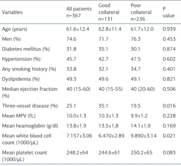
The mean age of the study population was 61.6±12.4, and 74% of the patients were male. 94 patients had a diagnosis of stable angina pectoris, 97 patients had unstable angina pectoris or non-ST elevation myocardial infarction, 176 had ST elevation myocardial infarction. Of the 367 patients, 131 patients (35.7 %) had good collateral development, and 236 patients (64.3 %) had poor collateral development. The two groups did not differ for age, gender, diabetes mellitus, hyperlipidemia, hyper-tension, and smoking history. The demographic and laboratory characteristics of the groups are shown in Table 1.
Mean MPV was 10.0±1.3 fL, and the mean platelet count was 248.2±64 (1000/μL). The mean MPV was 10.3±1.3 fL in the
Table 1. Clinical and laboratory characteristics among poor collateral and good collateral groups
Variables All patientsn=367 Good collateral n=131 Poor collateral n=236 P value Age (years) 61.6±12.4 62.8±11.4 61.7±12.0 0.939 Men (%) 74.6 71.7 76.3 0.453 Diabetes mellitus (%) 31.8 35.1 30.1 0.874 Hypertension (%) 45.7 42.7 47.5 0.602
Any smoking history (%) 33.8 32.1 34.7 0.401
Dyslipidemia (%) 49.3 49.6 49.1 0.821
Median ejection fraction (%)
40 (15-60) 40 (15-55) 40 (20-60) 0.506 Three-vessel disease (%) 25.1 35.1 19.5 0.016 Mean MPV (fL) 10.0±1.3 10.3±1.3 9.9±1.2 0.228 Mean heamoglobin (g/dl) 13.8±1.9 13.5±1,8 14.1±1,9 0.169 Mean white blood cell
count (1000/μL)
7.157±3.06 6.470±2.89 9.890±3.14 0.021 Mean platelet count
(1000/μL) 248.2±64 244.6±61 250.2±65 0.083 | Journal of Clinical and Analytical Medicine
24
| Journal of Clinical and Analytical Medicine Mean platelet volume and coronary collaterals
3
good collateral group and 9.9±1.2 fL in the poor collateral group. There was no difference regarding mean MPV between two groups (p=0.228). Platelet count was not different between two groups (p=0.083). White blood cell count was higher in the poor collateral group than good collateral group (p=0.021). There were more patients with 3-vessel disease among the pa-tients with good collateral development compared to papa-tients with poor collateral development (p=0.016). In multivariate analysis, three vessel disease was found to be a predictor of good collateral development (odds ratio 0.595, 95%CI: 0.380-0.933, p=0.024).
Discussion
In our study, we investigated whether MPV is related to collat-eral development in patients with CAD. We found that the MPV levels were not related to coronary collateral development. A well developed coronary collateral limits the ischemia, reduce the size of myocardial infarction, preserve left ventricle func-tion, and has a favorable impact on the prognosis of patients with coronary artery disease [8,14]. Collateral vessels are a valuable source for alternative blood supply to ischemic myo-cardium especially in case of unachievable revascularization. But, there is notable variation in the degree of coronary col-lateral development. Colcol-lateral development is a multifactorial process, and it is important to define the factors that facilitate collateral development.
Platelets play a crucial role in the pathogenesis of atheroscle-rotic complications, and they are important targets for the treatment of coronary artery disease. MPV is an indirect mark-er of platelet activity. Largmark-er, metabolically, and enzymatically more active platelets have greater prothrombotic features [5]. Elevated MPV was associated with worse clinical outcomes in patients with CAD [2]. Whether platelets with elevated MPV af-fect outcomes for worse by collateral formation or not hasn’t been explained clearly.
Previous studies about this issue are controversial. In a study of patients with the acute coronary syndrome, high MPV on admission was found to be associated with the presence of coronary collateral formation [15]. In contrast, in another study, elevated MPV was found to be a predictor of inadequate col-lateral development [16]. Some studies revealed that MPV lev-els were not related to coronary collateral development [17-19]. We also found that there wasn’t any significant relation between the collateral formation and MPV. Our study differed from the studies mentioned above in some aspects. Patients with >%50, >%80 or >%90 stenosis were enrolled in some of the previous studies. As the variations of the severity of the ste-nosis may affect collateral formation, we enrolled patients with at least one totally occluded major coronary artery. Also, there is a difference in classification of patients according to Rentrop classification. Rentrop 2-3 collaterals were accepted as good collateral formation in our study, whereas only Rentrop 3 was accepted as adequate collateral development in some studies. In a study by Tan et al. platelet activation was measured by soluble CD40 ligand, soluble P-selectin, and soluble glycopro-tein V. They reported that the correlation between the degree of collateralization and these platelets activation markers was not significant [20]. Thromboxane A2 and serotonin have been
shown to cause vasoconstriction of the collateral vessels [21]. Large platelets have a greater content of granules, increased thromboxane synthesis and serotonin release. It is possible that activated platelets within collateral vessels could cause vaso-constriction and decrease collateral flow. It has been reported that platelet activating factor caused a decrease in coronary collateral flow with the participation of thromboxane A2 [12]. It has been reported that serotonin blocker augments flow re-serve of the collateral circulation in anginal patients [22]. Col-lateral development is a multifactorial process, and the further studies are required to understand the role of activated plate-lets in collateral formation.
Conclusion
In conclusion, MPV levels were not related to coronary collateral development in a group of patients with either stable coronary artery disease or acute coronary syndrome.
Our study has some limitations. First, it is a retrospective study. Because of its retrospective design, there were no available data about previous antiplatelet drug use. It is possible that previous usage of antiplatelet drugs may have modulated MPV levels. Secondly, angiographically visible collaterals represent only a fraction of the total collateral vessel amount.
Animal and human rights statement
No animal studies were carried out by the authors for this arti-cle. The study was approved by the institutional ethics commit-tee. All procedures performed in studies involving human par-ticipants were in accordance with the ethical standards of the institutional and/or national research committee and with the 1964 Helsinki Declaration and its later amendments or compa-rable ethical standards.’
Scientific Responsibility Statement
The authors declare that they are responsible for the article’s scientific content including study design, data collection, analy-sis and interpretation, writing, some of the main line, or all of the preparation and scientific review of the contents and ap-proval of the final version of the article.
Animal and human rights statement
All procedures performed in this study were in accordance with the ethical standards of the institutional and/or national re-search committee and with the 1964 Helsinki declaration and its later amendments or comparable ethical standards. No ani-mal or human studies were carried out by the authors for this article.
Funding None
Conflict of interest
None of the authors received any type of financial support that could be considered potential conflict of interest regarding the manuscript or its submission.
References
1. Van der Loo B, Martin JF. A role for changes in platelet production in the cause of acute coronary syndromes. Arterioscler Thromb Vasc Biol. 1999(3); 19: 672-9. 2. Chu SG, Becker RC, Berger PB, Bhatt DL, Eikelboom JW, Konkle B, et al. Mean
Journal of Clinical and Analytical Medicine | 25
| Journal of Clinical and Analytical Medicine Mean platelet volume and coronary collaterals
4
platelet volume as a predictor of cardiovascular risk: a systematic review and meta-analysis. J Thromb Haemost. 2010; 8(1): 148-56.
3.Schoene NW. Design criteria: tests used to assess platelet function. Am J Clin Nutr. 1997; 65(5): 1665-8.
4.Bath PM, Butterworth RJ. Platelet size: measurement, physiology, and vascular disease. Blood Coagul Fibrinolysis. 1996; 7(2): 157-61.
5. Corash L, Tan H, Gralnick HR. Heterogeneity of human whole blood platelet subpopulations. I. The relationship between buoyant density, cell volume, and ul-trastructure. Blood. 1977; 49(1): 71-87.
6. Eisen A, Bental T, Assali A, Kornowski R, Lev EI. Mean platelet volume as a pre-dictor of long-term outcome after percutaneous coronary intervention. J Thromb Thrombolysis. 2013; 36(4): 469-74.
7. Kim EK, Choi JH, Song YB, Hahn JY, Chang SA, Park SJ, et al. A protective role of early collateral blood flow in patients with ST-segment elevation myocardial infarction. Am Heart J. 2016;171(1):56-63.
8. Hara M, Sakata Y, Nakatani D, Suna S, Nishino M, Sato H, et al.; OACIS In-vestigators. Impact of coronary collaterals on in-hospital and 5-year mortality after ST-elevation myocardial infarction in the contemporary percutaneous coro-nary intervention era: a prospective observational study. BMJ Open. 2016;6(7): e011105.
9. Pohl T, Seiler C, Billinger M, Herren E, Wustmann K, Mehta H, et al. Frequency distribution of collateral flow and factors influencing collateral channel develop-ment. Functional collateral channel measurement in 450 patients with coronary artery disease. J Am Coll Cardiol. 2001;38(7):1872-8.
10. Matsunaga T, Warltier DC, Weihrauch DW, Moniz M, Tessmer J, Chilian WM. Ischemia-induced coronary collateral growth is dependent on vascular endothelial growth factor and nitric oxide. Circulation. 2000;102(25):3098-103.
11.Schultz A, Lavie L, Hochberg I, Beyar R, Stone T, Skorecki K, et al. Interindividual heterogeneity in the hypoxic regulation of VEGF: significance for the development of the coronary artery collateral circulation. Circulation. 1999;100(5):547-52. 12. Kinn JW, Bache RJ. Effect of platelet activation on coronary collateral blood flow.
Circulation. 1998;98(14):1431-7.
13. Rentrop KP, Cohen M, Blanke H, Phillips RA. Changes in collateral channel filling immediately after controlled coronary artery occlusion by an angioplasty balloon in human subjects. J Am Coll Cardiol. 1985;5(3):587-92.
14. Habib GB, Heibig J, Forman SA, Brown BG, Roberts R, Terrin ML, et al. Influence of coronary collateral vessels on myocardial infarct size in humans. Results of phase I thrombolysis in myocardial infarction (TIMI) trial. The TIMI Investigators. Circulation. 1991;83(3):739-46.
15. Duran M, Gunebakmaz O, Uysal OK, Ocak A, Yilmaz Y, Arinc H, et al. Relation between mean platelet volume and coronary collateral vessels in patients with acute coronary syndromes. J Cardiol. 2013;61(4):295-8.
16. Ege MR, Acıkgoz S, Zorlu A, Sıncer I, Guray Y, Guray U, et al. Mean plate-let volume: an important predictor of coronary collateral development. Plateplate-lets. 2013;24(3):200-4.
17. Islamoglu Y, Ertas F, Acet H, Elbey MA, Evliyaogllu O, Tekbas E, et al. The as-sociation between mean platelet volume and coronary collateral circulation. Eur Rev Med Pharmacol Sci. 2013;17(2):276-9.
18. Akın F, Ayça B, Çelik Ö, Şahin C. Predictors of poor coronary collateral develop-ment in patients with stable coronary artery disease: neutrophil-to-lymphocyte ratio and platelets. Anatol J Cardiol. 2015;15(3):218-23.
19. Ayhan S, Ozturk S, Erdem A, Ozlu MF, Memioglu T, Ozyasar M, et al. Hema-tological parameters, and coronary collateral circulation in patients with stable coronary artery disease. Exp Clin Cardiol. 2013;18(1):12-5.
20. Tan KT, Tayebjee MH, Macfadyen RJ, Lip GY. Relation of platelet activa-tion to coronary angiographic severity and collateralizaactiva-tion. Am J Cardiol. 2005;96(2):208-10.
21. Wright L, Homans DC, Laxson DD, Dai XZ, Bache RJ. Effect of serotonin and thromboxane A2 on blood flow through moderately well developed coronary col-lateral vessels. J Am Coll Cardiol. 1992;19(3):687-93.
22. Tanaka T, Fujita M, Nakae I, Tamaki S, Hasegawa K, Kihara Y, et al. Improve-ment of exercise capacity by sarpogrelate as a result of augImprove-mented collateral circulation in patients with effort angina. J Am Coll Cardiol. 1998;32(7):1982-6.
How to cite this article:
Kurmus O, Aslan T, Kirbas O, Koseoglu C, Efe TH, Bilge M. Evaluation of Mean Platelet Volume in Patients with Different Degree of Coronary Collateral Develop-ment. J Clin Anal Med 2018;9(1): 23-6.
| Journal of Clinical and Analytical Medicine
26