Review
Hepatorenal syndrome
Selda Demırtas¸1, Murat Can2,* and Aysegu¨l¸
Yarpuzlu3
1University of Ufuk, Faculty of Medicine,
Department of Biochemistry, Ankara, Turkey
2University of Karaelmas, Faculty of Medicine,
Department of Biochemistry, Zonguldak, Turkey
3
University of Ankara, Faculty of Health Education, Ankara, Turkey
Abstract
This article summarizes the literature on current def-inition, suggested pathogenetic mechanisms and the role of laboratory assessment in the differential diagnosis of hepatorenal syndrome (HRS) from other causes of renal disease that may arise during hepatic cirrhosis and some diseases affecting both liver and kidney. It should be remembered that the main theory suggested for the pathogenesis of HRS is the arterial vasodilation hypothesis of portal hypertension, end-ing in type 1 and type 2 HRS, but there is no consen-sus supporting either mechanism as a solid theory for initiation of HRS pathogenesis to date. No laboratory test can firmly establish a diagnosis of HRS, which is mainly based on the absence of any specific cause of renal failure. Laboratory and ultrasonographic tests based on non-invasive techniques are being investi-gated as possible diagnostic approaches.
Keywords: cystatin C; hepatorenal syndrome;
labo-ratory assessment.
Introduction
Hepatorenal syndrome (HRS) is a serious complica-tion in the patient with cirrhosis and ascites, and is characterized by worsening azotemia with avid sodi-um retention and oliguria in the absence of identifia-ble specific causes of renal dysfunction (1). A more current definition of HRS has been established by an international consensus conference organized by the International Ascites Club (2). According to this definition, HRS is a clinical condition that occurs in patients with chronic liver disease, advanced hepatic failure, and portal hypertension characterized by impairment of renal function and marked abnormali-ties in the arterial circulation and activity of the
*Corresponding author: Murat Can, Karaelmas University, Faculty of Medicine, Department of Biochemistry, Zonguldak, Turkey
Phone: q90-372-2610169, Fax: q90-372-2610155, E-mail: [email protected]
endogenous vasoactive systems. There are two types of HRS, type 1 and 2.
Type 1 HRS involves an acute deterioration in renal function, as defined by doubling of the initial serum creatinine to a level greater than 225 mmol/L, or a 50% reduction in initial 24-h creatinine clearance to -20 mL/min over days or weeks, and occurs in an advanced stage of liver disease. The development of type 1 HRS has poor prognosis, with 80% mortality, and 50% of cases are precipitated by gastrointestinal bleeding, infection, dehydration from overt paracen-tesis or diuresis, surgery, or drug exposure, with the remaining 50% occurring spontaneously (1–5). Such patients are commonly oliguric or anuric.
Type 2 HRS occurs in patients with relatively pre-served liver function. These patients show a slow but progressive deterioration in glomerular filtration rate (GFR). This type of HRS usually occurs in patients with diuretic-resistant ascites. It is associated with poor prognosis, although the survival time is longer than that of patients with type 1 HRS (6). According to a recent study, among the possible etiologies of HRS, underlying alcohol-related liver disease may be more commonly associated with HRS compared to other etiologies (7). In vivo studies on the effects of alcohol on liver metabolism resulting in HRS are rare. Most studies are still based on experimental evalua-tions in the rat (3, 8).
Pathogenetic mechanisms and modulators
To date, several factors have been implicated in the pathogenesis of HRS (9, 10). In recent studies, the peripheral arterial vasodilation hypothesis has be-come generally accepted (11, 12). According to this hypothesis, a primary decrease in splanchnic and vascular resistance causes hyperdynamic circula-tion, with decreased systemic vascular resistance and increased cardiac output and arterial underfilling in the presence of portal hypertension (13, 14). The decrease in effective arterial blood flow is possibly the first hit on renal hemodynamics. The kidney is ready to protect itself by inducing intrarenal local vasodilatory substances. However; a break point occurs in renal compensatory mechanisms by the activation of vasoconstrictors, etc., which we call the second hit. Moreover, sinusoidal portal hypertension can induce increased renal sympathetic activity; this interaction is known as the hepatorenal reflex (15). Thus, a crucial imbalance between intra- and extra-renal vasoactive substances and complex neural interactions between the liver and kidneys lead to suitable conditions for the development of HRS (Fig-ure 1).
Figure 1 Pathogenesis of HRS according to arterial vaso-dilation theory.
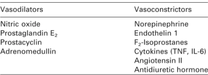
Table 1 Mediators that contribute to the pathogenesis of HRS.
Vasodilators Vasoconstrictors
Nitric oxide Norepinephrine
Prostaglandin E2 Endothelin 1 Prostacyclin F2-Isoprostanes Adrenomedullin Cytokines (TNF, IL-6)
Angiotensin II Antidiuretic hormone
The exact mechanism of the vasoconstriction is not well known. Several potential mediators play a con-tributory role in the pathogenesis and include the fac-tors listed in Table 1.
Nitric oxide (NO)
Endogenous production of NO has been found to be uniformly increased in cirrhotic patients (16–19) and inhibition of NO synthesis reverses some of the sys-temic and splanchnic circulatory changes in animal models or patients with liver cirrhosis (20, 21). Increased vascular production of NO has been pro-posed as the primary cause of arterial vasodilatation and the hyperdynamic circulation in cirrhosis. Cirrho-sis-associated endothelial dysfunction seems to in-validate the capability of intrarenal vasculature to produce NO (22) and deficient NO release in these vascular tissues might contribute to the development of HRS. Furthermore, renal vasoconstriction may occur in the presence of enhanced glomerular nitrite production, and this finding suggests that renal microcirculation in cirrhosis is less sensitive to NO (23). Splanchnic and systemic vasodilatation caused by the increased levels of NO leads to renal vasocon-striction by activation of endogenous vasoconstrictor systems. This phenomenon can be considered as the basis of the progressive renal failure that leads to HRS.
Prostaglandins
Renal prostaglandins play an important role in the preservation of renal function in all situations, such as dehydration, congestive heart failure, shock, or decompensated liver disease. In liver disease, urinary
excretion of prostaglandin E2 and prostacyclin meta-bolites (6-oxo-PGF 1a) are usually increased. The mechanism of increased synthesis is unknown, but is likely to be secondary to increased several vasocon-strictors that induce prostaglandin formation in vitro or in vivo (24–27). Non-selective inhibition of cyclo-oxgenase by non-steroidal anti-inflammatory drugs (NSAIDs) causes a significant decrease in renal blood flow and GFR in patients with cirrhosis and ascites, but in these patients selective cyclooxgenase-2 (COX-2) inhibition does not affect renal functions (28). These results suggest that COX-1-derived prostaglan-dins are involved in the homeostasis of renal func-tions in patients with cirrhosis and a decrease in vasodilator prostaglandin production may participate in renal vasoconstriction in HRS.
Adrenomedullin
Adrenomedullin is a peptide hormone that is highly expressed in cardiovascular tissues and has potent and long-lasting vasodilatory activity. Plasma levels of adrenomedullin were found to be increased in cir-rhotic patients and were inversely correlated with arterial pressure, GFR, renal plasma flow, and creati-nine clearance (29, 30). The pathophysiological role of adrenomedullin in the development of HRS is not clear.
Norepinephrine
The relation of renal vasoconstriction and increased sympathetic activity to cirrhosis and HRS has been shown by increased levels of circulating norepineph-rine (31) and increased release of norepinephnorepineph-rine in neuroeffector junctions (32). Furthermore, it has also been indicated that plasma norepinephrine levels, mean arterial pressure, urinary sodium excretion, and GFR are better predictors of survival than markers routinely used to assess hepatic function in cirrhotic patients (33, 34). The mechanisms of renal vasocon-striction have not yet been fully elucidated in the development of renal failure in patients with HRS (35, 36).
Endothelin 1
Endothelin 1 concentrations were significantly in-creased in HRS and well correlated with GFR in decompensated liver disease in several studies (37–39). After infusion of the endothelin antagonist BQ123, all patients with HRS showed a dose-related increase in both GFR and renal plasma blood flow (40). In contrast to this finding, there was no correla-tion between endothelin 1 and renal vasoconstriccorrela-tion assessed by duplex ultrasonography (41), suggesting that endothelin 1 may not be the only pressor agent responsible for renal vasoconstriction in HRS. Thus, the cause of increased plasma endothelin 1 levels needs to be investigated in further studies.
Table 2 Definition of hepatorenal syndrome. Major criteria
1. Chronic or acute liver disease with liver failure and portal hypertension
2. Low glomerular filtration rate as indicated by serum creatinine )1.5 mg/dL (133mmol/L) or a creatinine clearance -40 mL/min
3. Absence of shock, ongoing bacterial infection, or re-cent treatment with nephrotoxic drugs; absence of excessive fluid loss (including gastrointestinal loss) 4. No sustained improvement in renal function following
diuretic withdrawal and expansion of plasma volume with 1.5 L of isotonic saline
5. Proteinuria of -500 mg/day
6. No ultrasonagraphic evidence of obstructive uropathy or parenchymal renal disease
Minor criteria
1. Urine volume -500 mL/day 2. Urine sodium -10 mmol/day 3. Urine osmolality )plasma osmolality
4. Urine red blood cell count -50 per high-power field 5. Serum sodium -130 mmol/L
Adapted from Arroyo et al. (2).
F2-Isoprostanes
Increased synthesis of F2-isoprostanes, the products
of lipid peroxidation, in patients with HRS was found to be indicative of increased lipid peroxidation (42, 43). In a recent study in patients who were given a continuous infusion of the antioxidant N-acetylcys-teine for 5 days, creatinine clearance was approxi-mately doubled without any change in liver function or systemic hemodynamics (44). F2-Isoprostanes
should be further investigated to confirm if they are important mediators of renal vasoconstriction in HRS.
Cytokines
Recent studies have implicated increased circulating levels of several cytokines, including tumor necrosis factor (TNF) and interleukin-6 (IL-6), in patients with HRS. According to related studies, inflammatory re-sponse to infection, as estimated by increased levels of cytokines in plasma or ascitic fluid, leads to circu-latory dysfunction and concomitant renal impair-ment and increased mortality (45–47). Studies have shown that vasodilation was observed in cirrho-tic rats with portal hypertension on administration of anti-TNF antibodies, N-acetylcysteine and inhibitors of tyrosine kinase (48–50).
Renin-angiotensin-aldosterone (RAA) system
The RAA system is activated in most patients with decompensated cirrhosis and is further induced in patients with HRS (51–56). Increased plasma renin release followed by an increase in angiotensin II for-mation was found in refractory ascites and HRS, indi-cating a role of RAA in the development of HRS (57). Angiotensin II helps to maintain vascular tone in patients with advanced liver disease, but has no role in healthy controls or patients with compensated cir-rhosis, suggesting that this mediator contributes to vascular dysfunction in cirrhosis (58).
Antidiuretic hormone (ADH)
ADH or vasopressin causes vasoconstriction through V1 receptors and renal tubular water retention
through V2 receptors in the medullary collecting
ducts. This increases volume expansion by water retention and helps maintain arterial pressure. Inhi-bition of V1receptors in cirrhotic rats causes profound
hypotension. Vasopressin preferentially causes splan-chnic rather than renal vasoconstriction (59).
Vasopressin analogues (ornipressin and terlipres-sin) are used in HRS treatment for their vasocon-strictor effects. Administration of these drugs in combination with albumin improves arterial underfill-ing and renal function. Ornipressin is very effective in HRS treatment, but it is not widely used because it has serious ischemic complications such as ischemic colitis and myocardial ischemia (60). Terlipressin (triglycyl-lysine vasopressin) is cleaved in vivo by endothelial peptidases, releasing the active lysine-vasopressin. Treatment with terlipressin caused a sig-nificant decrease in serum creatinine concentrations,
an increase in arterial pressure, and suppression of the renin aldosterone system in HRS patients (61). It is the most commonly used drug in HRS therapy because it has fewer side effects and a prolonged duration of action (62).
Diagnosis
The diagnosis of HRS is based on major criteria for both clinical and laboratory aspects defined by the International Ascites Club in 1996 (2). Minor criteria are not necessary for the diagnosis of HRS, but these criteria are frequently present in HRS patients (Table 2).
Use of diagnostic tests for differential diagnosis
HRS may only be diagnosed after eliminating other potential causes of acute renal failure. Although chronic liver disease is easily diagnosed, diagnosis of the cause of renal failure may not be as easy. Prerenal causes of acute renal failure in patients with cirrhosis are gastrointestinal and renal fluid losses, hemor-rhage, shock, sepsis, congestive heart failure, NSAID use and HRS (63). Intrinsic causes include glomeru-lonephritis, interstitial nephritis and acute tubular necrosis (ATN), with postrenal causes due to the obstruction of urinary flow tract (63).
The differential diagnosis of HRS includes all these renal diseases. Generally, HRS is assumed to be a prerenal disease and distinguishing this condition from other disorders is clinically important because of the marked difference in prognosis. ATN and other causes of prerenal diseases are generally reversible, but prognosis is poor in HRS (64, 65).
Watt et al. reported that 40% of patients with advanced liver disease and renal failure are mistak-enly diagnosed as having HRS, suggesting that many
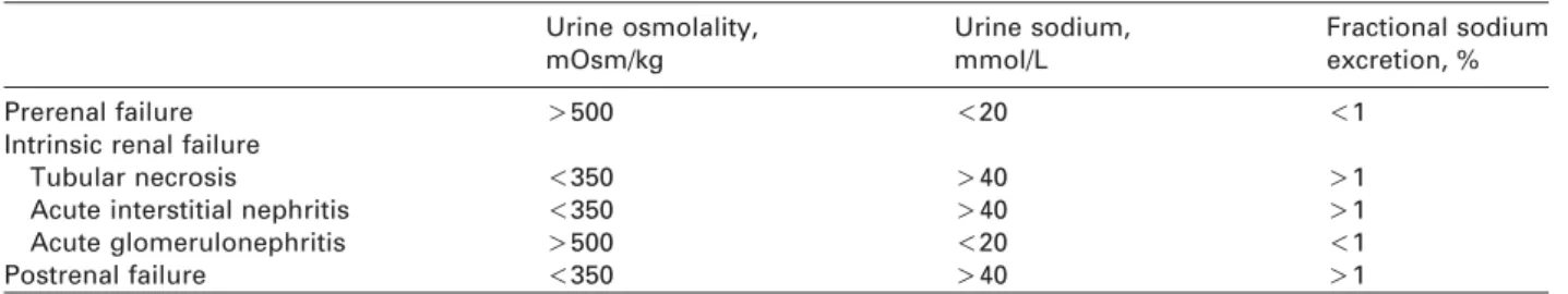
Table 3 Urinary parameters in different types of acute renal failure.
Urine osmolality, Urine sodium, Fractional sodium
mOsm/kg mmol/L excretion, %
Prerenal failure )500 -20 -1
Intrinsic renal failure
Tubular necrosis -350 )40 )1
Acute interstitial nephritis -350 )40 )1
Acute glomerulonephritis )500 -20 -1
Postrenal failure -350 )40 )1
Adapted from Moreau and Lebrec (72).
physicians are unaware of the criteria that exist for defining HRS (7). Some conditions that should be con-sidered that affect both the liver and the kidney are Weil’s disease and malaria.
Renal failure is common in cirrhotic patients with sepsis unrelated to spontaneous bacterial peritonitis and is associated with arterial underfilling and renal vasoconstriction (66). Therefore, diagnosis of HRS should always be ruled out with cultures, leukocyte count and C-reactive protein. The diagnosis of HRS can only be made if renal failure persists after com-plete resolution of the infection.
No laboratory test can firmly establish a diagno-sis of HRS, which is mainly based on the absence of any specific cause of renal failure (1). Laboratory and ultrasonographic tests based on non-invasive tech-niques are being investigated as possible diagnostic approaches.
Platt et al. prospectively studied the prognostic impact of renal duplex sonography in 180 cirrhotic patients with normal renal function at the time of first examination. They concluded that renal Doppler ultra-sonography non-invasively identified a subgroup of non-azotemic cirrhotic patients at significantly higher risk for renal dysfunction or HRS (67).
Despite intensive studies of non-invasive sonogra-phic techniques, routine laboratory tests are still com-monly used. Urinary examinations, plasma creatinine and blood urea nitrogen (BUN) assays and estimation of GFR are the most popular diagnostic tools. In addi-tion, bacterial cultures of blood, ascites, and urine should be evaluated in all patients with HRS to iden-tify occult infection before antibiotic therapy.
Plasma and urinary electrolytes and osmolality should be assessed in all patients to rule out other causes of renal failure when possible, because of their importance as minor diagnostic criteria (2). Some of these parameters are discussed in detail below.
Urinary examination
Urinanalysis may give valuable diagnostic informa-tion regarding HRS. Examinainforma-tion of urinary sediment is necessary, especially for the differential diagnosis of HRS from the other types of renal failure, such as the typical occurrence of pigmented granular casts and tubular epithelial cells alone or in casts in ATN and the red cell casts in glomerulonephritis (68–70). Proteinuria, which is a major component of the diag-nostic criteria, is typically mild and does not exceed 0.5 g/day in HRS (2).
Tubular function is usually well preserved at the time when HRS develops, but prolonged renal hypo-perfusion caused by progressive circulatory dysfunc-tion may eventually result in acute tubular necrosis by increasing sensitivity to other factors, such as radiographic contrast agents, nephrotoxic antibiotics, hemorrhage, endotoxemia, or any other cause of medullary hypoxia (71).
HRS can be difficult to distinguish clinically from acute tubular necrosis and other types of acute and chronic renal failure that may be handled in different ways (72). Most HRS patient have low urinary sodium (-10 mmol/L) and high urinary osmolality because of preserved tubular function and activation of tubular reabsorption of sodium. Some HRS patients also show high urinary sodium ()10 mmol/L) and low uri-nary osmalality, as in ATN (2). However, few cirrhotic patients with ATN have low urinary sodium (-10 mmol/L) and high urine osmolality (2) (Table 3). Therefore, urinary sodium and osmolality are not con-sidered major criteria for the diagnosis of HRS.
Measurement of the fractional excretion of sodium (FENa) has been recommended as a useful clinical tool in evaluating acute renal failure. FENa has been shown to be a reliable discriminatory test between prerenal failure and ATN (2, 74) (Table 3). However, some patients with ATN have FENa of -1% (73). In addition to this finding, some cases of prerenal failure, including HRS, have FENa of )1% (73). For these reasons, limited sensitivity of this parameter may make the interpretation of FENa difficult in this setting.
Assay of plasma urea and creatinine levels
Both urea and creatinine production may be highly reduced because of liver disease, reduced muscle mass and protein-meat intake. BUN tends to be vari-able in these patients. If urea production is markedly reduced, it may be lower than expected. The intense sodium avidity in this clinical setting can also raise BUN by accelerating sodium and water and eventu-ally passive urea reabsorption in the proximal tubule (74).
Although plasma creatinine is one of the major diagnostic criteria, there is still controversy over the diagnostic value of this test. In advanced liver dis-ease, because of muscle wasting and the insufficient conversion of creatine to creatinine, the net effect is a plasma creatinine concentration that appears to be
within the normal range, which leads to false values for creatinine clearance (64, 75).
Estimation of GFR
In HRS, the reduction in GFR is often masked clini-cally. Estimation of GFR by creatinine clearance will tend to overestimate the true GFR owing to increased tubular secretion of creatinine (64) or reduced pro-duction because of muscle wasting and incomplete urine collection (64, 76). In addition, because of the marked discrepancy between serum creatinine and GFR, this approach is not a valid parameter for assessing renal function in advanced cirrhosis (64, 77, 78).
Exogenously administered substances such as inu-lin or other radioisotopic agents can reflect GFR more precisely. However, these determinations are more invasive, requiring continuous intravenous infusion and urine sampling with a bladder catheter. These diagnostic limitations in identifying the true GFR have stimulated investigators to find more convenient and non-invasive techniques to assess the degree of renal conditions (67, 79, 80).
Cystatin C
Cystatin C is a non-glycosylated, basic protein of low molecular mass (13 kDa) that is a member of thecys-teine protease inhibitors. Cystatin C consists of 120 amino acids and is a new marker of GFR produced at a constant rate in all nucleated cells (81, 82). Its pro-duction is independent of gender and muscle mass (83). Since the first discovery of these beneficial fea-tures, it has been suggested as a better marker of GFR than creatinine (84–88). Reference ranges for cystatin C in children (89), adults (90) and the elderly (91) have already been determined.
Rosenthal et al. showed in a study of 226 patients with various nephropathies (53 patients with glome-rular and 26 patients with tubular impairment) that cystatin C and creatinine did not significantly differ with regard to efficacy. However, the efficacy of cys-tatin C as a screening test was superior to creatinine, with higher overall sensitivity and a higher negative predictive value (79). In a recent study, Gerbes et al., who identified separate cutoff concentrations for each of three analytes, reported a differential diagnostic advantage of cystatin C over creatinine and urea in patients with cirrhosis (92). In these patients, they found that serum cystatin C concentrations were sig-nificantly correlated with impaired renal function (creatinine clearance 40–69 mL/min) compared with patients with creatinine clearance )70 mL/min. The difference between these groups was less pro-nounced for serum creatinine and was not significant for serum urea concentrations. Subgroup analysis for various nephropathies indicated that neither glome-rular nor tubular impairment led to different cystatin C efficacy (79). We previously demonstrated that in patients with HRS, neither serum creatinine nor cre-atinine clearance were good indicators of HRS, because the mean value for creatinine clearance was
higher than Tc-DTPA clearance, and there was no cor-relation between these two parameters (rs0.059). In addition, the mean serum creatinine was within the normal range, whereas the mean Tc-DTPA clearance level was below the normal range. However, we found significant correlation between cystatin C and Tc-DTPA. Thus, we suggest that serum cystatin C assay, which shows good analytical performance, could replace or at least be added to creatinine meas-urement for GFR assessment in patients with cirrhosis (93). Orlando et al. found that creatinine, which show-ed sensitivity of only 23%, failshow-ed to detect rshow-educ- reduc-ed renal function, whereas cystatin C exhibitreduc-ed good diagnostic sensitivity of 88% (80).
Conclusions
This article summarizes the literature on the current definition, suggested pathogenetic mechanisms and the role of laboratory assessment in the differential diagnosis of HRS from other causes of renal disease that may arise during hepatic cirrhosis and some dis-eases affecting both liver and kidney. It should be remembered that the main theory suggested for the pathogenesis of HRS is the arterial vasodilation hypothesis of portal hypertension, ending in type 1 and type 2 HRS, but there is no consensus supporting either mechanism as a solid theory for the initiation of HRS pathogenesis to date. Thus, discussion of the humoral modulators originating from experimental models, as well as clinical data, is critical in support-ing either mechanism. Response to volume loadsupport-ing is useful in the differentiation of pre-renal failure from other forms of acute renal failure, but HRS rarely responds to volume loading, and even though a care-ful follow-up may lead to timely diagnosis of HRS, the treatment still does not yield positive progress. Final-ly, even though it is beyond the scope of this article to set criteria for the differential diagnosis of HRS, tests to identify etiologies underlying other causes of renal failure should be sufficient to differentiate renal failure due to hepatic disease accompanied by portal hypertension and ascites, given as a definition of HRS.
References
1. Dagher L, Moore K. The hepatorenal syndrome. Gut 2001;49:729–37.
2. Arroyo V, Gines P, Gerbes AL, Dudley FJ, Gentilini P, Laffi G, et al. Definition and diagnostic criteria of refractory ascites and hepatorenal syndrome in cirrhosis. Interna-tional Ascites Club. Hepatology 1996;23:164–76.
3. Gines A, Escorsell A, Gines P, Salo J, Jimenez W, Inglada L, et al. Incidence, predictive factors, and prognosis of the hepatorenal syndrome in cirrhosis with ascites. Gastro-enterology 1993;105:229–36.
4. Badalamenti S, Graziani G, Salerno F, Ponticello C. Hepa-torenal syndrome: new perspectives in pathogenesis and treatment. Arch Intern Med 1993;153:1957–67.
5. Rhodes J, Bosch J, Arroyo V. Clinical types and drug therapy of renal impairment in cirrhosis. Postgrad Med J 1975;51:492–7.
6. Bataller R, Gines P, Guevara M, Arroyo V. Hepatorenal syndrome. Semin Liver Dis 1997;17:233–47.
7. Watt K, Uhanova J, Minuk GY. Hepatorenal syndrome: diagnostic accuracy, clinical features, and outcome in a tertiary care center. Am J Gastroenterol 2002;97: 2046–50.
8. Bilotta J, Pazik M, Greizersen H, Acara M. Renal effect of ethanol in isolated perfused rat kidney. Eur J Pharmacol 1984;98:109–12.
9. Boyer TD, Zia P, Reynolds TB. Effect of indomethacin and prostaglandin A1 on renal function and plasma ren-in activity ren-in alcoholic liver disease. Gastroenterology 1979;77:215–22.
10. Epstein M. Renal prostaglandins and the control of renal function in liver disease. Am J Med 1986;80:46–55. 11. Schrier RW, Arroyo V, Bernardi M, Epstein M, Henriksen
JH, Rodes J. Peripheral arterial vasodilatation hypothe-sis: a proposal for the initiation of renal sodium and water retention in cirrhosis. Hepatology 1988;8:1151–7. 12. Arroyo V, Guevera M, Gines P. Hepatorenal syndrome:
pathogenesis and treatment. Gastroenterology 2002;122: 1658–76.
13. Colombato L, Albillos A, Groszmann RJ. Temporal rela-tionship of peripheral vasodilatation, plasma volume expansion and the hyperdynamic circulatory state in portal-hypertensive rats. Hepatology 1991;15:323–8. 14. Gines P, Martin PY, Niederberger M. Prognostic
signifi-cance of renal dysfunction in cirrhosis. Kidney Int 1997; 51(Suppl):S77–82.
15. Lang F, Tschernko E, Schulze E, Ottl I, Ritter M, Volkl H, et al. Hepatorenal reflex regulating kidney function. Hepatology 1991;14:590–4.
16. Vallance P, Moncada S. Hyperdynamic circulation in cir-rhosis: a role for nitric oxide. Lancet 1991;337:776–8. 17. Guarner C, Soriano G, Tomas A, Bulbena O, Novella MT,
Balazo J, et al. Increased serum nitrite and nitrate levels in patients with cirrhosis: relationship to endotoxemia. Hepatology 1993;18:1139–41.
18. Sogni P, Garnier P, Gadano A, Moreau R, Dall’Ava San-tucci J, Dinh-Xuan AT, et al. Endogenous pulmonary nitric oxide production measured from exhaled air is increased in patients with severe cirrhosis. J Hepatol 1995;23:471–3.
19. Moncada S, Higgs A. The L-arginine-nitric oxide path-way. N Engl Med 1991;329:2002–12.
20. Pizcueta MP, Pique JM, Fernandez M, Bosch J, Rodes J, Whittle BJ, et al. Modulation of the hyperdynamic cir-culation of cirrhotic rats by nitric oxide inhibition. Gas-troenterology 1992;103:1909–15.
21. Forrest EH, Jones AL, Dillon JF, Walker J, Hayes PC. The effect of nitric oxide synthase inhibition on portal pres-sure and azygos blood in patients with cirrhosis. J Hepa-tol 1995;23:254–8.
22. Hu LF, Sealey JE, Chen R, Zhou Y, Merali C, Shi YX, et al. Nitric oxide synthase inhibition accelerates the pres-sor response to low-dose angiotensin II, exacerbates tar-get organ damage, and induces renin escape. Am J Hypertens 2004;17:395–403.
23. Garcia-Estan J, Atucha NM, Sabio JM, Vargas F, Quesda T, Romero JC. Increased endothelium-dependent renal vasodilation in cirrhotic rats. Am J Physiol 1994;267: 549–53.
24. Moore K, Ward P, Taylor G, Williams R. Systemic and renal production of thromboxane A2 and prostacyclin in decompensated liver disease and hepatorenal syn-drome. Gastroenterology 1991;100:1069–77.
25. Guarner C, Colina I, Guarner F, Corzo J, Prieto J, Vilardell F. Renal prostaglandins in cirrhosis of the liver. Clin Sci 1986;70:477–84.
26. Laffi G, La Villa G, Pinzani M, Ciabattoni G, Patrignani P, Mannelli M, et al. Altered renal and platelet arachidonic acid metabolism in cirrhosis. Gastroenterology 1986; 90:274–82.
27. Rimola A, Gines P, Arroyo V, Camps J, Perez Ayuso RM, Quintero E, et al. Urinary excretion of 6-ketoprostaglan-din F1-alpha, thromboxane B2 and prostaglan6-ketoprostaglan-din E2 in cirrhosis with ascites: relationship to functional renal failure(hepatorenal syndrome). J Hepatol 1986;3:111–7. 28. Claria J, Kent JD, Lopez-Parra M, Escolar G,
Ruiz-del-Arbol L, Gines P, et al. Effects of celecoxib and naproxen on renal function in nonazotemic patients with cirrhosis and ascites. Hepatology 2005;41:579–87.
29. Guevera M, Gines P, Jimenez W, Sort P, Fernandez Esparrach G, Escorsell A, et al. Increased adrenomedul-lin levels in cirrhosis: relationship with hemodynamic abnormalities and vasoconstrictor systems. Gastroenter-ology 1998;114:336–43.
30. Fernandez-Rodriguez CM, Prada IR, Prieto J, Montuenga LM, Elssasser T, Quiroga J, et al. Circulating adreno-medullin in cirrhosis: relationship to hyperdynamic cir-culation. J Hepatol 1998;29:250–6.
31. Henriksen JH, Ring-Larsen H, Kanstrup IL, Fradsen E, Bendtsen F. Circulating norepinephrine and central hemodynamics in patients with cirrhosis. Scand J Gas-troenterol 1985;20:1185–90.
32. Bichet DG, Van Putten VJ, Schrier RW. Potential role of increased sympathetic activity in impaired sodium and water excretion in cirrhosis. N Engl J Med 1982;307: 1552–7.
33. Llach J, Gines P, Arroyo V, Rimola A, Tito L, Badalamenti S, et al. Prognostic value of arterial pressure, endoge-nous vasoactive systems, and renal function in cirrhotic patients admitted to the hospital for the treatment of ascites. Gastroenterology 1988;94:482–7.
34. Solis-Herruzo JA, Duran A, Favelza V, Castellano G, Madrid JL, Munoz-Yague MT, et al. Effects of lumbar sympathetic block on kidney function in cirrhotic patients with hepatorenal syndrome. J Hepatol 1987; 5:167–73.
35. Wong F, Blendis L. New challenge of hepatorenal syn-drome: prevention and treatment. Hepatology 2001;34: 1242–51.
36. Moreau R. Hepatorenal syndrome in patients with cir-rhosis. J Gastroenterol Hepatol 2002;17:739–47. 37. Moller S, Emmeluth C, Henricksen JH. Elevated
circulat-ing plasma endothelin-1 concentrations in cirrhosis. J Hepatol 1993;19:285–90.
38. Uchihare M, Izumi N, Sato C, Marumo F. Clinical signif-icance of elevated plasma endothelin concentration in patients with cirrhosis. Hepatology 1992;16:95–9. 39. Moore K, Wendon J, Frazer M, Karani J, Williams R, Badr
K. Plasma endothelin immunoreactivity in liver disease and the hepatorenal syndrome. N Engl J Med 1992; 327:1774–8.
40. Soper CP, Latif AB, Bending MR. Amelioration of hepa-torenal syndrome with selective endothelin-A antago-nist. Lancet 1996;347:1842–3.
41. Kitamura H, Shimada R, Kobayashi A, Nomura K, Noike T, Harada H, et al. Plasma concentration of endothelin-1 does not reflect renal vasoconstriction as estimated by duplex ultrasonography in cirrhosis. Dig Dis Sci 1997; 42:542–5.
42. Morrow JD, Moore KP, Award JA, Ravenscraft MD, Mari-ni G, Badr K, et al. Marked overproduction of non-cyclo-oxygenase derived prostanoids (F2-isoprostane) in the hepatorenal syndrome. J Lipid Mediators 1993;6:417–20. 43. Holt S, Marley R, Goodier D, Harry D, Fernando B, Moore K. Oxidant stress and renal dysfunction in cholestasis. Gut 1998;43:153.
44. Holt S, Marley R, Fernando B, Harry D, Anand R, Goodier D, et al. Acute cholestasis-induced renal failure: effects
of antioxidants and ligands for the thromboxane A2 receptor. Kidney Int 1999;55:271–7.
45. Akriviadis E, Botla R, Briggs W, Han S, Reynolds T, Sha-kil O. Pentoxifylline improves short-term survival in severe acute alcoholic hepatitis: a double-blind, placebo-controlled trial. Gastroenterology 2000;119:1637–48. 46. Sheron N, Bird G, Koskinas J, Portmann B, Ceska M,
Lindley I, et al. Circulating and tissue levels of the neu-trophil chemotaxin, interleukin 8 are elevated in severe acute alcoholic hepatitis, and tissue levels correlate with neutrophil infiltration. Hepatology 1993;18:41–6. 47. Sheron N, Bird G, Goka J, Alexander G, Williams R.
Elevated plasma interleukin 6 and increased severity and mortality in alcoholic hepatitis. Clin Exp Immunol 1991;84:449–53.
48. Fernando B, Marley R, Holt S, Anand R, Harry D, San-derson P, et al. Acetylcysteine prevents development of the hyperdynamic circulation in the portal hypertensive rat. Hepatology 1998;28:689–94.
49. Lopez-Talavera JC, Levitzki A, Martinez M, Gazit A, Este-ban R, Guardia J. Tyrosine kinase inhibition ameliorates the hyperdynamic state and decreases NO production in cirrhotic rats with portal hypertension and ascites. J Clin Invest 1997;100:664–70.
50. Lopez-Talavera JC, Merrill WW, Groszman RJ. Tumor necrosis factor alpha: a major contributor to the hyper-dynamic circulation in prehepatic portal hypertensive rats. Gastroenterology 1995;108:761–7.
51. Wilkinson SP, Willams R. Renin-angiotensin-aldosterone system in cirrhosis. Gut 1980;21:545–54.
52. Schroeder ET, Eich RH, Smulyan H, Gould AB, Gabuzda GJ. Plasma renin level in hepatic cirrhosis: relation to functional renal failure. Am J Med 1970;49:189–91. 53. Cobden I, Shore A, Wilkinson R, Record CO. Captopril in
the hepatorenal syndrome. J Clin Gastroenterol 1985; 7:354–60.
54. Schroeder ET, Anderson GH. Effect of blockade of angi-otensin II on blood pressure, renin and aldosterone in cirrhosis. Kidney Int 1976;9:511–9.
55. Helmy A, Jalan R, Newby DE, Hayes PC, Webb DJ. Role of angiotensin II in regulation of basal and sympatheti-cally stimulated vascular tone in early advanced cirrho-sis. Gastroenterology 2000;118:565–72.
56. Schneider AW, Kalk JF, Klein CP. Effect of losartan, an angiotensin II receptor antagonist, on portal pressure in cirrhosis. Hepatology 1999;29:334–9.
57. Debarnardi-Venon D, Baretti C, Marzano A, Marzano A, Baronio M, Todros L, et al. Efficacy of irbesartan, an angiotensin II receptor selective antagonist, in the treat-ment of portal hypertension. Dig Dis Sci 2002;47:401–4. 58. Garcia-Tsao G. Angiotensin receptor antagonist in the pharmacological therapy: a caution. Gastroenterology 1999;117:740–2.
59. Claria J, Jimenez W, Arroyo V, La Villa G, Lopez C, Asbert M, et al. Effect of V1-vasopressin receptor block-ade on arterial pressure in conscious rats with cirrhosis and ascites. Gastroenterology 1991;100:494–501. 60. Guevara M, Gines P, Fernandez-Esparrach G, Sort P,
Sal-meron JM, Jimenez W, et al. Reversibility of hepatorenal syndrome by prolonged administration of ornipressin and plasma volume expansion. Hepatology 1998; 27:35–41.
61. Rodriguez-Perez F, Groszmann RJ. Pharmacologic treat-ment of portal hypertension. Gastroenterol Clin North Am 1992;21:15–40.
62. Ortega R, Gines P, Uriz J, Cardenas A, Calahorra B, De Las Heras D, et al. Terlipressin therapy with and without albumin for patients with hepatorenal syndrome: results of a prospective, nonrandomized study. Hepatology 2002;36:941–8.
63. Podolsky DK, Isselbacher KJ. Cirrhosis. In: Braunwald E, Isselbacher KJ, Petersdorf RG, Wilson JD, Martin JB,
Fauci AS, editors. Harrison’s principles of internal med-icine, 11th ed. New York: McGraw-Hill, 1987.
64. Caregaro L, Menon F, Angeli P, Amodio P, Merkel C, Bor-toluzzi A, et al. Limitations of serum creatinine level and creatinine clearance as filtration markers in cirrhosis. Arch Intern Med 1994;154:201–5.
65. Linas SL, Schaefer JW, Moore EE, Good JT Jr, Giansi-racusa R. Peritoneovenous shunt in the management of the hepatorenal syndrome. Kidney Int 1986;30:736–40. 66. Terra C, Guevara M, Torre A, Gilabert R, Fernandez J,
Martin Llahi M, et al. Renal failure in patients with cir-rhosis and sepsis unrelated to spontaneous bacterial peritonitis: value of MELD score. Gastroenterology 2005;129:1944–53.
67. Platt JF, Ellis JH, Rubin JM, Merion RM, Lucey MR. Renal duplex Doppler ultrasonography: a noninvasive predic-tor of kidney dysfunction and hepapredic-torenal failure in liver disease. Hepatology 1994;20:362–9.
68. Abuelo JG. Diagnosing vascular causes of renal failure. Ann Intern Med 1995;123:601–14.
69. Brady HR, Singer GG. Acute renal failure. Lancet 1998; 346:1533–40.
70. Thadhani R, Pascual M, Bonventre JV. Acute renal fail-ure. N Engl J Med 1996;334:1448–60.
71. Heyman SN, Darmon D, Goldfarb M, Bitz H, Shina A, Rosen S, et al. Endotoxin-induced renal failure. A role for altered renal microcirculation. Exp Nephrol 2000; 8:266–74.
72. Moreau R, Lebrec D. Acute renal failure in patients with cirrhosis: perspectives in the age of MELD. Hepatology 2003;37:233–43.
73. Klahr S, Miller SB. Acute oliguria. N Engl J Med 1998; 338:671–5.
74. Porcel A, Diaz F, Rendon P, Marcias M. Dilutional hypo-natremia in patients with cirrhosis and ascites. Arch Intern Med 2002;162:323–8.
75. Papadakis MA, Arieff A. Unpredictability of clinical eval-uation of renal function in cirrhosis. Prospective study. Am J Med 1987;82:945–52.
76. De Santo NG, Anastasio P, Loguercio C, Spitali L, Del Vecchio BC, Corvinelli M, et al. Creatinine clearance: an adequate marker of renal filtration in patients with early posthepatic cirrhosis (child A) without fluid retention and muscle wasting. Nephron 1995;70:421–4.
77. Woitas RP, Stoffel-Wagner B, Flommersfeld S, Poege U, Schiedermaier P, Klehr HU, et al. Correlation of serum concentrations of cystatin C and creatinine to inulin clearance in liver cirrhosis. Clin Chem 2000;46:712–5. 78. Shemesh O, Golbetz H, Kriss JP, Myers BD. Limitations
of creatinine as a filtration marker in glomerulopathic patients. Kidney Int 1985;28:830–8.
79. Herget-Rosenthal SH, Trabold S, Pietruck F, Holtmann M, Philipp T, Kribben A. Cystatin C: efficacy as screening test for reduced glomerular filtration rate. Am J Nephrol 2000;20:97–102.
80. Orlando R, Musap M, Plebani M, Piccoli P, De Martin S, Florenai M, et al. Diagnostic value of plasma cystatin C as a glomerular filtration marker in decompensated liver cirrhosis. Clin Chem 2002;48:850–8.
81. Newman DJ, Price CP. Renal function and nitrogen metabolites. In: Burtis CA, Ashwood ED, editors. Tietz textbook of clinical chemistry, 3rd ed. Philadelphia, PA: Saunders, 1999:1251–4.
82. Grubb A. Diagnostic value of analysis of cystatin C and protein HC in biological fluids. Clin Nephrol 1992;38: 20–7.
83. Randers E, Erlandsen EJ. Serum cystatin C as an endo-genous marker of the renal function – a review. Clin Chem Lab Med 1999;37:389–95.
84. Randers E, Erlandsen EJ, Pedersen OL, Hasling C, Danielsen H. Serum cystatin C as an endogenous para-meter of the renal function in patients with normal to
moderately impaired kidney function. Clin Nephrol 2000; 54:203–9.
85. Kyhse-Andersen J, Schimidt C, Nordin G, Andersson B, Nilsson-Ehle P, Lindstrom V, et al. Serum cystatin C, determined by a rapid automated particle-enhanced turbidimetric method, is a better marker than serum creatinine for glomerular filtration rate. Clin Chem 1994; 40:1921–6.
86. Newman DJ, Thakkar H, Edwards RG, Wilkie M, White T, Greubb AO, et al. Serum cystatin C measured by auto-mated immunoassay: a more sensitive marker of chang-es in GFR than serum creatinine. Kidney Int 1995; 47:312–8.
87. Bo¨kenkamp A, Domanetzki M, Zinck R, Schumann G, Byrd D, Brodehl J. Cystatin C: a new marker of glome-rular filtration rate in children independent of age and height. Pediatrics 1998;101:875–81.
88. Helin I, Axenram M, Grubb A. Serum cystatin C as a determinant of glomerular filtration rate in children. Clin Nephrol 1998;49:221–5.
89. Stickle D, Cole B, Haock K, Hruska KA, Scott MG. Cor-relation of plasma concentrations of cystatin C and cre-atinine to inulin clearance in a pediatric population. Clin Chem 1998;44:1334–8.
90. Burkhardt H, Bojarsky G, Gretz N, Gladish R. Creatinine clearance, Cockcroft-Gault formula and cystatin C: esti-mation of true glomerular filtration rate in the elderly. Gerontology 2002;48:104–6.
91. Burkhardt H, Bojarsky G, Gladish R. Diagnostic efficiency of cystatin C and serum creatinine as markers of reduced glomerular filtration rate in the elderly. Clin Chem Lab Med 2002;40:1135–8.
92. Gerbes AL, Gu¨lberg V, Bizler M, Vogester M. Evaluation of serum cystatin C concentration as a marker of renal function in patients with cirrhosis of the liver. Gut 2002;50:106–10.
93. Demirtas S, Bozbas A, Akbay A, Yavuz Y, Karaca L. Diag-nostic value of serum cystatin C for evaluation of hepa-torenal syndrome. Clin Chim Acta 2001;311:81–9. Received July 7, 2005, accepted January 3, 2006