Address for correspondence: Dr. Gamze Babür Güler, Tem Avrupa Otoyolu Göztepe Çıkışı, Medipol Üniversitesi, No:1 Bağcılar/İstanbul-Türkiye
Fax: +90 212 460 70 70 E-mail: [email protected] Accepted Date: 12.02.2016 Available Online Date: 25.04.2016
©Copyright 2016 by Turkish Society of Cardiology - Available online at www.anatoljcardiol.com DOI:10.14744/AnatolJCardiol.2016.6862
Gamze Babür Güler, Ekrem Güler, Suzan Hatipoğlu
1, Hacı Murat Güneş,
Çetin Geçmen
2, Gültekin Günhan Demir, İrfan Barutçu
Department of Cardiology, Faculty of Medicine, Medipol University; İstanbul-Turkey
1Clinic of Cardiology, Ersoy Hospital; İstanbul-Turkey
2Department of Cardiology, Kartal Koşuyolu Heart Center; İstanbul-Turkey
Assessment of 25-OH vitamin D levels and abnormal blood pressure
response in female patients with cardiac syndrome X
Introduction
The causes and consequences of vitamin D deficiency are widespread public health concerns that are still being investi-gated. Vitamin D deficiency is associated with coronary artery disease, hypertension, heart failure, endothelial dysfunction, and metabolic syndrome (1). The receptors for vitamin D are expressed on smooth muscle cells, endothelium, and myocytes (2). The mechanisms underlying the potential cardioprotective effects of vitamin D have been investigated previously, where the most impressive finding was that the plasma vitamin D levels were inversely associated with renin and angiotensin II levels (3, 4). Although vitamin D replacement therapy can reportedly result in suppressed systemic inflammation, decreased blood pressure levels, and enhanced endothelial function, the mechanisms
un-derlying its clinical effects are not clear (5).
Cardiac syndrome X (CSX), also named as microvascular an-gina, is defined as effort angina with detectable ischemia on non-invasive tests; however, no evidence of stenosis or vasospasm of epicardial coronary arteries is present (6). Impaired coronary microcirculation, inflammation, and insulin resistance resulting in endothelial dysfunction are accepted as the etiology for CSX; in addition, a decrease in coronary flow reserve and autonomic dysfunctions are believed to be associated with the onset of symptoms (7–9). This phenomenon has been detected in about 10%–20% of coronary angiographies (9).
Further, hypertensive response to exercise is associated with future hypertension, cardiovascular diseases, and end organ damage (10). However, to the best of our knowledge, its significance in patients with CSX and relationship with plasma
Objective: Vitamin D deficiency is associated with coronary artery disease, hypertension, heart failure, endothelial dysfunction, and metabolic syndrome. The pathophysiology of cardiac syndrome X (CSX) involves many pathways that are influenced by vitamin D levels. This study aimed to investigate the relationship between vitamin D deficiency and abnormal blood pressure response to exercise in patients with CSX.
Methods: This was a cross-sectional and observational study. Fifty females with normal epicardial coronary arteries who presented with typical symptoms of rest or effort angina and 41 healthy age-matched female controls, were included. Patients with cardiomyopathy, severe valvular disease, congenital heart disease, and left ventricular hypertrophy were excluded. All patients underwent stress electrocardiography examina-tion and 25-hydroxy (OH) vitamin D level measurements.
Results: Levels of 25-OH vitamin D were significantly lower in CSX patients (9.8±7.3 ng/mL vs. 18.1±7.9 ng/mL; p<0.001). Systolic blood pressure (SBP) (188±15 mm Hg vs. 179±17 mm Hg; p=0.013) and diastolic blood pressure (DBP) (98±9 mm Hg vs. 88±9 mm Hg; p<0.001) during peak exercise were higher in CSX patients. Levels of 25-OH vitamin D were negatively correlated with peak SBP (r=–0.310, p=0.004) and peak DBP (r=–0.535, p<0.001) during exercise. To discard the multicollinearity problem, two different models were used for multivariate analyses. In the first model, metabolic equivalents (METs) (p=0.003) and 25-OH vitamin D levels (p=0.001) were independent predictors. METs (p=0.007), 25-OH vitamin D levels (p=0.008), and peak DBP were determined as independent predictors in the second multivariate model.
Conclusion: In patients with CSX, 25-OH vitamin D levels were lower than those in controls; moreover, 25-OH vitamin D deficiency was also as-sociated with higher levels of peak DBP during exercise. (Anatol J Cardiol 2016; 16: 961-6)
Keywords: 25-OH vitamin D, cardiac syndrome X, abnormal blood pressure response
vitamin D levels has not yet been studied.
In this study, to define the pathophysiological significance of vitamin D deficiency in patients with CSX, we aimed to evaluate the following: 1) 25-hydroxy (OH) vitamin D (vitamin D) levels in patients with CSX compared with those in healthy controls, 2) the relation-ship between abnormal blood pressure response to exercise and vitamin D levels in patients with CSX, and 3) plasma vitamin D lev-els and exercise electrocardiography (ECG) data in combination.
Methods
Study designThe study was designed as a cross-sectional and obser-vational study. For this purpose, we included 50 females diag-nosed with CSX at our clinic and 41 healthy age-matched fe-males as the control group.
Female patients with normal epicardial coronary arteries who presented with symptoms of typical rest or effort angina and were referred to coronary angiography because of exercise ECG test positivity constituted the patient group. The control group was composed of females with similar demographic properties, atypical angina, and negative exercise ECG test. We enrolled fe-male patients who were in their perimenopausal period because prevalence of CSX has been reported to increase in this popula-tion in previous studies that included patients with CSX (6, 7, 9). None of the subjects in the patient or control group were involved in any type of professional sports. Patients with cardiomyopathy, severe valvular disease, congenital heart disease, left ventricu-lar hypertrophy, right ventricuventricu-lar failure, and pulmonary arterial hypertension on echocardiographic examination and those with a history of peripheral arterial disease, chronic hepatitis, renal failure, endocrinal disorders, and osteoarthritis or inflammatory polyarthritis were excluded. During the visual evaluation of the angiographic views, if any atheromatous plaques, slow flow, ec-tasia, myocardial bridge, or vasospasm were detected, then that patient was not included in the study. This study was approved by our institutional ethical committee; oral and written informed consents were obtained from all study participants.
Exercise electrocardiography
All patients underwent exercise ECG test (GE T2100, USA) with Bruce protocol as a stress test to identify inducible ischemia. Heart rate and blood pressure were monitored. Patients receiving anti-hypertensive therapy underwent exercise ECG stress test without drug discontinuation. Twelve-lead ECG was recorded at rest, with 1 min intervals during exercise, at peak exercise, and at the re-covery phase. Test results were categorized as positive (≥1.0 mm horizontal or down-sloping ST depression, ≥1.5 mm up-sloping ST depression, or ≥1.0 mm up-sloping ST depression if associated with anginal symptoms) or negative exercise ECG test. Test results with submaximal heart rate during exercise or new onset ventric-ular or supraventricventric-ular arrhythmias were excluded. The test time, metabolic equivalent (MET), and exercise blood pressure were
recorded using standard display methods. After the first measure-ment at rest, systolic blood pressure (SBP) and diastolic blood pressure (DBP) measurements were recorded at the end of each 3-min steps and during peak exercise. Any abnormal response to exercise was described as follows: (i) SBP at peak exercise ≥230 mm Hg, (ii) DBP at peak exercise ≥120 mm Hg, or (iii) increase in DBP ≥12 mm Hg, provided that DBP was >100 mm Hg.
Coronary angiography
Coronary angiography was performed using the femoral or radial access, whenever it was available, with standard Judkins catheters and iohexol (Omnipaque) contrast material. Coronary arteries were visualized in multiple projections in 15 frames per second. Each coronary artery was evaluated for irregularities on its luminal surface and local or diffuse stenosis, and no evidence of luminal irregularities by visual inspection was regarded as a normal coronary artery.
Blood samples
Blood samples were collected to evaluate hemoglobin, plate-let, urea, creatinine, alkaline phosphatase (ALP), calcium, and low-density lipoprotein cholesterol (LDL-C) levels. Additionally, vi-tamin D levels were measured using electrochemiluminescence protein binding assay. Normal values of vitamin D were accepted as >20 ng/mL using Cobas® kit.
Statistical analysis
Continuous variables are expressed as mean±standard de-viation and median (maximum–minimum). Categorical data are shown as frequencies and percentages. Continuous variables were tested using the Kolmogorov–Smirnov test and histograms. The correlation coefficients were presented using Pearson’s cor-relation analysis. Unpaired t-test was employed to determine dif-ferences in continuous variables that had normal distribution be-tween the patient and control groups. Mann–Whitney U test was used to test non-normally distributed continuous variants. Fisher’s exact test was used to compare categorical variables. Because variables such as, peak SBP, peak DBP, test duration, MET, and vi-tamin D levels were correlated with each other, we tried to ignore multicollinearity. Therefore, we considered two different multiple logistic regression models, which used significant independent variables at 10% level from univariate analyses. The results of the models were reported as odds ratio (OR) with 95% confidence interval, β-, and p-values. A p value of <0.05 was considered sig-nificant for all tests. Statistical Package for the Social Sciences (SPSS version 11.0, SPSS Inc., Chicago, IL, USA) was used.
Results
The demographic characteristics of 50 female with CSX and 41 control patients (age, body mass index, hypertension, hyper-lipidemia, diabetes mellitus, and smoking) were matched. The frequencies of antihypertensive (all of them were receiving ACE
inhibitors), statin, and oral antidiabetic drug use were similar for both the groups. Echocardiographic parameters such as ejec-tion fracejec-tion and left atrial diameter did not differ significantly between the two groups. ALP, calcium, creatinine, and hemoglo-bin levels were similar between the patient and control groups, whereas vitamin D levels were less than normal in both the groups. In addition, these levels were also significantly lower in CSX patients (9.8±7.3 ng/mL vs. 18.1±7.9 ng/mL; p<0.001).
Abnormal response to exercise test was not different when the two groups were compared [16 (32%) vs. 6 (14.6%); p=0.084]; however, when the components of abnormal response were ana-lyzed, SBP (188±15 mm Hg vs. 179±17 mm Hg, p=0.013) and DBP during peak exercise (98±9 mm Hg vs. 88±9 mm Hg, p<0.001) were detected to be significantly higher in CSX patients. Increase in the DBP was also similar between the two groups (10±3 mm Hg vs. 9±1.8 mm Hg; p=0.124). Increase in the DBP was not significant between the two groups (10±3 mm Hg vs. 9±1.8 mm Hg; p=0.124). Moreover, exercise duration was shorter (7.6±1.5 min vs. 8.7±1.5 min; p=0.002) and METs were lower (8.7±1.5 vs. 9.9±1.3, p<0.001) in patients with CSX. Clinical characteristics of both the groups are displayed in Table 1 in a comparative manner. The correlation coefficients calculated using Pearson’s correlation analysis are presented in Table 2. Vitamin D levels negatively correlated with peak SBP (r=–0.310, p=0.004) and peak DBP (r=–0.535, p<0.001) during exercise. To remove the multicollinearity problem between the variables with correlation coefficients as shown in Table 3, two different models were used for the multivariate analysis.
First, the logistic regression model used the significant vari-ables (p<0.10) such as METs, vitamin D levels, and abnormal blood pressure response to exercise from univariate analyses. In the second multiple model, we have used METs, vitamin D levels, and peak DBP during exercise as covariates. The results of these models are presented in Table 3. First model revealed that METs [β=–0.652, p=0.003, OR=0.521, 95% CI (0.340–0.797)] and vitamin D levels [β=–0.129, p=0.001, OR=0.879, 95% CI (0.813–0.949)] were independent predictors of CSX (Hosmer and Lemeshow test: p=0.095, Nagelkerke R Square: 0.433). METs [β=–0.571, p=0.007, OR=0.565, 95% CI (0.372–0.859)], vitamin D levels [β=–0.104, p =0.008, OR=0.901, 95% CI (0.834–0.973)], and peak DBP [β=0.075, p=0.027, OR=1.078, 95% CI (1.009–1.151)] were all determined as independent predictors in the second multiple model (Hosmer and Lemeshow test: p=0.755, Nagelkerke R Square: 0.480).
Discussion
To the best of our knowledge, this is the first study evaluating the relationship between abnormal blood pressure response to exercise and vitamin D levels in patients with CSX. In this study, we observed that in patients with CSX, vitamin D levels and METs were lower, exercise duration was shorter, and peak SBP and DBP were higher those that in the control group. Addition-ally, METs, vitamin D levels, and peak DBP during exercise were independent predictors of CSX.
Vitamin D receptors are expressed on various tissues and cells, where they participate in gene regulation associated with cell proliferation, differentiation, apoptosis, and angiogenesis (1). Vitamin D deficiency was reported to be associated with endo-thelial dysfunction resulting in atherosclerosis, cardiovascular morbidity, and mortality (11). Different mechanisms have been introduced to explain the protective effects of vitamin D against atherosclerosis and vascular calcification as described below: (i) vitamin D inhibits the proliferation of vascular smooth muscle cells via vitamin D receptor (12), (ii) vitamin D deficiency results in increased levels of parathyroid hormone, which causes myo-cardial calcification (13), (iii) vitamin D downregulates inflamma-tory cytokines that have a role in endothelial dysfunction (14),
Table 1. Demographic and clinical characteristics of patients and controls Patients Control P (n=50) (n=41) Age, years 50±6.4 50±6 0.869* BMI, kg/m2 31±3.42 30±3.38 0.428* HT, n (%) 12 (24) 8 (19.5) 0.800** HL, n (%) 7 (14) 6 (14.6) 1** DM, n (%) 6 (12) 3 (7.3) 0.506** Smoking, n (%) 5 (10) 4 (9.8) 1** Drugs Antihypertensive use, n (%) 11 (22) 5 (12.2) 0.275** Statin use, n (%) 5 (10) 6 (14.6) 0.535** OAD use, n (%) 4 (8) 2 (4.9) 0.687** Treadmill test MET 8.7±1.5 9.9±1.3 <0.001* PSBP, mm Hg 188±15 179±17 0.013* PDBP, mm Hg 98±9 88±9 <0.001* IDBP, mm Hg 10±3 9±1.8 0.124* ARE, n (%) 16 (32) 6 (14.6) 0.084** Test duration, minutes 7.6±1.5 8.7±1.5 0.002* Laboratory ALP, IU/L 51±19 45±20 0.278* 25-OH-D vitamin, ng/mL 9.8±7.3 18.1±7.9 <0.001* 6.7, [39–3] 18, [35.8–4.64] <0.001*** Calcium, mg/dL 8.7±0.72 8.6±0.69 0.731* Creatine, mg/dL 0.75±0.13 0.80±0.18 0.147* Hg, mg/dL 12.6±1.1 12.6±1 0.972* EF (%) 60 [65–55] 60 [65–60] 0.415*** LA diameter, cm 3.65±0.18 3.64±0.14 0.834* *Unpaired t-test was used; **Fisher’s exact test was used; ***Mann-Witney U test was used. ALP - alkaline phosphatase; ARE - abnormal response to exercise; BMI - body mass index; DM - diabetes mellitus; EF - ejection fraction; IDBP - increase in diastolic blood pressure; Hg - hemoglobine; HL - hyperlipidemia; HT - hypertension; LA - left atrium; MET - metabolic equivalent; OAD - oral anti-diabetic; PDBP - peak diastolic blood pressure; PSBP - peak systolic blood pressure; 25-OH-D vitamin - 25-hydroxy vitamin D
and (iv) vitamin D has important roles in insulin sensitivity (11). Low levels of vitamin D were reported to be associated with ear-ly atherosclerosis, increase in carotid intima-media thickness, and atherosclerotic plaque burden (15). In experimental studies, it was postulated that vitamin D increases nitric oxide produc-tion and inhibits macrophages that transform into foam cells (16, 17). Although there are some studies that claim that vitamin D deficiency is not related to cardiovascular diseases, vitamin D deficiency was reported to be associated with coronary artery disease, myocardial infarction, stroke, increased incidence of cardiovascular events, and mortality (18, 19). In a meta-analysis by Wang et al. (20), 19 independent studies with 65994 patients with 6123 cardiovascular events were evaluated. An inverse and linear association was detected between cardiovascular event incidence and vitamin D levels of 20 to 60 nmol/L.
Etiological causes of CSX may include impaired coronary microvascular circulation, insulin resistance, and endothelial dysfunction (21, 22). With the development of endothelial
dys-function, decrease in microvascular vasodilatation, increase in intracoronary pressure, and decrease in subendocardial per-fusion occur, and the patient experiences angina pectoris (23). Moreover, decreased endogenous nitric oxide and adiponectin, increased plasma endothelin-1, and lipoprotein (a) were ob-served in CSX patients, and these are all thought to be indicators of endothelial dysfunction and premature atherosclerosis (24, 25). We believe that the relationship between vitamin D deficien-cy and endothelial dysfunction is an explanation to our finding that vitamin D is associated with CSX and the abnormal response to exercise in patients with CSX. Additionally, in our study vitamin D levels were found to be an independent predictor of CSX.
It is well known that hypertension is associated with endo-thelial dysfunction, atherosclerosis, left ventricular hypertrophy, cardiovascular morbidity, and mortality. Furthermore, abnormal blood pressure response to exercise was suggested to be asso-ciated with future hypertension and cardiovascular disease (26, 27). In the Framingham Heart Study, 1026 male and 1284 female patients were evaluated for the consequences of exercise-in-duced hypertension, and DBP was reported to be a predictor of future hypertension in both females and males (28). Gupta et al. (29) investigated the prognostic value of exercise-induced SBP in 6145 patients. Maximal exercise SBP had prognostic value for the prediction of cardiovascular mortality, independent of age, ST segment deviation, and exercise capacity. The patients were followed up for more than six years, and an increase in SBP, i.e., ≥44 mm Hg, was associated with 23% improvement in survival. In some studies on CSX, patient risk factors such as hypertension and diabetes were defined as exclusion criteria and myocardial perfusion scintigraphy was used for noninvasive monitoring of ischemia. Therefore, it was difficult to evaluate the effect of hy-pertensive response to exercise in the patient group. Because we excluded patients with prominent left ventricular hypertrophy and uncontrolled hypertension, the exact definition of “abnormal hypertensive response to exercise” (SBP >260 mm Hg and DBP >130 mm Hg) was not met in any of our participants (30). However, abnormal blood pressure response to exercise was observed in normotensive patients and in hypertensive patients with normal blood pressure levels who were on antihypertensive drugs. In our study, the initial blood pressure levels of the patients and controls were not different from each other; however, exercise-induced peak SBP and DBP were higher in CSX patients, and peak DBP was an independent predictor of CSX.
Autonomic dysfunction has been thought to be one of the etiologic reasons for the angina and ischemia detected in pa-tients with CSX. The major mechanism underlying abnormal blood pressure response to exercise is the inability to obtain necessary hyperemia because of autonomic dysfunction, endo-thelial dysfunction, and disordered systemic cytokine balance (31, 32). Furthermore, some studies reported that hypertension was associated with vitamin D deficiency, and some stated that although there is no direct relationship, vitamin D replacement therapy in pre-hypertensive and grade 1 hypertensive patients
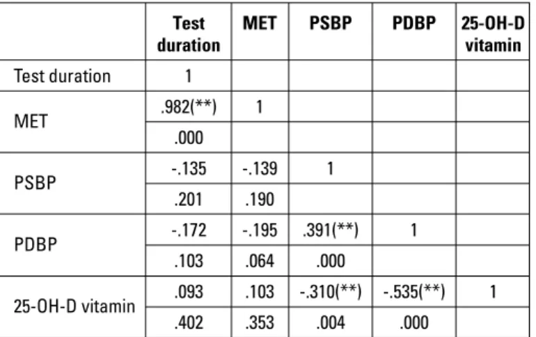
Table 2. Correlations among significant variants in univariate analyses Test MET PSBP PDBP 25-OH-D
duration vitamin Test duration 1 MET .982(**) 1 .000 PSBP -.135 -.139 1 .201 .190 PDBP -.172 -.195 .391(**) 1 .103 .064 .000 25-OH-D vitamin .093 .103 -.310(**) -.535(**) 1 .402 .353 .004 .000
**Pearson's correlation test was used. MET - metabolic equivalent; PDBP - peak dia-stolic blood pressure; PSBP - peak sydia-stolic blood pressure; 25-OH-D vitamin - 25-hydroxy vitamin D
Table 3. Two different multivariate logistic regression analyses for the predictors of cardiac syndrome X
First model (Hosmer and Lemeshow Test: P:0.095, Nagelkerke R Square: 0.433)*
β P OR 95% CI
MET -.652 0.003 0.521 0.340–0.797 ARE 0.749 0.295 2.114 0.813–0.949 25-OH-D vitamin -.129 0.001 0.879 0.813–0.949
Second model (Hosmer and Lemeshow Test: P:0.755, Nagelkerke R Square: 0.480)*
MET -.571 0.007 0.565 0.372–0.859 25-OH-D vitamin -.104 0.008 0.901 0.834–0.973 PDBP 0.075 0.027 1.078 1.009–1.151 *Multivariate logistic regression test was used. ARE - abnormal response to exercise; MET - metabolic equivalent; PDBP - peak diastolic blood pressure; 25-OH-D vitamin - 25-hydroxy vitamin D
resulted in decreased blood pressure levels (33, 34). In our pa-tients, the abnormal blood pressure response to exercise may be related to autonomic dysfunction. Apart from specific pathways such as endothelial dysfunction and autonomic dysregulation, the relationship between CSX, vitamin D, and abnormal blood pressure response to exercise may be multifactorial.
Study limitations
One of the limitations of our study is the relatively small number of patients involved. Another concern is that we did not use intravascular imaging methods such as optical coher-ence tomography or intravascular ultrasound to describe normal coronary arteries. Coronary angiography could be performed for better documentation of normal coronary anatomy. Moreover, intracoronary ergonovine was also not applied because we had concerns about severe and persistent vasospasms. Similarly, adenosine-induced coronary flow reserve could be tested us-ing echocardiography. Hyperventilation and cold presser tests, which are used for the same purpose, have lower sensitivity. Ad-ditionally, vitamin D levels of the patient group were measured in different seasons, although we tried to correct the seasonal dif-ferences in vitamin D levels by collecting blood samples from the control group in a short period of time. The levels of inflammatory markers such as CRP or ESR and their relationship with vitamin D levels could have been tested. The effect of abnormal blood pressure response on future hypertension could not be evalu-ated in this study because a long-term follow-up of the patients was not issued.
Conclusion
Patients with CSX had lower levels of vitamin D than healthy controls; in addition, vitamin D deficiency was associated with abnormal response to exercise. Further studies are required to understand the pathophysiological mechanisms and effects of vitamin D supplementation in these patients.
Conflict of interest: None declared. Peer-review: Externally peer-reviewed.
Authorship contributions: Concept – G.B.G., E.G., İ .B.; Design – G.B.G., E.G.; Supervision – G.G.D., E.G.; Fundings – İ.B.; Data collection &/ or processing – H.M.G., Ç.G., E.G.; Analysis &/or interpretation – G.B.G., S.H., G.G.D.; Literature search – İ.B., C.G., G.B.G.; Writing – G.B.G., S.H., G.G.D.; Critical review – Ç.G., H.M.G., G.B.G., E.G.
References
1. Holick MF. Vitamin D deficiency. N Engl J Med 2007; 357: 266-81. 2. Merke J, Milde P, Lewicka S, Hügel U, Klaus G, Mangelsdorf DJ, et
al. Identification and regulation of 1,25-dihydroxyvitamin D3 recep-tor activity and biosynthesis of 1,25-dihydroxyvitamin D3. Studies in cultured bovine aortic endothelial cells and human dermal
capillar-ies. J Clin Invest 1989; 83: 1903-15. Crossref
3. Judd SE, Tangpricha V. Vitamin D deficiency and risk for cardiovas-cular disease. Am J Med Sci 2009; 338: 40-4. Crossref
4. Li YC, Kong J, Wei M, Chen ZF, Liu SQ, Cao LP. 1,25-Dihydroxyvitamin D(3) is a negative endocrine regulator of the renin-angiotensin sys-tem. J Clin Invest 2002; 110: 229-38. Crossref
5. Sokol SI, Srinivas V, Crandall JP, Kim M, Tellides G, Lebastchi AH, et al. The effects of vitamin D repletion on endothelial function and inflammation in patients with coronary artery disease. Vasc Med 2012; 17: 394-404. Crossref
6. Lanza GA, Parrinello R, Figliozzi S. Management of microvascular angina pectoris. Am J Cardiovasc Drugs 2014; 14: 31-40. Crossref
7. Jones E, Eteiba W, Merz NB. Cardiac syndrome X and microvascu-lar coronary dysfunction. Trends Cardiovasc Med 2012; 22:161-8. 8. Kaski JC, Elliott PM. Angina pectoris and normal coronary
arterio-grams: clinical presentation and hemodynamic characteristics. Am J Cardiol 1995; 76: 35D–42D. Crossref
9. Kemp HG, Kronmal RA, Vlietstra RE, Frye RL. Seven year survival of patients with normal or near normal coronary arteriograms: a CASS registry study. J Am Coll Cardiol 1986; 7: 479-83. Crossref
10. Sharabi Y, Ben-Cnaan R, Hanin A, Martonovitch G, Grossman E. The significance of hypertensive response to exercise as a predictor of hypertension and cardiovascular disease. J Hum Hypertens 2001; 15: 353-6. Crossref
11. Ku YC, Liu ME, Ku CS, Liu TY, Lin SL. Relationship between vitamin D deficiency and cardiovascular disease. World J Cardiol 2013; 5: 337-46. Crossref
12. Wu-Wong JR, Nakane M, Ma J, Ruan X, Kroeger PE. Effects of Vi-tamin D analogs on gene expression profiling in human coronary artery smooth muscle cells. Atherosclerosis 2006; 186: 20-8. 13. Rostand SG, Drüeke TB. Parathyroid hormone, vitamin D and
car-diovascular disease in chronic renal failure. Kidney Int 1999; 56: 383-92. Crossref
14. Lavie CJ, Lee JH, Milani RV. Vitamin D and cardiovascular disease will it live up to its hype? J Am Coll Cardiol 2011; 58: 1547-56. 15. Reis JP, von Mühlen D, Michos ED, Miller ER 3rd, Appel LJ, Araneta
MR, et al. Serum vitamin D, parathyroid hormone levels, and carotid atherosclerosis. Atherosclerosis 2009; 207: 585-90. Crossref
16. Molinari C, Uberti F, Grossini E, Vacca G, Carda S, Invernizzi M, et al. 1α, 25 dihydroxycholecalciferol induces nitric oxide production in cultured endothelial cells. Cell Physiol Biochem 2011; 27: 661-8. 17. Oh J, Weng S, Felton SK, Bhandare S, Riek A, Butler B, et al.
1,25(OH)2 vitamin D inhibits foam cell formation and suppresses macrophage cholesterol uptake in patients with type 2 diabetes mellitus. Circulation 2009; 120: 687-98. Crossref
18. Karakaş M, Thorand B, Zierer A, Huth C, Meisinger C, Roden M, et al. Low levels of serum 25-hydroxyvitamin D are associated with increased risk of myocardial infarction, especially in women: re-sults from the MONICA/KORA Augsburg case-cohort study. J Clin Endocrinol Metab 2013; 98: 272-80. Crossref
19. Sun Q, Pan A, Hu FB, Manson JE, Rexrode KM. 25-Hydroxyvitamin D levels and the risk of stroke: a prospective study and meta-anal-ysis. Stroke 2012; 43: 1470-7. Crossref
20. Wang L, Song Y, Manson JE, Pilz S, März W, Michaëlsson K, et al. Circulating 25-hydroxy-vitamin D and risk of cardiovascular dis-ease: a meta-analysis of prospective studies. Circ Cardiovasc Qual Outcomes 2012; 5: 819-29. Crossref
21. Jadhav S, Ferrell W, Greer IA, Petrie JR, Cobbe SM, Sattar N. Ef-fects of metformin on microvascular function and exercise toler-ance in women with angina and normal coronary arteries: a
ran-domized, double-blind, placebo-controlled study. J Am Coll Cardiol 2006; 48: 956-63. Crossref
22. Kaski JC, Elliott PM, Salomone O, Dickinson K, Gordon D, Hann C, et al. Concentration of circulating plasma endothelin in patients with angina and normal coronary angiograms. Br Heart J 1995; 74: 620-4. 23. Panting JR, Gatehouse PD, Yang GZ, Grothues F, Firmin DN, Collins
P, et al. Abnormal subendocardial perfusion in cardiac syndrome X detected by cardiovascular magnetic resonance imaging. N Engl J Med 2002; 346: 1948-53. Crossref
24. Piatti P, Fragasso G, Monti LD, Setola E, Lucotti P, Fermo I, et al. Acute intravenous L-arginine infusion decreases endothelin-1 levels and improves endothelial function in patients with angina pectoris and normal coronary arteriograms: correlation with asym-metric dimethylarginine levels. Circulation 2003; 107: 429-36. 25. Güler E, Güler GB, Kızılırmak F, Batgerel U, Demir GG, Günes HM,
et al. Evaluation of adiponectin and lipoprotein(a) levels in cardiac syndrome X. Herz 2015; 40: 291-7. Crossref
26. Nakashima M, Miura K, Kido T, Saeki K, Tamura N, Matsui S, et al. Exercise blood pressure in young adults as a predictor of future blood pressure: a 12-year follow-up of medical school graduates. J Hum Hypertens 2004; 18: 815-21. Crossref
27. Tzemos N, Lim PO, Mackenzie IS, MacDonald TM. Exaggerated ex-ercise blood pressure response and future cardiovascular disease.
J Clin Hypertens (Greenwich) 2015; 17: 837-44. Crossref
28. Singh JP, Larson MG, Manolio TA, O'Donnell CJ, Lauer M, Evans JC, et al. Blood pressure response during treadmill testing as a risk factor for new-onset hypertension. The Framingham heart study. Circulation 1999; 99: 1831-6. Crossref
29. Gupta MP, Polena S, Coplan N, Panagopoulos G, Dhingra C, Myers J, et al. Prognostic significance of systolic blood pressure increases in men during exercise stress testing. Am J Cardiol 2007; 100: 1609-13. Crossref
30. Griffin BP, Callahan TD, Menon V, Wu WM, Cauthen CA, Dunn JM. Manual of cardiovascular medicine. Lippincott, Williams and Wilkins, Philadelphia, PA Edition 4,2013. P785.
31. Gillian DM, Panza JA, Kilcoyne CM, Waclawiw MA, Casino PR, Quyyumi AA. Contribution of endothelium- derived nitric oxide to exercise-induced vasodilation. Circulation 1994; 90: 2853-8. 32. Bitigen A,Türkyılmaz E, Özdemir N. Blood pressure response to
treadmill exercise testing. Arch Turk Soc Cardiol 2006; 34: 376-81. 33. Forman JP, Giovannucci E, Holmes MD, Bischoff-Ferrari HA,
Tworoger SS, Willett WC, et al. Plasma 25-hydroxyvitamin D levels and risk of incident hypertension. Hypertension 2007; 49: 1063-9. 34. Arora P, Song Y, Dusek J, Plotnikoff G, Sabatine MS, Cheng S, et al.
Vitamin D therapy in individuals with prehypertension or hyperten-sion: the DAYLIGHT trial. Circulation 2015; 131: 254-62. Crossref