73
Hepatic artery aneurysm: Imaging findings
Hepatik arter anevrizması: görüntüleme bulgularıAysel Türkvatan, R. Sarper Ökten, Esra Kelahmet, Ensar Özdemir, Tülay Ölçer
Türkiye Yüksek İhtisas Hastanesi, Radyoloji Bölümü Hepatic artery aneurysm is uncommon with an estimated incidence of less than 0.25%. Because most patients are asymptomatic, the diagnosis is usually made as an incidental finding on imag-ing studies performed for other reasons. Because of their propensity to rupture with potential catastrophic intraperitoneal hemorrhage, early diognosis is important. Herein, relatively asymp-tomatic two aneurysms of the hepatic artery of atherosclerotic etiology is presented. The impor-tance of imaging findings in the diagnosis of this condition is discussed and relevant literature is reviewed.
Key words: Hepatic artery aneurysm, computed tomography, angiography
Hepatik arter anevrizmaları ender olup, insidansı % 0.25’den daha azdır. Hastaların çoğu asemp-tomatik olduğu için tanı genellikle başka nedenlerle yapılan görüntüleme tetkikleri sırasında tesadüfen konulur. Hepatik arter anevrizmaları, potansiyel katastrofik intraperitoneal kanamayla rüptüre olmaya eğilimli oldukları için erken tanı önemlidir.
Burada, etyolojisinde aterosklerozun rol oynadığı iki adet HAA’sı bulunan bir olgu sunulmaktadır. Tanıda görüntüleme bulgularının önemi tartışılmış ve ilgili literaür gözden geçirilmiştir.
Anahtar kelimeler: Hepatik arter anevrizması, bilgisayarlı tomografi, anjiografi
T
he hepatic artery is the fourth common site of intraabdominal aneurysm from any cause following infrarenal aorta, iliac artery and splenic artery (1,2). Hepatic artery aneurysm (HAA) represent approximately 20% of all visceral aneurysms (3). 80% HAAs are extrahepatic and 20% are intrahepa-tic. 63% of HAAs involve the common hepatic artery, 28% involve the right hepatic artery, 5% involve the left hepatic artery, and 4% both the left and right hepatic arteries (1,2).Case report
A 53 year-old female patient presented with 6-month history of right upper abdominal pain. The physical examination was unremarkable. Routine hemato-logical and biochemical profiles were normal.
Gray scale abdominal ultrasonography revealed two well-defined, rounded cystic masses measuring 20 mm and 10 mm in diameter in the region of the porta hepatis.
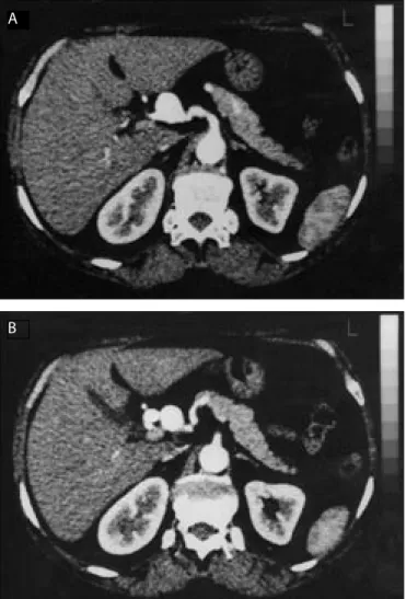
Doppler sonography, performed to evaluate the vascularity associated with the lesions revealed pulstatile flow within the lesions with a vascular origin. The findings suggested HAA and helical computed tomography (CT) was subse-quently performed. Unenhanced CT scan showed two well-circumscribed le-sions at the porta hepatis (Figure 1a, 1b). After intravenous administration of 100 ml. of bolus of contrast medium and scanning in early arterial phase
(15-Corresponding Author
Aysel Türkvatan
Türkiye Yüksek İhtisas Hastanesi Radyoloji Bölümü, Ankara Tel : (312) 306 16 37
Faks : (312) 312 4120 E-mail : [email protected]
Received: 02.06.2003 • Accepted: 10.11.2004
Ankara Üniversitesi Tıp Fakültesi Mecmuası 2005; 58:73-75
DAHİLİ BİLİMLER / MEDICAL SCIENCES
74 Hepatic artery aneurysm: Imaging findings Ankara Üniversitesi Tıp Fakültesi Mecmuası 2005; 58(2)
40 seconds) marked enhancement of the lumen of the le-sions was noted along the course of the hepatic artery from its origin from the coeliac artery. The diagnosis was con-firmed by angiography. An angiogram of the coeliac artery showed two hepatic arterial aneurysms, the one aneurysm was situated at the common hepatic artery and the other one was situaled at the bifurcation of the gastroduodenal and proper hepatic artery (Figure 2).
The patient underwent surgical correction of the aneu-rysms. Histopathology of the specimen was suggestive of atheromatous affection. At follow-up 6 months later the patient was asymptomatic.
Discussion
There are various etiologies for HAAs. Mycotic aneu-rysms historically accounted for most HAAs but accounted for only 4% in a recent review (3). HAAs are most fre-quently caused by atherosclerosis, which is present in as many as 30% of affected patients, and by medial
degenera-tion, which is present in 24% (1,4). Less common causes are polyarteritis nodosa, tuberculosis, periarterial inflam-mation (caused by cholecystitis or pancreatitis), fibromus-cular dysplasia, trauma, surgery (orthotopic liver trans-plantation or hepatic tumor embolization) and diagnostic instrumentation (1-4).
HAAs is not initially diagnosed in many cases because the majority of patients with HAAs are asymptomatic prior to rupture (3). In 80 % of the cases rupture of the aneurysms is the first clinical manifestation (5,6). The an-eurysms can rupture with equal frequency into the biliary tree or abdominal cavity (3). Of the patients who present with clinical symptoms, abdominal pain is found in 55% and gastrointestinal haemorrhage occured in up to 46% of symptomatic patients (4). The classic triad of epigastric pain, haemobilia and obstructive jaundice is only present in up to 33% of cases (1-4).
Physical examination is usually normal, although large aneurysms may be associated with a pulsatile mass or an abdominal bruit (1,2,7).
Combining the appropriate imaging techniques makes the definitive of HAAs.
Plain film of the abdomen may occasionally show a curvilinear calcification represently the wall of aneurysm in the right upper quadrant of the abdomen (4,8-10). Con-trast studies of the upper gastrointestinal tract may show a deformity of the duodenal curve from external compres-sion (4,8,9).
Figure 1 a,b. CT scan at the level of the porta hepatis demonstrates two aneurysms of the common hepatic artery
Figure 2. Selective coeliac axis arteriogram showing two saccular aneurysms of the common hepatic artery (CA: coeliac axis, HA: common hepatic artery, A:aneurysm)
A
75
Aysel Türkvatan, R. Sarper Ökten, Esra Kelahmet et al. Journal of Ankara University Faculty of Medicine 2005; 58(2)
Endoscopic retrograde cholangiopancreatography or percutaneous transhepatic cholangiography may show biliary dilatation and filling defects, especiallly in patients with melena (4,8,9).
Ultrasound is an excellent noninvasive method in the evaluation of the liver and porta hepatis for the presence of these lesions. The aneurysm can appear as a mixed echogen-ic mass with varying proportions of cystechogen-ic and solid com-ponents, depending on the extent of thrombosis. Calcifica-tions can occasionally be seen in the wall (11-13). Doppler ultrasound can aid in differentiating vascular from other types of masses. Color Doppler shows arterial or turbulent flow in the lesion suggestive of it being a mass of vascular origin (1-3). Furthermore, color Doppler ultrasound can differentiate aneurysms from other vascular abnormali-ties, such as arteriovenous fistulas or malformations. Dop-pler sonography plays a significant role in the follow-up patients who undergo embolization, allowing unnecessary follow-up angiography to be avoided (1-3,14).
Abdominal CT is often requested in trauma patients. Vessel wall calcification on unenhanced scans usually in-dicates arteriosclerotic change. Thrombotic deposits in the vessel lumen can be seen as ring-shaped or semilunar inter-nal areas of hypodensity. Images after intravenous contrast medium clearly demonstrate the vessel lumen. An attenu-ation of 70 HU in the tissues surrounding the aneurysm is a sign of the fresh hemorrhage. While conventional CT can demonstrate an aneurysm, the artery of origin is not always clear. Three dimentional spiral CT may allow a de-finitive diagnosis to be made prior to angiography in some cases (4,14-17).
Appropriate management of HAA requires detailed angiography. This can confirm the diagnosis, identify any other aneurysms (%20 are multiple), delineate feeding ves-sel, demonstrate any arterioportal fistula and provide the anatomical information needed for surgery or emboliza-tion (1,3,5,17).
Treatment of HAAs is usually in the form of ligation and surgical correction for the extrahepatic aneurysms and transcatheter embolization for the intrahepatic ones (2,4,18).
In conclusion, hepatic artery aneurysms are uncommon lesions that have varied clinical presentations. Early diag-nosis is essential because the natural tendency of the lesion is to rupture into peritoneal cavity or surrounding organs. Our case is notable, because two atherosclerotic aneurysms of the hepatic artery are extremely rare with very few cases reported so far and to diagnose a hepatic artery aneurysm before rupture is also unusual.
References
1. Bachar GN, Belenky A, Lubovsky L et al. Sonographic diagnosis of a giant aneurysm of the common hepatic artery. J Clin Ultrasound
2002; 30:300-2.
2. Parmar H, Shah J, Shah B et al. Imaging findings in a giant hepatic artery aneurysm. J Postgrad Med 2000; 46:104-5.
3. Chandramohan C, Khan AN, Fitzgerald S et al. Sonographic diagnosis and follow-up of idiopathic hepatic artery aneurysm, an unusual cause of obstructive jaundice. J Clin Ultrasound 2001; 29: 466-71.
4. O’Driscoll D, Olliff SP, Olliff JF. Hepatic artery aneurysm. Br J Radiol 1999; 72: 1018-25.
5. Komori K, Sonoda T, Ikeda Y et al. Demonstration of hepatic artery aneurysm by substraction angiography. Am J Gastroenterol 1991; 86:1650-3.
6. Sunthornlekhla B, Chandaragga S, Sundusadee K et al. Hepatic artery aneurysm: a case report. J Med Assoc Thai 1996; 79: 60-4. 7. Hubleue I, Keymeulen B, Delvaux G et al. Hepatic artery
aneurysm. Case Report with review of the literature. Acta Clin Belg 1993; 48: 246-52.
8. Psathakis D, Muller G, Noah M et al. Present management of hepatic artery aneurysms. Vasa 1992; 21: 210-5.
9. Countryman D, Norwood S, Register S et al. Hepatic artery aneurysm. Report of an unusual case and review of the literature. Am Surg 1983; 49:51-4.
10. Ibach EG, O’Halloran MJ, Prendergast FJ. Hepatic artery aneurysm: Two case reports and review of the literature. Aust N Z J Surg 1997; 67: 143-7.
11. Paolella LP, Scola FH, Cronan JJ. Hepatic artery aneurysm:an ultrasound diagnosis.J Clin Ultrasound 1985; 13:360-2.
12. Rigaux A, Vossen P, Van Baarle A et al. A.Hepatic artery aneurysm: ultrasonic diagnosis. J Clin Ultrasound 1986; 14:401-3.
13. Stokland E, Wihed A, Ceder S et al. Ultrasonic diagnosis of an aneurysm of the common hepatic artery. J Clin Ultrasound 1985; 13:360-2.
14. Warshauer DM, Keefe B, Maura MA. Intrahepatic hepatic artery aneurysm: computed tomography and color-flow Doppler ultrasound findings. Gastrointest Radiol 1991; 16:175-7. 15. Howling SJ, Gordon H, McArthur T et al. Hepatic artery
aneurysms: Evaluation using three dimensional spiral CT angiography. Clin Radiol 1997; 52:227-30.
16. Zalcman M, Matos C, Van Gansbeke D et al. Hepatic artery aneurysm: CT and MR features. Gastrointest Radiol 1987; 12:203-5.
17. Barkin JS, Potash JB, Hernandez M et al. Hepatic artery aneurysm simulating a cystic mass of the pancreas. Dig Dis Sci 1987; 32:1196-200.
18. Cooper SG, Richman AH. Spontaneous rupture of a congenital hepatic artery aneurysm. J Clin Gastroenterol 1988; 10:104-7.