Ankara Üniv Yet Fak Derg, 50, 97-102, 2003
EvaIuation of kidney abnormalities in mongrel dogs using clinical,
ultrasonographical and biochemical examinations*
.
Ramazan GÖNEN Cİ
i,Ramazan DURGUT
2,Suat ERDOGAN3, M. Enes ALTUGı, Ramazan BAL
4lDepartments of Surgery, "Internal Medicine, 3Biochemistry, 4Physiology, Faculty of Yeterinary Medicine, University of Mustafa Kemal, Antakya
Summary: In this study, 25 female and 15 male mongrel dogs, aged ranging from i to 7 years, were used. They were subjected to clinical, ultrasonographical and biochemical examinations. Ultrasonographically in 18 of the 40 do gs (45%), different kidney abnormalities were observed, whereas the other 22 dogs were found normaL. Likewise, serum urea and creatinine levels were measured to be higher in the dogs with kidney abnormalities. Serum creatinine elearance estimated using the Cockroft-Gault equation was exceeding the physiological range. In conelusion, for mongrel dogs ultrasonographic examination, measurements of serum urea nitrogen (BUN) and creatinine, and calculation of serum creatinine elearance using the Cockroft-Gault equation might be useful to diagnose early kidney abnormalities.
Key words: Biochemical analysis, dog, kidney, ultrasonography
Melez köpeklerde böbrek bozukluklarının
ultrasonografi ve biyokimyasal muayenelerle
değerlendirilmesi
Özet: Bu çalışmada yaşları i-7 arasında değişen 25 dişi ve 15 erkek melez köpek kullanıldı. Köpekler her iki böbrek yönünden klinik, ultrasonografik ve biyokimyasal yönden muayene edildi. Ultrasonografide kırk köpeğin i8'inde (%45) farklı böbrek anormallikleri gözlenirken diğer 22 köpeğin normalolduğu belirlendi. Ayrıca, serum üre ve kreatinin düzeyi ile Cockroft-Gault denklemini kullanarak belirlenen serum kreatinin klirensi, böbrek anormalliği belirlenen bu köpeklerde fizyolojik sınırların üstündeydi. Sonuç olarak, erken böbrek anormalliklerini tanımak için rutin olarak ultrasonografik muayenenin yapılması, serum üre ve kreatinin seviyelerinin ölçülmesi yanısıra Cockroft-Gault formülünü kullanarak serum kreatinin klirensinin düzenli olarak hesaplanmasınında faydalı olabilleceği kanısına varıldı.
Anahtar kelimeler: Biyokimyasal analiz, böbrek, köpek, ultrasonografi
Introduction
The kidneys are two of the most important organs in
the body. Their essential function is to maintain the
homeostasis, for instance, excreting waste products and
toxic substances into urine, controlling the balance of
acids or bases in the body (6, 16). On:ee started, a kidney
disease is unstoppable; kidneys may eventually lose the
ability to remove waste products and excess nutrients
from the blood, which can ultimately lead to death
(2,3,7,15,16). Kidney diseases are not easily detectable,
especially in its early stages. Serum creatinine and urea nitrogen levels are not changed until about three-fourths
of kidney function is lost (15,16). Ultrasonography is the
most common modes for visualizing aberration in the
number, size, and texture and position of the kidneys
(1,1 1,15,18) and related structures as well as the presence
of mineralizing densities (4,10,13,14). The elearance of
endogenous creatinine has been used as a measure of
glomerular filtration rate (GFR) since the 1940s (5,8,12),
which can be cakulated with different method s using
different parameters. Using Cockroft-Gault equation, it is
possible to calculate creatinine elearance with sufficient
accuracy, provided serum creatinine and the patients
weight and age, as follows: creatinine elearance=(140-age
[year] x (patient's weight [kg])172x (serum creatinine
[mg/dl]), correcting serum creatinine to 1 mg/dı (5,8,12). The aim of this study was to investigate the incidence of kidney problems in mongrel dogs in Antakya province
from March 2001 to March 2002 using elinical and
ultrasonographical exarninations, and calculating creatinine elearance.
Materials and Methods
For this study, 25 females and 15 males mongrel
dogs, aged between i and 7 years, were evaluated
between March 2001 and March 2002. They were
t
98 Ramazan Gönenci - Ramazan Durgut - Suat Erdoğan - M. Enes Altuğ - Ramazan Bal
subjected to clinical, ultrasonog~aphical and biochemical
examinations for the presence of kidney diseases or any
other abnormalities. Following the generel examination,
dogs were restrained in dorsal, and lateral recumbency
and aleohool and ultrasonic coJpling gel applied to the
clipping skin for ultrasonography.
Ultrasonographie evaluation of kidneys and ureters
was performed using a scannerı 100 LC Yet ultrasound
machine (Pie Medical Equipmetıt B.Y., Philipsweg 6227
AI Maastricht, The Netherlands) with a 5.0/7.5
mega-hertz (MHz) LA DF Yyt TRD (41460, 41518)
ultrasonic transducer. KidneYs were examined in
longitudinal, sagittal, and transversal plan~s. Images were recorded on a 1.44 MB standard computer disc. Results were recorded for each dog at tı:ıe time of examination as
either negative or positive fdr the disease based on
identifiable structure within the kidneys.
For biochemical evaluations, 10 ml of blood
samples were taken from the ju~ular vein, and sera were
separated by centrifugation. Kidney function was
evaluated by testing serum blood urea nitrogen (BUN), creatinine albumin, total protein and phosphate in AMS
Autolab analyser using BMSI kits. Serum creatinine
clearance was caleulated by the Cockroft-Gault equation
as follows: Creatinine clearance=(140-age [year] x
(patient's weight [kg])/72x(serum creatinine [mg/dl])
(5,8,12).
Results
i
In ultrasonography of 22 dogs, the longitudinal
plane appeared bean-shaped. In the transverse section, the kidney was rounded. The renal medulla was anechoic and had several segments. The me1dulla and cortex were of
~ !
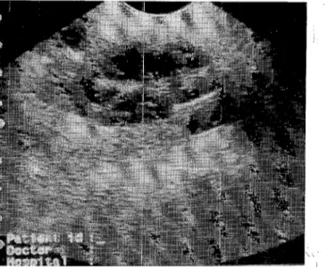
Figure i.Ultrasonographic appearance of a mass with increased ıneduııary echogenicity and reducedıclarity of the corticoıneduııary junction.
equal thickness. Blood urea nitrogen, cFeatinine, total
protein, albumin, and phosphate were in ndrmal reference ranges.
Clinical, ultrasonographical and biochemical
examinations showed the presence of kidney
abnormalities in 18 of the 40 dogs. Dorsal ultrasound
(case no 1) image of right kidney in a dog ~howed a mass
with increased medullary eehogenieity
i
and redueedclarity of the cortieomedullary junction (Figure 1). In two
dogs (ease no 2,3) a dilated aneehoie pelvik with a dilated ureter was observed (Figure 2). A bright, lechogenic line
casting aeoustic shadows was present at the
eortieomedullary junction in a dog (ease Jo 4). Diserete,
i
round, anechoic structures with clcep aeoustic
enhancements were seen, which were ran~~ng in diameter from 1.8 mm to 3.5 mm in two dogs (Fif,ure 3; case no
i
5,6). In a dog (case no 7), on the right kidney there were
variable degrees of aeoustic enhaneemebts with more
echogenieity, with a thick irregular wall(Figure 4). In
two animals (case no 8,9), left kidneys ere small and
hypereehoie, loss of normal arehiteeture ith thinning of
eortex were striking (Figure 5).
i
In four dogs (case no 10,11,12,13), t~iffuse increase
in eehogenieity were detected. In adJlition, los s of
distinction between cortex and medulla wJre seen (Figure 6). Urinalyses of these animals indieated dıild proteinuria,
and low speeific gravity (1005- 1010). S/iff-legged gait,
painful kidneys on pal pa tion, less ur~ne production,
weakness and exereise intoleranee and pale mucous
membranes were observed in these three dbgs.
i
In two animals (case no 14,15), pelvic dilatation and
parti al eehogenicity in their left kidneyJ with enlarged
and irregular arehiteeture were the major hndings (Figure
i
Figure 2. A dilated anechoic pelvis and ure~rs in ultrasonog-raphy.
Ankara Üniv Vet Fak Derg, SO, 2003 99
Figıırc 3. Ulırdsuııographie appearanee of adiscrete, round and dııcl'iı,)ic slnıcııırc in a kidncy.
FiglllT 5. Smail and hypcreehoic appearances of a kidney with luss ol normal arehileetme and ıhinning of eortex in
Liitrd" ııı,ıgraphy.
Figure 7. Pelvie dilatation and parti al eehogenicity wiıh enlarged and irregular arehiteetme in ultrasonography.
Figme 4. A variable degrees of aeoustie enhaneemenıs, a clearly dcfined internal septa. and thiek irregular wal1.
Figııre 6. Ultrasonographie appearanee of a diffuse inerease in eehogenieity. Note loss of distinetion between eortex and medulla.
7). Dilatation of renal pelvis resulted in a C-shaped
echogenicity with a central anechogenic area (Figure 8).
Strongly retlective structures producing deep
acoustic shadowing in both kidneys were detected in
another two do gs (case no 16,17), and they remained
constant even with changes in transducer angulations
(Figure 9).
Dorsal ultrasound image s of the right kidneys
showed a heterogeneous mass, which is located in the
cranial pole of the kidney in another dog (case no 18)
with hematuria and weight los s (Figure lOA). Oblique
ultrasound image of the left kidney showed circular
anechoic cavitary lesion and ureteral dilatation (Figure
lOB),
Serum concentrations of urea and creatinine were
ınn
Ramazan Gö~enci - Ramazan Durgut - Suat Erdoğan - M. Enes Altuğ - Ramazan Bali
Dİscussİon and Conclusİon
Many pet owners are unaware of the high incidence of kidney diseases in dogs. It is reported that a kidney
disease can be the leading cause of non-accidental death
in dogs (10,16), because 20% of dogs with more than
75% reduction in kidney function cal' liye without
showing any symptoms. Upon further reductions
resulting in total kidney failure theyare no longer able to
remove the waste products and toxins, aceumulate in the blood and the n show clinical signs of kidney diseases. All
i
breeds of any age can be affeeted. Therefore, routine
ultrasonographie examination should be applied to dogs
Figurc iIl. Ultrasonographic appearance of a heterogenous mass loeated in the cranİal pole of the left kidney (A). OllliQUe ultrasound image of the same kİdney with anec,hoic cavitary lesİons and meteral dilatation (B).
i
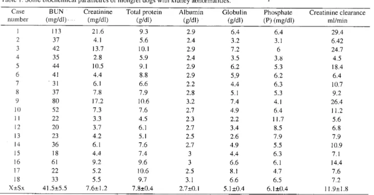
the normal referenee ranges (9). Results of some periodieally for early diagnosis of kidney abnormalities
bioehemieal parameters and creatinine clearance of the (1,4,18).
i
18 dogs were shown in Table 1. it is reported that diagnostic ultrasound is an
exeellent technique for demonstratinJ the internal
architecture of parenchymal organs a1d fluid filled
struetures (4,13,18). it is seen in this study that five
mega-hertz (MHz) transducer was a~propriate for
visualization of urinary tract struetures. Itlproduced good
resolution with aeceptable depth of peretration. It is
therefore ideally suited for the investiigation of dog
urinary tract diseases. It is non-invasive a/ıd safe for both
i
patient and operator. Ultrasound is also of more use than
intravenous urography when renal funetilon is poor and
contrast medium excretion is redieed and is
complementary for almost definite diagnosis of kidney
Ankara Üniv Yet Fak Derg, 50, 2003 101
Table
ı.
Some bioehemieal parametres of mongrel dogs with kidney abnormalities.Case BUN Creatinine Total protein Albumin Globulin Phosphate Creatinine elearance
number (mg/dı) (mg/dı) (gldı) (gldı) (gldı) (P) (mg/dı) ml/min 1 113 21.6 9.3 2.9 6.4 6.4 29.4 2 37 4.1 5.6 2.4 3.2 3.1 6.42 3 42 13.7 10.1 2.9 7.2 6 24.7 4 35 2.8 5.9 2.4 3.5 3.8 4.5 5 44 10.5 9.1 2.9 6.2 5.3 18.4 6 41 4.4 8.8 2.9 5,9 6.2 6.4 7 . 31 6.1 6.6 2.2 4.4 6.3 10.7 8 37 7.8 7.9 2.8 5.1 5.3 9.2 9 80 17.2 10.6 3.2 7.4 4.1 26.4 LO 52 7.3 7.6 2.7 4.9 6.4 11.2 1i 22 3.3 4.5 2.3 2.2 11.7 5.6 12 20 3.7 6.1 2.7 3.4 8.5 6.8 13 23 4.2 5.1 2.5 2.6 7.9 7.9 14 36 6.1 7.6 2.7 4.9 5.5 10.9 15 18 4.4 7.4 3 4.4 6.3 7.1 16 61 9.2 9.6 3 6.6 6.1 14.4 17 22 5.2 10.6 2.5 8.1 4.7 7.6 18 33 5.5 9.7 3.1 6.6 6.5 7.2 X:tSx 41.5:t5.5 7.6:t1.2 7.8:t0.4 2.7:tO.1 5.1 :t0.4 6.1:t0.4 11.9:t 1.8
useful information about the internal architeeture of the
kidneys with focal lesions. Although it is difficult to
visualize the normal canine ureters ultrasonographically,
meteral dilatations were able to be detected along with
various abnormalities in two dogs with dilated anechoic
pelvis in this study. Therefore, there was consisteney
with previous reports (4,10,13).
InCl'eased cortical echogenicity has been show n to
correlate with renal diseases of many etiologies, but it is
generalI y considered as nonspecific cı,4, 16). Therefore,
BUN and creatinine levels, which are two most
significant parameters for diagnosis of kidney
abnormalities, were measured in this study, Creatinine is
thought to be a more reliable indicator of kidney
function, as it well shows that the kidneys are filtering
out the toxins, and is less dependent on dietary factors
and hydration status of the dog. Serum creatinine is freely
filtered İn the glomeruli and there is normally 10-40%
excretion in the tubules (5,8,12). In the study presented
here, serum creatinine and urea nitrogen values were
high, implying renal function disorders and consistent
with diagnosis by ultrasonography. For this reason, it
could be suggested that high serum creatinine and urea
nitrogen values may be indexes for the poor glomerular filtration rate in dogs.
Phosphate is a bone mineral, which is freely filtered
through the glomerulus and reabsorbed in the proximal
tubules, but excess amounts are actively secreted to the
mine. The phosphate concentration in serum increases in
renal insufficiency due to intestinal over absorption of
phosphate and decreased secretion in the tubules (3,17).
In this study, serum phosphate level was higher than the
normal physiological reference range in the animals with
kidney abnormalities, indicating that there is consisteney
with the previous reports.
The 24-h period of urine collection for measuring
creatinine clearance, being a time-consuming procedure
may be impractical for day-to-day monitoring of renal
function (3,5,8,12). We, therefore, believe that
measurement of creatinine clearance using the
Cockroft-Gault equation is very easy with sufficient
accuraey, if the dog's weight and age are known.
In conclusion, the incidence of kidney problems was
found 45% in mongrel dogs in the present study.
Convincing evidence presented here suggest that dogs
should be regularly checked for diagnosis of possible
kidney abnormalities using ultrasonography, laboratory
parameters, including urea, creatinine, phosphate, and
creatinine clearance.
References
I. Bahr A, Wrigley R, Salman M (2000): Quantitative eva-luation of imagent as an abdominal ultrasound contrast medium in dogs. Yet Radiol Ultrasound, 41,50-55. 2. Barlough JE, Osborne CA, Steven JB (1981): Canine
and feline urinalysis: Value of macroscopic and mic-roscopic examinations. JAYMA, 178, 61-67.
3. Barsanti JA, Finco DR (1979): Laboratory findings in
uri-nary tract infections. Yet Clin Nort Am,9,729-735. 4. Cartee RE, Selcer BA, Patton CS (1980).
Ult-rasonographic diagnosis of renal disease in smail animals.
J Am Yet Med Assoc, 76, 426-430.
5. Cockcroft DW, Gault MH (1976): Prediction of cre-atinine clearance from serum crecre-atinine. Nephron, 16, 31-41.
102 Ramazan Gönenci - Ramazan Durgut - Suat Erdoğan - M. Enes Altuğ - Ramazan Bal
6. Cowgill LO, Spangler WL (1981): Renal insufficiency in geriatric dogs. Yet Clin North lAm Small Anim Pract, 11,
727-748.7. Finco DR, Crowell WA, Barsanti JA (1985): Effects of three diets on dogs with induced chronic renal failure. Am J Yet Res, 46, 646-653.
8. Herget-Rosenthal S, Kribberl A, Pietruck F, Ross B, Philhipp T (1999): Two hy two hour creatinine
elearance-repeatahle and valid. Clin Nephro1, 51, 348-354.
9. Kaneko
n,
Harvey JW, Bruss ML (1997): ClinicalBi-ochemistry of Domestic Animall Academic Press, London.
ı
O. Kruger JM, Osborne CA, Nachreiner RF, Refsal KR(1996): Hypercalcemia and renal failure. Etiology, pat-hophysiology, diagnosis, and treatment. Yet Clin North
i
Am Small Anim Pract, 26,1411-1445.
iJ. Lamb CR (1998): Vltrasonography of the ureters. Yet
Clin North Am Small Anim Pract, 28, 823-848.
12. Mcligeyo SO (1993): Calculation of creatinine elearance
ji'O/11 plasma elearance. E Afr ~ed J, 70, 3-5.
ı
3. Miles K (1997): Imaging ahdominal masses. Yet Clin Norıh Am Small Anim Pract, 27,1403-1431.14. Ruiz de Gopegui R, Espada
y,
Majo N (1999): Bilateralhydroureter and hydronephrosis in a nine-year-oldfemale German shepherd dog. J SmalllAnim Prac, 40, 224-226.
15. Seeds JW (1998): Borderline genitourinary tract ah-normalities. Semin Ultrasound CT MR, 19, 347-354.
16. Uechi M, Nogami Y, Terui H, Nakayama T, Ishikawa R, Wakao Y, Takahashi M (1994): Evaılıtion ofurinary enzymes in dogs with early renal disorder. J Yet Med Sci,
56, 555-556.
17. Verbeek XA, Willigers JM, Prinzen FJN, Peschar M, Ledoux LA, Hoeks AP (2001): High-resolution functional
imaging with ultrasound contrast agents hbsed on RF pro-i
cessing in an in vivo kidney experiment. Ultrasound Med
Biol, 27, 223-233.
J
18. Watson AD, McDona1d PJ (1981): Comp~rison ofseveral
lahoratory tests for quant(fying azotaemJı in dogs. Aust
Yet J, 57, 407-410.
Geliş tarihi: 16.4.2002/ Kahul tarihi: 13.5. 002
Correspondence address:
Yrd. Doç. Dr. Ramazan Gönenci Mustafa Kemal Üniversitesi Veteriner Fakültesi Cerrahi Anahilim Dalı 31040 Antakya/Hatay