Thelazia callipaeda (Railliet and Henry, 1910) Case in a Dog: First
Record in Turkey
Mustafa ESER
1,aÖzlem MİMAN
2,bAbuzer ACAR
3,c
1 Anadolu University Open Education Faculty Health Programs, TR-26470 Tepebaşı, Eskişehir - TURKEY
2 Dokuz Eylül University, Faculty of Medicine, Department of Medical Parasitology, TR-35210 Alsancak, İzmir - TURKEY 3 Afyon Kocatepe University, Faculty of Veterinary Medicine, Department of Internal Medicine, TR-03200
Afyonkarahisar - TURKEY
a ORCID: 0000-0003-1542-2989; b ORCID: 0000-0003-3415-4959; c ORCID:0000-0002-4235-2763
Article ID: KVFD-2018-20487 Received: 05.07.2018 Accepted: 01.11.2018 Published Online: 01.11.2018
How to Cite This Article
Eser M, Miman Ö, Acar A: Thelazia callipaeda (Railliet and Henry, 1910) case in a dog: First record in Turkey. Kafkas Univ Vet Fak Derg, 25 (1): 131-134,
2019. DOI: 10.9775/kvfd.2018.20487
Abstract
A 2.5 years old male Golden Retriever breed dog with the itching, runny eyes, and continuous unease complaints was brought to a private veterinary medical centre in the Thrace region of Turkey, in September 2017. It was observed that there was a purulent conjunctivitis in the left eye and there was a mobile structure under the membrane nictitans after the examination. A drop of local anesthetic was dripped into the eye, and the mobile structure was removed with the help of a forceps. This removed structure was taken into the solution of 70% alcohol on suspicion of parasite. Both the extracted material and the blood samples were sent to the Department of Internal Medicine, Afyon Kocatepe University, Faculty of Veterinary Medicine for evaluation and it was evaluated with parasitologist. The parasite was cleared by taken into a 0.9% physiological saline and kept in the lactophenol for two days for transparency. Then, the transparent parasite was determined as Thelazia callipaeda after microscopic examination. The ocular form of thelaziasis caused by T. callipaeda in a dog has been reported for the first time in Turkey with this case report. By this report, first case of ocular thelaziasis reported seen a dog in Turkey and it was aimed to point out that this parasite can lead to significant eye problems in animals.
Keywords: Dog, Ocular thelaziasis, Thelazia callipaeda, Golden Retriever
Bir Köpekte Thelazia callipaeda (Railliet ve Henry, 1910) Olgusu:
Türkiye’de İlk Kayıt
Öz
Türkiye’nin Trakya Bölgesi’nde bir özel veteriner tıp merkezine 2017 yılı Eylül ayında gözde kaşıntı, akıntı ve sürekli huzursuzluk şikâyetleri ile 2.5 yaşında erkek Golden Retriever ırkı bir köpek getirilmiştir. Yapılan muayene sonrasında sadece sol gözde purulent bir konjunktivitisin olduğu ve membrana nictitansın altında hareketli bir yapının olduğu gözlenmiştir. Göze bir damla lokal anestezik damlatılarak hareketli yapı gözden bir pens yardımıyla çıkarılmıştır. Çıkarılan bu yapı parazit olması şüphesi ile %70’lik alkol içine alınmıştır. Alınan numune ve kan numuneleri değerlendirilmek üzere Afyon Kocatepe Üniversitesi Veteriner Fakültesi İç Hastalıkları Anabilim Dalı’na gönderilmiş ve parazitolog eşliğinde değerlendirilmiştir. Parazit %0.9’luk fizyolojik tuzlu suya alınarak temizlenmiş ve şeffaflaşması için iki gün laktofenolde bekletilmiştir. Şeffaflaştırılan parazitin mikroskobik muayenesi sonrasında Thelazia callipaeda olduğu belirlenmiştir. T. callipaeda’nın köpekte oluşturduğu oküler thelaziasis vakası Türkiye’de ilk olarak bu olgu sunumu ile bildirilmektedir. Bu rapor ile Türkiye’de bir köpekte rastlanılan ilk oküler thelaziasis vakası bildirilmiş ve parazitin hayvanlarda önemli göz problemlerine sebep olabileceğine dikkat çekmek istenmiştir.
Anahtar sözcükler: Köpek, Oküler thelaziazis, Thelazia callipaeda, Golden Retriever
INTRODUCTION
Thelazia species, which are important nematode parasites
that can be inoculated in the eye, are on the Spirurida order and in Thelaziidea family [1]. There are different types of Thelazia in domestic and wild animals such as in cattle:
Thelazia rhodesii, T. gulosa (Syn. T. alfortensis), T. skrjabini, in
buffalo: T. bubalis, in sheep, cat, dog, human: T. californiensis, in camel: T. leesei, in horse: T. lacrymalis, in dog, rabbit, human:
T. callipaeda, and in pig: T. erschowi [2]. It has been reported that these parasites could be found in the eyelids, membrana nictitans, and lacrimal channels, and sometimes in the nose and pharynx [3]. Eye involvement is high and infection induces from mild (conjunctivitis, epiphora and
İletişim (Correspondence)
+90 272 2182833/2716
abuzeracar@hotmail.comKafkas Universitesi Veteriner Fakultesi Dergisi
ISSN: 1300-6045 e-ISSN: 1309-2251 Journal Home-Page: http://vetdergikafkas.org Online Submission: http://submit.vetdergikafkas.org
Case Report
Kafkas Univ Vet Fak Derg 25 (1): 129-132, 2019
130
Thelazia callipaeda (Railliet and Henry, 1910) Case ... ocular discharge) to severe (keratitis and corneal ulcers) ocular manifestation in animals as well as humans [4]. Thelaziasis is known a zoonosis. Dog’s thelaziasis caused mainly by T. californiensis and T. callipaeda. The two species are also important for humans [3]. However, T. californiensis has been reported also in sheep, deer, jackals and bears [5]. Adult worms look like creamy white threads [6]. Male adults are 4.5-13 mm in length and 0.25 to 0.85 mm in diameter, while the females are longer, from 6.2 to 17 mm and from 0.3 to 0.85 mm in diameter [7]. There are fine lines and protrusions in the cuticle of Thelazia species. There is a hexagonal mouth capsule and six festons on the inside edge of the mouth capsule. The presence of tail wings and lengths of spiculations in males varies according to species [6]. The vulva position is used as the diagnostic criterion. T.
callipaeda’s vulva is located at the front of the esophagus
region vulva also has a short cover [6]. While other Thelazia species are vivipar, T. callipaeda is ovovivipar [3].
In the life cycle of T. callipaeda, flies in the Diptera order act as vectors. Although Shi et al.[8] have suggested that
Musca domestica may seldom be a vector, Otranto et al.[9] reported that Phortica spp. in the family Drosophilidae. (P.
variaegata and P. okadai) should be the important vector
for T. callipaeda, and that M. domestica could not be vector by both the natural infections they encountered and the experimental studies they performed.
In the parasite life cycle, the first period of larvae in the lacrimal secretion of the infected eye (very short life span, 1-2 hours) is taken during the feeding of the vector flies. They pass through the intestines of the flies to their abdomen and stay there for 1-2 days. On the third day, in the female flies they are moving to the adipose tissue and in the male flies to the testicles. They change moult twice and become L3 in 14-21 days. They reach the mouth organelles through the body cavity of the flies. The flies transfer larvae to the environment during feeding with lacrimal secretions around the eyes of the last hosts. There is no migration period in the last host. By changing moult twice (in 35 days) they become adults [10]. Prepatent time is 3-6 weeks. Infection is seen in seasons when flies are active, so it is depending on the season. Infections peak in two periods, beginning of the summer and the ending of the summer [11,12].
By this case report, it was aimed to point out that this parasite can lead to significant eye problems in animals.
CASE HISTORY
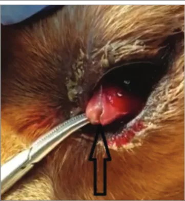
A 2.5 years old male Golden Retriever breed dog with the itching, runny eyes, and continuous unease complaints was brought to a private veterinary medical centre in the Thrace region of Turkey, in September 2017. It was observed that there was a purulent conjunctivitis in the
left eye (Fig. 1) and there was a mobile structure under the membrane nictitans after the examination.
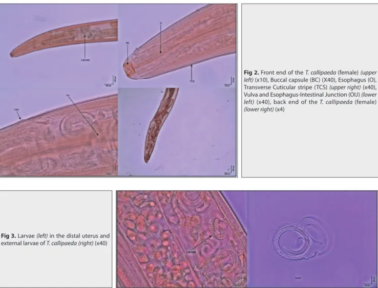
A drop of local anesthetic was dripped into the eye, and the mobile structure was removed with the help of a forceps. There was only one worm-parasite. This parasite was taken into the solution of 70% alcohol. Both the extracted material and the blood samples were sent to the Department of Internal Medicine, Afyon Kocatepe University, Faculty of Veterinary Medicine for evaluation and it was evaluated with parasitologist. The parasite was cleared by taken into a 0.9% physiological saline and kept in the lactophenol for two days for transparency. After this processes, morphological examinations were carried out on a light microscope (Olympus CX31) by parasitologist and pictures were taken (Olympus Imaging System Olympus LC30). The morphological features of the parasite were determined and the species was diagnosed by using the related literature [3,6,7,10]. There was no pathological result in the blood test. After cleaning and clarification, the front, and back of the parasite examined in the light microscope separately and measured. The size of the parasite was 11.34 mm, and the width was 0.3 mm. It was noted that it had a hexagonal buccal capsule on the front end. The large part (upper part) of the buccal capsule (upper part) was 0.04, and the narrow part (base) is 0.03 mm. A prominent esophageal structure, esophagus, and intestinal junction were observed. It has been observed that vulva is localized in the anterior part of the esophagus-intestine junction (OIJ). It is noted also that the parasitic cuticle is the transverse stripe (TCS). This transverse stripe structure was also measured as 0.02 mm (Fig. 2).
131 ESER, MİMAN, ACAR
In the parasite’s uterus, grown larvae and larvae in the development phase were observed (Fig. 3, left). The egg-shell in the sheath style was determined outside of the freed larvae (Fig. 3, right).
Upon diagnosis of thelaziasis, the dog was treated with ivermectin (200 µg/kg, S.C. injection, Ivomec®, Merial, Turkey) two times with an interval of two weeks. It was stated that the eyes of two other dogs belonging to the animal owner have similar clinical symptoms and brought to veterinary center for treatment. It has also been mentioned that there was conjunctivitis in animal owner for about 6 months. For the possibility of zoonosis, the owner was informed and suggested to consult by a specialist physician.
DISCUSSION
Railliet and Henry first identified T. callipaeda in 1910 in the eyes of a dog in Pakistan [11]. Then Evans and Rennie reported in Myanmar, while Stuckey reported T. callipaeda in dogs in China. The medical records were followed by other countries such as Far East countries (Former Soviet Countries, India, China, Thailand, Taiwan, Indonesia, South Korea, and Japan) [3,13] and Europe [4,14,15]. The first human
cases (4 cases) reported from Italy and France [16]; and
T. callipaeda reported found in Italy in cat and foxes [17].
T. callipaeda is endemically present in poor, rural areas,
and communities with low health and socio-economic standards as in this case.
Since information on T. callipaeda is rare and less known, the diagnosis of infections of this zoonotic species is omitted [7]. To the best of our knowledge, in Turkey, there is not a case report about this species. There are only prevalence studies on cattle, sheep, and horses related to
Thelazia species in Turkey [18-22]. The prevalence in different regions of Turkey was reported as in cattle 5.5% and 22% [18,21] and in buffaloes 1.2% [19] where the causative agent was T. rhodesii. Doganay and Oge [22] have done studies on the prevalence of sheep.
Literature reported that adult female T. callipaeda`s length may be 6.2-17 mm and width may be 0.3-0.85 mm [7]. The parasite’s measure has been determined 11.34 mm (lenght), 0.3 mm (width) in this case. These measurements coincide with the values given in the literature. It has been reported this species in both sexes has a serrated cuticle [16] and the buccal capsule has a hexagonal profile [3,7,16] 0.036 mm width and 0.030 mm depth on the front end [12]. It has
Fig 2. Front end of the T. callipaeda (female) (upper
left) (x10), Buccal capsule (BC) (X40), Esophagus (O),
Transverse Cuticular stripe (TCS) (upper right) (x40), Vulva and Esophagus-Intestinal Junction (OIJ) (lower
left) (x40), back end of the T. callipaeda (female) (lower right) (x4)
Fig 3. Larvae (left) in the distal uterus and
132
Thelazia callipaeda (Railliet and Henry, 1910) Case ... been observed in findings, this parasite cuticle’s serrated and the buccal capsule has hexagonal profile (Fig. 2, upper
right). It was determined upper part of length 0.04 mm and
0.03 mm the base part of length of the buccal capsules of the parasite. This finding supports the information of the shape of the cuticle and the buccal capsule in the literature. The most important diagnostic criteria for the identification of the adult female T. callipaeda is the position of the vulva. Vulva located anterior to the oesophagus region [3,6,10]. Localisation of the vulva at the anterior of the oesophagus-intestinal junction separates this species from other species [12,16]. It was observed that the parasite is located at the anterior part of the vulva and in front of the oesophagus-intestinal junction in this case (Fig. 2, lower
left). These findings have been corroborated T. callipaeda
of this parasite.
According to Naem [3] T. callipaeda was ovoviviparous; Otranto ve Dutto [16] reported that mature female nematodes had embryonated eggs in the proximal uterus and larvae in the distal uterus. It has also been reported that a shell membrane around the first period larvae of the parasite is seen [12]. In the taken pictures, the parasite had larvae in the distal uterus and freed first-stage larvae outside a shell membrane were seen (Fig. 3, right).
The researches on the vectors of T. callipaeda have been determined that the most important vector was Phortica
varieagata. T. callipaeda infections are encountered in
environments where vector flies are suitable for ecological living conditions. Phortica species, also known as fruit flies, are living in forest habitats with relative humidity of 50-70%. They prefer tree shades (Central Europe, Austria, the Czech Republic, Ukraine, Poland, Slovakia and Hungary) [15]. In two different studies conducted in Turkey, it was reported that these flies live in Zonguldak [23] and in Pehlivankoy/ Kırklareli [24]. In this case report, the subject lives on the European side of Turkey at latitude 41.16° and longitude 27.79° (Tekirdag, Corlu district). This city is a neighbor to Kırklareli where previous researches on the vector have been made. In Corlu, the summers are warm and dry, and the winters are warm and rainy. In winter, there is more precipitation than in summer months. The average annual rainfall is 577 mm, and the average temperature is 12.7°C. No cases reported from Turkey formerly to be explained by two reasons: the vector flies rarity and misdiagnosis of zoonotic eye complaints. In addition, it is needed to include this pathogen in the differential diagnosis of bacterial and allergic conjunctivitis. The clinical manifestations usually occur in the form of a single-eye infection [7] as in this case. As a conclusion, the ocular form thelaziasis caused by an adult female T. callipaeda in a dog has been reported firstly in Turkey, despite it is a widespread zoonotic pathogen in the animal world. By this case report, it was aimed to raise awareness of the eye problems, in order to control of its spread within population of domestic animals.
REFERENCES
1. Tınar R, Umur U, Köroğlu E, Güçlü F, Ayaz E, Şenlik B: Veteriner
Helmintoloji. 1. Baskı, 349-352, Dora Basım-Yayın-Dağıtım, Bursa, 2011.
2. Taylor MA, Coop RL, Wall RL: Parasites of dogs and cats. In, Taylor MA,
Coop RL, Wall RL (Eds): Veterinary Parasitology. 3rded., 427-428, Blackwell Publishing, 2007.
3. Naem S: Thelazia species and conjunctivitis. In, Pelkan Z (Ed): Conjuctivitis a
complex and multifaceted disorder. InTech, 201-232, 2011. DOI: 10.5772/28335
4. Otranto D, Dantas-Torres F, Brianti E, Traversa D, Petrić D, Genchi C, Capelli G: Vector-borne helminths of dogs and humans in Europe. Parasit
Vectors, 6:16, 2013. DOI: 10.1186/1756-3305-6-16
5. Anderson RC: Nematode Parasites of Vertebrates. Their Development and
Transmission. 2nd ed., 404-405, CABI Publishing, Guilford, UK, 2000.
6. Güralp N: Helmintoloji. Ankara Üniversitesi Veteriner Fakültesi Yayınları. No:
307: 496-500, Ankara Üniversitesi Basımevi, Ankara, 1974.
7. Otašević S, Božinović MT, Tasić A, Petrović A, Petrović V: Thelazia
callipaeda and eye infections. Acta Fac Med Naiss, 31 (3): 171-176, 2014. DOI:
10.2478/afmnai-2014-0021
8. Shi YE, Han JJ, Yang WY, Wei DX: Thelazia callipaeda (Nematoda: Spirurida):
Transmission by flies from dogs to children in Hubei, China. Trans R Soc Trop
Med Hyg, 82 (4): 627, 1988. DOI: 10.1016/0035-9203(88)90535-4
9. Otranto D, Lia RP, Cantacessi C, Testini G, Troccoli A, Shen JL, Wang ZX:
Nematode biology and larval development of Thelazia callipaeda (Spirurida, Thelaziidae) in the drosophilid intermediate host in Europe and China.
Parasitology, 131 (6): 847-855, 2005. DOI: 10.1017/S0031182005008395
10. Şimşek S, Balkaya İ: Gözlerde görülen helmint hastalıkları thelaziosis. In,
Özcel MA, Dumanlı N (Eds): Veteriner Hekimliğinde Parazit Hastalıkları. Türkiye Parazitoloji Derneği Yayını No: 24, Cilt: 2, 1275-1282, İzmir, 2013.
11. Faust EC: Studies on Thelazia callipaeda Railliet and Henry, 1910. J
Parasitol, 15 (2): 75-86, 1928. DOI: 10.2307/3271341
12. Otranto D, Lia RP, Buono V, Traversa D, Giangaspero A: Biology
of Thelazia callipaeda (Spirurida, Thelaziidae) eyeworms in naturally infected definitive hosts. Parasitology, 129 (5): 627-633, 2004. DOI: 10.1017/ S0031182004006018
13. Colwell DD, Dantas-Torres F, Otranto D: Vector-borne parasitic zoonoses:
Emerging scenarios and new perspectives. Vet Parasitol, 182 (1): 14-21, 2011. DOI: 10.1016/j.vetpar.2011.07.012
14. Colella V, Kirkova Z, Fok É, Mihalca AD, Tasić-Otašević S, Hodžić A, Otranto D: Increase in eyeworm infections in Eastern Europe. Emerg Infect Dis,
22 (8): 1513-1515, 2016. DOI: 10.3201/eid2208.160792
15. Čabanová V, Kocák P, Víchová B, Miterpáková M: First autochthonous
cases of canine thelaziosis in Slovakia: A new affected area in Central Europe.
Parasit Vectors, 10: 179, 2017. DOI: 10.1186/s13071-017-2128-2
16. Otranto D, Dutto M: Human thelaziasis, Europe. Emerg Infect Dis, 14 (4):
647-649, 2008. DOI: 10.3201/eid1404.071205
17. Otranto D, Ferroglio E, Lia RP, Traversa D, Rossi L: Current status and
epidemiological observation of Thelazia callipaeda (Spirurida, Thelaziidae) in dogs, cats and foxes in Italy: A “coincidence” or a parasitic disease of the Old Continent?. Vet Parasitol, 116 (4): 315-325, 2003. DOI: 10.1016/j. vetpar.2003.07.022
18. Oytun HŞ: Anadolu sığırlarında görülen thelezia neviler ve yaptıkları göz
hastalıkları. YZE Derg, 2, 601-603, 1944.
19. Güralp N, Oğuz T: Türkiye’de mandalarda (Bubalus bubalis) thelaziose. Ankara
Univ Vet Fak Derg, 17 (2): 109-113, 1970. DOI: 10.1501/Vetfak_0000001729
20. Merdivenci A: Türkiye Parazitleri ve Parazitolojik Yayınları. İÜ Cerrahpaşa
Tıp Fak Yayın No. 1610/9. Kurtulmuş Matbaası. İstanbul, 1970.
21. Taşcı S, Toparlak M, Yılmaz H: Van mezbahasında kesilen sığırlarda
Thelaziose’un yayılışı. Ankara Univ Vet Fak Derg, 36 (2): 352-357, 1989. DOI: 10.1501/Vetfak_0000001311
22. Doğanay A, Öge S: Türkiye’de koyun ve keçilerde görülen helmintler.
Kafkas Univ Vet Fak Derg, 3 (1): 97-114, 1997.
23. Máca J: Amiota (Phortica) goetzi sp. n. (Diptera, Drosophilidae) with
faunistic notes to Drosophilidae, Odiniidae and Periscelididae from south-eastern Europe and Turkey. Acta Ent Mus Nat Pra, 42, 311-320, 1987.
24. Pârvu C, Popescu-Mirceni R: Faunistic data on some dipteran families
(Insecta: Diptera) from West Turkey. Trav Mus Natl Hist Nat Grigore Antipa, 49, 283-295, 2006.

