https://doi.org/10.1007/s13760-019-01125-3
ORIGINAL ARTICLE
Management of acute mitochondriopathy and encephalopathy
syndrome in pediatric intensive care unite: a new clinical entity
Şükrü Arslan1 · Alaaddin Yorulmaz2 · Ahmet Sert3 · Fatih Akin4 Received: 6 January 2019 / Accepted: 12 March 2019 / Published online: 18 March 2019 © Belgian Neurological Society 2019
Abstract
Acute mitochondriopathy and encephalopathy syndrome (AMES) is described differently by different authors in the litera-ture. As a new clinical entity, we aimed to present the clinical signs and symptoms, diagnosis and treatment algorithm of our patients with AMES. 56 patients aged between 2 months and 18 years who were followed up in pediatric intensive care units of Konya Training and Research Hospital and Selcuk University Medical Faculty Hospital, between January 2010 and June 2017 were included. Patients’ data were obtained retrospectively from the intensive care unit patient files. 34 (60.7%) of the patients were male and 22 (39.3%) were female. The median age of our patients was 10.0 months. At the time of admis-sion, 42 (75%) of the patients had fever, 35 (62.5%) vomiting, 27 (48.2%) abnormal behaviour and agitation and 28 (50%) convulsion. The etiological classification of patients with AMES was divided into four groups as infection, metabolic disor-der, toxic, and hypoxic-ischemic. 39 (69.6%) patients were found to have infection, 10 (17.9%) patients hypoxia, 7 (12.5%) patients metabolic disorders. AMES occurs rarely, but should be kept in mind in the differential diagnosis of patients with any encephalopathy of unknown origin especially in those with a history of ingestion of drugs, previous viral infection and vomiting. Early recognition and treatment is imperative to reduce morbidity and mortality in children with AMES.
Keywords Children · Encephalopathy · Mitochondrial dysfunction · Treatment protocol
Introduction
Reye’s syndrome (RS) is an acute, rare, severe pediatric ill-ness characterized by encephalopathy, cerebral edema, and fatty degeneration and infiltration of liver [1]. Although this syndrome was clinically characterized in 1963 by Reye et al., patients with these clinical signs had been sporadically described since 1929 [1, 2].
From an etiological point of view, it is suggested that there is a relationship between the use of aspirin during viral infections and Reye’s syndrome, but this point is
controversial [3]. The frequency of the syndrome was sig-nificantly reduced gradually and this decrease was attributed to the recommendation to restrict aspirin use. RS, however, had gradually disappeared from countries such as Australia, where aspirin was not used since the 1950s [4]; and France and Belgium, where use of aspirin had been continued in children without any change [5].
By the late 1980s, inborn errors of metabolism that could mimic RS clinically, biochemically, and pathologically were discovered. Then the term RS was replaced by the term Reye like syndrome (RLS), while possible causes were attributed as viral infections, metabolic disorders, salicylates uses and other exogenous agents including a number of drugs, toxins or other chemicals [6].
Although the exact etiology is unknown, the syndrome is generally preceded by a viral infection episode, with an intermediate free interval of 3–5 days [6]. Influenza A or B and chicken pox are found to be involved frequently, but also other previous viral agents, especially those affecting the res-piratory tract may also be responsible. Recently it has been supposed that rotavirus infection can cause RLS too [6, 7]. * Alaaddin Yorulmaz
1 Department of Pediatric Rheumatology, Selçuk University Medical School, Konya, Turkey
2 Department of Pediatrics, Selçuk University Medical School, Konya, Turkey
3 Department of Pediatric Cardiology, Selçuk University Medical School, Konya, Turkey
4 Department of Pediatrics, Meram Medical Faculty, Necmettin Erbakan University, Konya, Turkey
The onset of RLS occurs during the early febrile period of a viral infection, and proceeds to a fulminant course with rapid development of coma. RLS does not only affect the brain, but also affects organs including liver, kidneys, heart and skeletal muscles, as well as bone marrow. Thus, the clinical presentation includes encephalopathy, multi-ple organ failure, disseminated intravascular coagulation and hemophagocytic syndrome [8].
Mitochondria is the organelle which is found in the cytoplasm of almost all cells. ATP is produced by oxi-dative phosphorylation of fatty acids, carbohydrates and proteins in mitochondria. The number of mitochondria found in a cell can vary from several hundred to several thousand. The amounts of mitochondrias in any cell are directly proportional to energy need of that cell. More than 90% of the energy need of the body is provided by mitochondria. These mitochondria vary in size and shape. They can be spherical with a few hundred microns in diameter, or they can even be filamentous with only a micron diameter. The structure of mitochondria includes twofold inner and outer membrane, a gap between mem-branes, matrix, and cristae [9].
Mitochondrial diseases occur as a result of mito-chondrial insufficiency in all organs of the body. When mitochondrias are damaged energy production gradually reduces. If this mitochondrial damage continues in the body, the whole system will collapse and a life-threaten-ing situation occurs. The genetic defect in mitochondrial diseases include nuclear DNA and mitochondrial DNA mutations. Decreased ATP levels due to the mutations affecting oxidative phosphorylation result in multisys-temic symptoms and findings. The nervous system, retina, heart, skeletal muscle, liver and kidneys which require higher aerobic energy, are most affected from mitochon-drial disorders.
We chose to use the term of AMES instead of RLS because we think that multisystemic diseases are associ-ated with an acute damage of mitochondria. Information about mitochondriopathy is very limited and clinically it is diagnosed as RLS by many physicians. Our clinical observations over many years show that the term AMES is descriptive for these patients. We think that informa-tion on this topic will increase gradually in the literature. Clinical signs and symptoms of acute mitochondriopa-thy and encephalopamitochondriopa-thy syndrome (AMES) are unknown by many centers and AMES is misdiagnosed as life-threatening multiorgan dysfunction syndrome in sepsis. These patients often die due to inadequate treatment. AMES is described distinctly by different authors in the literature. As a new clinical entity, we aimed to present the clinical signs and symptoms, diagnosis, and treatment algorithm of our patients with AMES.
Materials and methods
Study population
Patients who were followed up in pediatric intensive care units of Konya Training and Research Hospital and Selcuk University Medical Faculty Hospital between Jan-uary 2010 and June 2017 with the diagnosis of AMES were included into this retrospective observational study. Patients’ data were obtained from the intensive care unit files.
AMES was determined as one in which there is acute, non-inflammatory encephalopathy, manifested clinically by alterations in the level of consciousness and docu-mented, when such results are available, by the presence of cerebral edema with imaging of the brain as well as hepatic dysfunction. The exclusion criteria were inflam-matory encephalopathy, malignancies and intracranial haemorrhage.
Following criteria were used to choose patients eligible for participation in the study: (1) children aged under 18 years, (2) during or while recovering from gastroenteri-tis or viral illness (most commonly influenza) or hypoxic conditions, (3) presence of any symptom including, repeti-tive vomiting, altered behaviour, lethargy, confusion, irri-tability, aggressiveness, (4) unexplained non-inflammatory encephalopathy, (5) elevated serum hepatic transaminases, plasma ammonia, and creatinine kinase with normal serum bilirubin levels, (6) normal cerebrospinal fluid examina-tions. All the criteria need to be met for inclusion.
Laboratory investigation
In addition to radiological neuroimaging analysis, follow-ing clinical characteristics were reviewed: demographic features (age at clinical onset, gender), preceding infec-tions, family history of metabolic disorders, and dura-tion of follow-up. Routine laboratory tests on admission included complete blood count, erythrocyte sedimenta-tion rate (ESR), C-reactive protein (CRP), electrolytes, liver enzymes, creatinine, urea, glucose, viral serology, serum bilirubin, albumin, alanine aminotransferase (ALT), aspartate aminotransferase (AST), alkaline phosphatase, gamma-glutamyl transferase, creatinine kinase, lactate dehydrogenase, blood gas analysis and plasma ammonia concentration, blood culture. Cerebrospinal fluid (CSF) examinations included cell count, protein, glucose, gram stain and cultures. These analyses were obtained immedi-ately after admission.
The complete blood count analyses were performed in the same Coulter analyzer (Sysmex XE-2100, Sysmex
Corporation, Kobe, Japan) in the central laboratory of two institutions. Standard tubes with constant amount of eth-ylenediaminetetraacetic acid were used. ESR was mined by the Westergren method. CRP titres were deter-mined using standard reagents Beckman-Coulter DXC 800 systems analyzer. Standard liver function tests (ALT, AST) were measured with an auto analyzer.
Treatment protocol for patients with AMES
Our treatment protocol of patients with AMES is given in Table 1. This protocol is developed according to our clinical experience.
Statistical analysis
The results were given in mean ± standard deviation (SD) (minimum–maximum). The data are shown as percent-age values. The statistical analysis was performed through SSPS Windows version 21.0.
Results
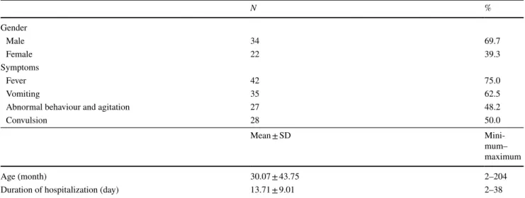
56 patients aged between 2 months and 18 years were included in the study. 34 (60.7%) of the patients were male and 22 (39.3%) female (M/F = 1.54). Demographic charac-teristics of patients are given in Table 2. The median age of our patients was 10.0 months (min–max 2-204 months). Mean hospital stay of the patients was 13.71 ± 9.01 days (min–max 2–38). At the time of admission, 42 (75%) of the patients had fever, 35 (62.5%) vomiting, 27 (48.2%) abnormal behaviour and agitation and 28 (50%) convulsion. While six of the patients had a history of epilepsy (10.71%), 9 (16.07%) had cerebral palsy. Physical examination showed acute encephalopathy in all the cases. Consciousness level ranged from a somnolent state to coma. Mechanical ventila-tion was required for respiratory support in 35 (62.5%) of our patients. The time period between the initiation of the symptoms and admission was an average of 6 days (range 3–11 days). None of the children had taken salicylates or any other medications.
The etiological classification of patients with AMES was divided into four groups as infection, metabolic disor-der, toxic, and hypoxic-ischemic. 39 (69.6%) patients were found to have infection, 10 (17.9%) patients hypoxia, 7 (12.5%) patients metabolic disorders and 1 (1.8%) patient had toxic cause. In patients with hypoxia the reasons were drowning in 4, foreign body aspiration in 3, carbon mon-oxide poisoning in 2, and cyanide poisoning in 1 patient. Paracetamol use was responsible for toxic AMES in one patient. 33 (58.92%) patients had viral upper respiratory tract infection and/or lower respiratory tract infection, and 15 (26.8%) patients had viral gastroenteritis. There was no history of rash or other signs of any exanthematous viral illness. 22 patients (39.28%) underwent lumber puncture
Table 1 Treatment protocol for patients with AMES 1. Fluid and electrolyte therapy
2 Intravenous administration of 25% human albumin (0.5–1 g/kg/ dose, 30 min intravenous infusion) followed by furosemide (2 mg/kg/dose, intravenous bolus)
3 Dexamethasone; 1–3 days 2 mg/kg/day 4–7 days 0.5 mg/kg/day 4 Semi-Fowler’s position
5 Brain hypothermia
6 Phenobarbital; 5 mg/kg/day, 2 dose/day 7 If needed vitamin K and fresh frozen plasma
Table 2 Demographic characteristics of patients with AMES
N % Gender Male 34 69.7 Female 22 39.3 Symptoms Fever 42 75.0 Vomiting 35 62.5
Abnormal behaviour and agitation 27 48.2
Convulsion 28 50.0
Mean ± SD
Mini-mum– maximum
Age (month) 30.07 ± 43.75 2–204
and the results revealed normal. Disseminated intravascu-lar coagulation (DIC) was detected in 2 patients. 3 patients had status epilepticus, 2 had dilated cardiomyopathy, and 2 had congenital heart disease. One patient had myocarditis, one patient had pericardial effusion. AMES developed in follow-up period after cardiac surgery in those with con-genital heart disease.
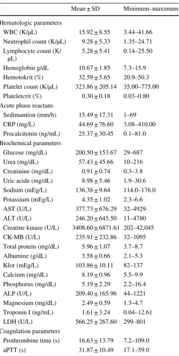
Results of the laboratory analysis of the patients are given in Table 3.
Follow‑up
Blood urea levels were high (> 40 g/dL) in 30 (53.57%) patients and creatinine levels (> 0.8 mg/dL) in 20 patients (35.71%). 12 (21.42%) patients underwent peritoneal dial-ysis due to acute renal failure. Uric acid level was found high in 39 (69.64%) patients (> 7.2 mg/dL). Hypona-tremia (< 136 mEq/L) was found in 29 patients (51.78%) and hypernatremia (> 145 mEq/L) in 7 patients (12.5%). Two patients were diagnosed as inappropriate ADH hor-mone syndrome. 10 (17.85%) patients were found to have hypokalemia (< 3.5 mEq/L) and 12 (21.42%) hyperkalemia (> 5.1 mEq/L). Creatinine phosphokinase (CPK) levels were high in all patients. 17 (30.35%) patients had tenfold increased CPK level than normal. While serum AST and ALT levels were high in all patients bilirubin levels were normal in all patients. Alkaline phosphatase levels were normal in all patients. 26 (46.42%) patients had hypoalbu-minemia (< 3.8 g/dL). Hypoglycemia was detected in 12 patients (21.42%). While 34 (60.71%) patients had hyper-glycaemia (> 105 mg/dL), six patients had received insulin therapy. Prothrombin time was high in 23 patients (41.07%). Serum ammonia levels were determined in 52 cases and was elevated (normal range 27.2–102 µg/dl). Blood gas analysis showed metabolic acidosis in 52 patients (92.85%).
Neuroimaging
Brain MRIs were performed on admission or shortly after admission. Brain MRIs showed brain edema in 52 (86.7%) patients. Brain MRIs were not available in two patients because of unstable conditions. These two patients also had clinically diagnosed brain edema.
Morbidity and mortality
The mortality rate was 30.35% (n = 17) in the study group. Five patients who received dialysis due to acute renal failure, one patient with cyanide poisoning, two patients with car-bon monoxide poisoning, and two patients with drowning in water had died. All these cases were late applications. Five patients who underwent surgery due to congenital malfor-mation were lost. No neurological deficit was seen in the children who had survived.
Discussion
We described AMES as a new clinical entity for the first time in the literature. AMES can occur during the course of viral infections as well as due to congenital metabolic
Table 3 Laboratory characteristics of patients with AMES
Mean ± SD Minimum–maximum Hematologic parameters WBC (K/µL) 15.92 ± 8.55 3.44–41.66 Neutrophil count (K/µL) 9.28 ± 5.33 1.35–24.71 Lymphocyte count (K/ µL) 5.28 ± 5.41 0.14–25.50 Hemoglobin g/dL 10.67 ± 1.85 7.3–15.9 Hemotokrit (%) 32.59 ± 5.65 20.9–50.3 Platelet count (K/µL) 323.86 ± 205.14 35.00–775.00 Plateletcrit (%) 0.30 ± 0.18 0.03–0.80 Acute phase reactans
Sedimantion (mm/h) 15.49 ± 17.31 1–69 CRP (mg/L) 44.69 ± 76.60 3.08–410.00 Procalcitonin (ng/mL) 25.37 ± 30.45 0.1–81.0 Biochemical parameters Glucose (mg/dL) 200.50 ± 153.67 29–687 Urea (mg/dL) 57.43 ± 45.66 10–216 Creatinine (mg/dL) 0.91 ± 0.74 0.3–3.8 Uric acide (mg/dL) 8.98 ± 5.46 1.9–30.6 Sodium (mEg/L) 136.38 ± 9.64 114.0–176.0 Potassium (mEg/L) 4.35 ± 1.02 2.3–6.6 AST (U/L) 377.73 ± 676.29 32–4929 ALT (U/L) 246.20 ± 645.50 11–4780 Creatine kinase (U/L) 3408.60 ± 6871.61 202–42,045 CK-MB (U/L) 235.91 ± 232.86 32–1095 Total protein (mg/dL) 5.96 ± 1.07 3.7–8.7 Albumine (g/dL) 3.58 ± 0.66 2.1–5.3 Klor (mEg/L) 103.86 ± 10.11 82–137 Calcium (mg/dL) 8.19 ± 0.96 5.5–9.9 Phosphorus (mg/dL) 5.19 ± 2.29 2.2–16.4 ALP (U/L) 209.40 ± 165.96 44–1221 Magnesium (mg/dL) 2.49 ± 0.59 1.3–4.7 Troponin I (ng/mL) 1.61 ± 3.24 0.04–12.61 LDH (U/L) 566.25 ± 267.60 299–801 Coagulation parameters Prothrombine time (s) 16.63 ± 13.79 7.2–109.0 aPTT (s) 31.87 ± 10.49 17.1–59.0
diseases, hypoxic-anoxic conditions, drugs, toxins and rare bacterial infections. Although the etiology may be different, the clinical and laboratory findings are surprisingly similar.
AMES is a rare syndrome, which is characterised by an acute, life-threatening, non-inflammatory encephalopathy with the absence or minimal clinical signs of liver involve-ment. Various factors including infectious, toxic, drug and metabolic etiologies have been considered in the pathogen-esis of RS, but only aspirin intake during the acute phase of a viral process has been proven to be associated with its onset [3, 10]. In classic RS, mitochondrial structure shows a severe but self-limiting defect and concomitant enzymatic failures occur. This process accompanies by an intense, acute catabolic state associated with cerebral edema, in the absence of encephalitis or meningitis [11, 12].
The onset of AMES’ occurs during early febrile period of a viral infection, and runs a fulminant course with the rapid development of coma. Mitochondrias are found in different numbers in all of our body cells, and mostly in myocar-dial and CNS cells. Thus, its clinical presentation includes encephalopathy, multiple organ failure, disseminated intra-vascular coagulation, and hemophagocytic syndrome [8].
It has been reported that cytokine storm could play role in the pathogenesis of RLS [8]. Concentrations of pro-inflammatory cytokines, such as tumour necrosis factor-a (TNF-a) and interleukin-6 (IL-6) in serum and cerebrospi-nal fluid, are found to be abnormally high in many cases of RLS. The cytokine levels usually tend to be higher in serum than in cerebrospinal fluid [13, 14]. Pathologic studies of the brains of patients with RLS at necropsy have showed severe brain edema and perivascular plasma exudation. These find-ings indicate that vascular edema with the damage in the
blood–brain barrier, is the pathologic substrate of encepha-lopathy in RLS. Although vascular injury is thought to be caused by endothelial damage from inflammatory cytokines, the exact mechanism is unknown [8]. Our opinion is that cytokine storm (Fig. 1) and oxidative stress related to acute mitochondrial dysfunction can also play an important role in AMES similar to RLS as mentioned by Morishima et al. [15].
Viral infection associated AMES usually occurs in a child, a few days after the recovery phase of the viral illness. This illness is generally an upper respiratory tract infection, varicella infection, or gastroenteritis. The patient abruptly deteriorates, about 3 days after apparent improvement. Initially protracted vomiting (rarely, this may be absent) occurs which is followed by apathy, lethargy and drowsi-ness 24–48 h later. Use of anti-emetics may complicate the presentation by masking vomiting. Patients under the age of 2 years may present with initial signs of diarrhea and tachypnea. Subsequently the patient continues to deteriorate, neurological symptoms become prominent. Initially this may be a withdrawn state which is followed by delirium, confu-sion, aggressive behaviour and stupor. Visual hallucinations may also be seen. Pupillary responses may be affected, but ophthalmological features of increased intracranial pres-sure are not always present. Seizures and coma may occur if clinical deterioration continues. Decerebrate posturing, opisthotonus, dilated/unequal pupils, deep rapid respirations, variations in pulse and, finally, a flaccid apneic state precede death. Liver function also impairs, mild hepatomegaly may be seen but jaundice is not a usual finding. The course of the illness varies (4–60 h) and the neurological status may start to improve spontaneously or with therapy or proceed
Fig. 1 Pathogenesis of acute encephalopathy caused by cytokine storm: AMES
to brain death. In infants, the presentation may be with pre-dominant respiratory symptoms (tachypnea, respiratory dis-tress, hyperinflation, and apnea) and temperature instability. Hypoglycaemia and hepatomegaly are more common, and vomiting and history of a preceding viral infection is usually absent in infants [16].
Differential diagnosis must include other causes of coma, congenital metabolic disorders of the urea cycle, fatty acids, gluconeogenesis, and glucogenolysis [17], and some toxic and infectious conditions such as multiple organ dysfunction syndrome in sepsis [11, 12, 18, 19].
Therapeutic approaches are commonly symptomatic and prognosis depends on the stage of the syndrome, timing and adequacy of intensive care treatment [19, 20]. Monitoring the neurologic, respiratory, cardiovascular, coagulation, metabolic, fluid and electrolyte balance status is necessary [21]. However, there is a mortality rate of 40% despite these therapeutic approaches. Severe neurologic sequelae can be present in 30% of the survived cases [8, 22].
Although AMES is associated with complex metabolic impairments, both death and neurological sequelae are attributed to the degree of central nervous system involve-ment [23, 24]. Therefore, maintaining an adequate cerebral perfusion pressure is the mainstay of the treatment. All patients with AMES should be monitored in an intensive care unit, because the clinical condition may deteriorate rapidly even in mild cases. The targets of the treatment of patients with AMES include providing fluid-electrolyte bal-ance, ensuring the adequacy of blood circulation and res-piration, and normalizing intracranial pressure and body temperature. Correction of any metabolic abnormality (i.e., hypoglycemia, metabolic acidosis, and hyperammonemia) and coagulation disorder is essential. Our treatment proto-col included high dose dexamethasone, brain hypothermia, and phenobarbital and to keep patient in semi-Fowler’s posi-tion. We did not use mannitol to reduce intracranial pres-sure because of its negative effects on electrolyte balance and renal side effects. In our study, clinical course of three patients who showed renal failure with elevated creatinine levels were very severe and they finally died. Four patients had died in the first week of admission. The rest of the patients had improved and discharged in good condition. No neurological deficit was seen in the children who survived. Lemberg et al. reported a mortality rate of 41.7% in patients with RLS [25]. The mortality rate was lower (31.8%) in our study group when compared with other reports in the lit-erature. The reason why our mortality rate is low is due to immediate starting of the treatment protocol which we have developed over time with our 30 years of experience and careful follow-up.
Successful management of the AMES depends on early diagnosis. It includes preventing, correcting and minimis-ing metabolic abnormalities and controllminimis-ing increased ICP.
Early recognition and treatment are essential to decrease the rates of death and neurological sequeles. Treatment strategies are outlined here but it is not possible to apply them in primary care. A general practitioner should recog-nise the condition early and refer the patient urgently to a centre where intensive care unit is available. It is important to note that the patient should be kept as stable as possible, since unnecessary processing or warning will raise the ICP. Patient’s airway, breathing and circulation should be established and maintained. Glucose level should also be checked and dextrose be administered to manage hypo-glycemia. On admission, some of the steps taken in ini-tial management should include: (1) maintaining airway and brain oxygenation; (2) correction of hypoglycaemia with IV dextrose infusion. Blood glucose levels should be between 11 and 22 mmol/L (ie higher than normal); (3) if blood pressure is normal, the head and upper trunk should be elevated at a 40° angle and overhydration should be avoided; (4) monitoring blood biochemistry and avoid dehydration; (5) ondansetron may be cautiously used to control the vomiting; (6) treatment of hyperammonaemia with sodium phenylacetate/sodium benzoate or haemodi-alysis; (7) obtaining normal body temperature; (8) treating seizures with anticonvulsants (e.g., phenytoin); (9) correc-tion of coagulacorrec-tion defects (FFP and/or vitamin K); (10) consulting the patient with expert colleagues and refer the patient to ICU. Stage 2 or worse needs referral to a tertiary centre where ICP can be monitored.
In conclusion, our treatment protocol is safe and effective in children with AMES. AMES occurs rarely, but should be kept in mind in the differential diagnosis of patients with any encephalopathy of unknown origin especially in those with a history of ingestion of drugs, previous viral infection or vomiting. Early recognition and treatment is imperative to reduce morbidity and mortality in children with AMES.
Author contributions All authors contributed to revising critically the final version of the manuscript. Interpretation of data and revising of manuscript: ŞA, FA. Study conception and design: AY. Preparation of manuscript: AY, FA. Literature review: ŞA, Review and supervision of manuscript: AS.
Funding None.
Compliance with ethical standards
Conflict of interest All authors declare that they have no conflict of interest.
Ethical approval The ethics committee approval for the study was received from the ethical committee of Selcuk University (2018/01).
Informed consent Informed consent was obtained from all individual participants included in the study.
References
1. Reye RDK, Morgan G, Baral J (1963) Encephalopathy and fatty degeneration of the viscera: a disease entity in childhood. Lancet 2:749–752
2. Brain WR, Hunter D, Turnbull HM (1929) Acute meningo-encephalomyelitis of childhood: report of 6 cases. Lancet 1:221–227
3. Waldman RJ, Hall WN, McGee H, van Amburg G (1982) Aspirin as a risk factor in Reye’s syndrome. J Am Med Ass 247:3089–3094
4. Orlowski JP, Gillis J, Kilham HA (1987) A catch in the Reye. Pediatrics 80:638–642
5. Smith TCG (1996) Reye’s syndrome and the use of aspirin. Scott Med J 41:4–8
6. Schror K (2007) Aspirin and Reye syndrome: a review of the evidence. Pediatr Drugs 9:195–204
7. Devulapalli CS (2000) Rotavirus gastroenteritis possibly causing Reye syndrome. Acta Pediatr 89:613–619
8. Mizuguchi M, Yamanouchi H, Ichiyama T, Shiomi M (2007) Acute encephalopathy associated with influenza and other viral infections. Acta Neurol Scand Suppl 186:45–56
9. Leonard JV, Schapira AHV (2000) Mitochondrial respira-tory chain disorders I: mitochondrial DNA defects. The Lancet 355:299–304
10. Starko KM, Ray CG, Dominguez LB, Stromberg WL, Woodall DF (1980) Reye’s syndrome and salicylate use. Pediatrics 66:859–864 11. Baldellou Va`zquez A (2003) Reye’s syndrome. Forty years later.
An Pediatr 59:319–322
12. Glasgow JFT, Middelton B (2001) Reye syndrome-insights on causation and prognosis. Arch Dis Child 85:351–353
13. Togashi T, Matsuzono Y, Itakura O, Narita M (1999) IL-6 and TNF-a in cerebrospinal fluid from infantile encephalitis encepha-lopathy patients during influenza seasons. J Jpn Pediatr Soc 103:16–19
14. Ichiyama T, Endo S, Kaneko M, Isumi H, Matsubara T, Furukawa S (2003) Serum cytokine concentrations of influenza associated acute necrotizing encephalopathy. Pediatr Int 45:734–736
15. Morishima T (2003) Studies on the epidemiology and patho-genesis of encephalitis/encephalopathy occurring during the clinical course of influenza. In: 2000–2002 General Report of the Research Committee for Research on Emerging and Re-emerg-ing Diseases. Ministry of Health, Labour and Welfare of Japan, Tokyo, pp 1–23 (in Japanese)
16. Weiner DL et al (2012) Medscape, Reye Syndrome
17. Mohnike K, Starke I, Bannert N, Heise HR (1993) Differential diagnosis and therapy of hypoglycemia in childhood. Kinderarztl Prax 61:192–201
18. Chang PF, Huang SF, Hwu WL, Hou JW, Ni YH, Chang MH (2000) Metabolic disorders mimicking Reye’s syndrome. J For-mos Med Assoc 99:295–299
19. Visentin M, Salmona M, Tacconi MT (1995) Reye’s and Reye-like syndromes, drug-related diseases? (causative agents, etiol-ogy, pathogenesis, and therapeutic approaches). Drug Metab Rev 27:517–539
20. Gill RQ, Sterling RK (2001) Acute liver failure. J Clin Gastroen-terol 33:191–198
21. Wulur H, Kho LK (1990) Treatment of Reye’s syndrome at Sum-ber Waras Hospital. Acta Paediatr Jpn 32:435–442
22. Brunner RL, O’Grady DJ, Partin JC, Partin JS, Schubert WK (1979) Neuropsychologic consequences of Reye syndrome. J Pediatr 95:706–711
23. Devivo DC (1978) Reye syndrome: a metabolic response to an acute mitochondrial insult? Neurology 28:105–108
24. Devivo DC, Keating JP (1976) Reye’s syndrome. Adv Pediatr 22:175–229
25. Lemberg A, Fernández MA, Coll C, Rosello DO, Romay S, Per-azzo JC et al (2009) Reyes’s syndrome, encephalopathy, hyperam-monemia and acetyl salicylic acid ingestion in a city hospital of Buenos Aires, Argentina. Curr Drug Saf 4:17–21
Publisher’s Note Springer Nature remains neutral with regard to jurisdictional claims in published maps and institutional affiliations.