Eurasian J Vet Sci, 2015, 31, 1, 60-62
60
CASE REPORT
Surgical treatment of a calf with jejunum intussusception: Case report
Muharrem Erol
1*, Yılmaz Koç
2, Semih Altan
3, Hanifi Erol
11Department of Surgery, Faculty of Veterinary Medicine, University of Erciyes, 38030, Kayseri, 2Department of Surgery, Faculty of Veterinary Medicine, University of Selcuk, 42075, Konya, 3Department of Surgery, Faculty of Veterinary Medicine, University of Dicle, 21280, Diyarbakır, Turkey
Received: 17.07.2014, Accepted: 15.09.2014 *[email protected]
Öz
Erol M, Koç Y, Altan H, Erol H. Buzağıda jejenum
invaginas-yonunun operatif tedavisi: Olgu sunumu.
Bu vaka sunumunda 5 aylık bir erkek buzağıda teşhis edilen jejunum invaginasyonunun klinik, laboratuar ve operasyon bulguları değerlendirildi. Tanımlanan klinik vakada hayva-nın 3 gündür iştahsız olduğu ve defekasyon yapmadığı sahi-binden öğrenildi. İç Hastalıkları kliniğinde yapılan klinik ve laboratuar muayenelerinden sonra hayvan cerrahi kliniğine sevk edildi. Sol açlık çukurluğundan laporatomi operasyonu yapıldı. Operasyonda bağırsaklar patolojik vaziyet değişikliği yönünden kontrol edildi. Operasyon esnasında yapılan mu-ayenede ventral hattın hafif sağında katı kıvamlı bağırsak segmenti palpe edildi. Palpe edilen bağırsak segmenti karın boşluğundan çıkarıldığında invagine olmuş jejunum olduğu görüldü. İnvagine kısım ödematöz, siyanotik ve fibröz adez-yonların bulunmasından dolayı rezeksiyonu ve uç uca anos-tomozu yapıldı. Taburcu edilen buzağı hakkında hasta sahi-binde bilgi alındı. Sonuç olarak bağırsak invaginasyonunun 5 aylık buzağılarda nadiren görüldüğü, teşhisinin experimen-tal laparotomi ile koyulabileceği ve bu sürede rutin cerrahi operasyon ile tedavisinin yapılabileceği kanısına varıldı. Anahtar kelimeler: Buzağı, invaginasyon, jejenum
Abstract
Erol M, Koc Y, Altan H, Erol H. Surgical treatment of a calf
with jejunum intussusception: Case report.
The purpose of the present case report was to describe the clinical, laboratory and surgical findings of jejunum intus-susception in a five-month old male calf. In this clinical case history, anorexia and lack of defecation for three days were learned from the owner. After clinical and laboratory exa-mination the calf was referred to the Surgery Clinic. A left-flank laparotomy was performed. Intestines were examined for pathologic situation changes. During examination, solid consistency intestine segment was palpated at slight right of the ventral line. During macroscopic examination, invagina-ted intestine segment was detecinvagina-ted as jejunum. Invaginainvagina-ted parts of intestine were edematous, cyanotic and fibrous ad-hesive, resection and end-to-end anastomosis were perfor-med. The calf discharged was followed by owner. In conclusi-on, intussusceptions are rarely seen in five month old calves. Its diagnosis could be made by experimental laparotomy and intestine intussusceptions treated with routine surgical in-terventions in this period.
Keywords: Calf, intussusception, jejunum
Eurasian J Vet Sci, 2015, 31, 1, 60-62
DOI: 10.15312/EurasianJVetSci.201518479
Eurasian Journal
of Veterinary Sciences
http://ejvs.selcuk.edu.tr www.eurasianjvetsci.org
Eurasian J Vet Sci, 2015, 31, 1, 60-62
61
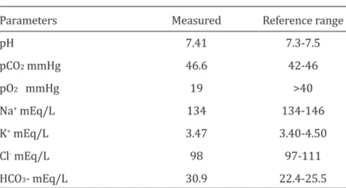
Intussusception is the invagination of a portion of the gastro-intestinal tract into the lumen of an adjacent segment of bo-wel due to peristaltic movement and is often a cause of intes-tinal obstruction in cattle (Constable et al 1997, Turgut and Ok 1997). Jejunojejunal intestine intussusception is the most frequent form observed in cattle. Ileoileal, ileocecocolic, ce-cocolic and colocolic forms may be seen in cattle, as well (Ha-milton and Tulleners 1980, Constable et al 1997). Intestine intussusception is observed in calves less than two months of age (Karapınar and Köm 2007). Operative treatment is the only treatment method for intestine intussusception (Turgut and Ok 1997). In this case presentation, it is aimed to evalu-ate the clinical, laboratory and operative findings of jejunum intussusception determined in a five-month male calf. The presented clinical case composed of a five-month Hols-tein male calf referred to the Department of Internal Medici-ne, Veterinary Faculty of Selcuk University due to three-day anorexia and defecation difficulty. Following the clinical and laboratory examinations, this calf was sent to the surgery cli-nic due to the possibility of passing difficulty in the ostium reticulo-omasicum. Hearth-respiration rate and body tempe-rature of the calf were determined as 86/min, 27/min and 38.3/min in the clinical examination, respectively. Capillary filling time was calculated as 3 sec. No significant change was detected in skin elasticity. The calf was observed at ease and no indication of abdominal pain like the displacement of rear legs or kicking the lower abdomen was observed. No difference was observed between the right and left abdomi-nal walls in the inspection. There was no sensibility in ab-dominal palpation and abab-dominal distention. The calf was immediately subjected to surgical intervention due to the possibility of passing problem in ostium reticulo-omasicum. Laboratory results are given in Table 1.
Local infiltration anesthesia was made on the left paralum-bar fossa region of the calf using 2% of lidocaine (L-Anestin® amp, Alke, Istanbul, Turkey) following the routine laparo-tomy preparations. Abdominal cavity was reached with left paralumbar laparotomy, and rumen was observed to be tight and filled with gas and other contents. Rumenotomy was
performed after the transperitoneal exploration. Gas and contents in the rumen were discharged, and rumen was eva-luated for ostium reticulo-omasicum and omasum obstruc-tion. No obstruction was detected in this region. Rumen was routinely closed. Subsequently, intestines were examined for pathologic changes. In the ventral of abdominal cavity, vis-cous and bulky intestine segment different from the normal intestinal viscosity was palpated, and this segment taken out of abdominal cavity (Figure 1-A). This segment was deter-mined as around 20-25 cm invaginated jejunum. Intestine segments in the cranial of invaginated part were filled with gase (Figure 1-B). Invaginated segment of the intestine was hyperemic and rather viscous and was tried to be opened starting from the rear side (Figure 1-C). As the invaginated parts were highly edematous and adhesive, opening applica-tion was not performed (Figure 1-D). Invaginated intestine segment was determined cyanotic in the incision made on its antimesenteric side. Therefore, resection of the intussuscep-tioned segment was considered (Figure 1-E).
Invaginated part was removed by putting intestine pincers on its front and rear sides. The bleeding on mesenterium was controlled, two layers end-to-end anastomosis perfor-med on intestine ends with absorbable monofilament suture (0-Polydioxanone®, PDS, Ethicon). Teared mesenterial part was closed by 0-Polydioxanone, and thus the enterectomy completed. Peritoneum was closed with 0-number and ab-dominal muscles closed also with 1-number Polydioxanone; eventually, skin was closed with 2-number silk suture, and operation completed. In the macroscopic examination of re-sected intestine segment, 50-60 cm length of rere-sected intes-tine with necrosis was detected (Figure 1-F).
Parenteral fluid treatment was applied on postoperative 1st and 2nd days. IM combination of 10.000 IU benzyl procaine penicillin G and 10 mg dihydrostreptomycin (Reptopen-S® inj, Ceva-DIF, Istanbul, Turkey) was applied during posto-perative five days. 1.1 mg/kg flunixin meglumine (Flumed® inj., Alke, Istanbul, Turkey) IM was used for postoperative analgesia. Defecation was started at postoperative 2nd day. Intussusception cases are not frequently observed related to the gastrointestinal system in cattle; however, they are among the common causes for intestinal obstruction. In ad-dition, intestinal obstruction could be caused by volvulus dilatation, torsion, tumoral formations and foreign substan-ces in the structures of gastrointestinal system (Archer et al 1988, Kemble et al 1994).
Intussusception cases were frequently reported for calves of 0-2 months of age, but they were also detected in older ages (Strand et al 1993, Fubini and Trent 2004). Consistent with this knowledge, in the present case the calf was five-months old. Diarrhea was stated as the most important reason for in-tussusception in calves. Peristaltic movements increased by
Reference range 7.3-7.5 42-46 >40 134-146 3.40-4.50 97-111 22.4-25.5 Parameters pH pCO2 mmHg pO2 mmHg Na+ mEq/L K+ mEq/L Cl- mEq/L HCO3- mEq/L Measured 7.41 46.6 19 134 3.47 98 30.9
Table 1. Results of blood gas evaluation performed before surgical intervention.
Erol et al Jejunum intussusception
Eurasian J Vet Sci, 2015, 31, 1, 60-62
62
certain factors like enteritis, foreign substances in intestineand sudden change of feed were reported to cause intussus-ception in cattle of older ages (Dabak et al 2001, Abutarbush and Naylor 2006).
There were no relationship between anamnesis, clinical and laboratory examinations for this case in the present study. Intussusception was reported for different segments of catt-le intestine, but the cases are mostly in jejunojejunal type as the mesenterium part, adhered by the jejunum, is rather long and active (Constable et al 1997). Localization of the inva-gination in the case is compatible with the literature. Pain symptoms in the intestinal intussusception of cattle are not as severe as in horses; however, they could differ depending on the severity of ischemia formed in the disease. No such symptoms were observed in the case. Alkalosis is present in the intussusception as in many other passing problems in the intestine (Smart et al 1997). In the presented case, ab-sence of alkalosis and pain symptoms, cyanotic structure of the resected intestine segment, and incomplete formation of necrosis were attributed to the early stage of the disease. Right paralumbar region was reported as the most suitable region for intestinal operations (Anderson et al 1993, Fubini and Trent 2004). Due to the primary possibility of passing problem in ostium reticulo- omasicum, laparotomy was per-formed in the left paralumbar region. On the other hand, ope-ration was easily completed as the animal was young. Small intestinal intussusceptions are surgically repaired by resec-tion and end-to-end anastomosis of intestine in cattle beca-use end-to-end anastomosis cabeca-uses less chance of stricture formation and leakage (Constable et al 1997, Karapınar and Köm 2007, Baird 2013). This technique was preferred in this calf and no complications were observed.
In conclusion, intestine intussusceptions in calf could be mistaken with passing problems in early period; therefore, definitive diagnosis could be made with experimental lapa-rotomy and intestine intussusceptions could be treated with routine surgical interventions in this period.
References
Abutarbush SM, Naylor JM, 2006. Obstruction of the small intestine by a trichobezar in cattle: 15 cases (1992-2002). JAVMA, 229, 1627-1630.
Anderson DE, Constable PD, St. Jean G, Hull BL, 1993. Small-intestinal volvulus in cattle: 35 cases (1967-1992). JAVMA, 203, 1178-1183.
Archer RM, Cooley AJ, Hinchcliff KW, Smith DF, 1988. Jejuno-jejunal intussusception associated with a transmural ade-nocarcinoma in an aged cow. JAVMA, 192, 209-211. Baird AN, 2013. Bovine Gastrointestinal Surgery, in: Turner
and McIlwraith’s Techniques in Large Animal Surgery, Eds: Hendrickson DA, (Nickie) Baird AN, Wiley Blackwell, USA, pp: 211-234.
Constable PD, St. Jean G, Hull BL, Rings DM, Morin DE, Nel-son DR, 1997. Intussusception in cattle: 336 cases (1964-1993). JAVMA, 210, 531-536.
Dabak M, Unsaldi E, Gul Y, 2001. Diagnosis and treatment of intussusception in a cow. Turk J Vet Anim Sci, 25, 387-389. Fubini SL, Trent AM, 2004. Small intestine surgery in cattle, in: Farm Animal Surgery, Eds: Fubini SL, Ducharme N, Else-vier, USA, pp: 242-244.
Hamilton GF, Tulleners EP, 1980. Intussusception involving the spiral colon in a calf. Can Vet J, 21, 32.
Karapınar T, Köm M, 2007. Transrectal ultrasonographic diagnosis of jejunoileal intussusception in a cow. IVJ, 60, 422-424.
Kemble T, Gardiner M, May SA, 1994. An unusual case of cae-cal volvulus in a cow. Vet Rec, 134, 521-522.
Smart ME, Fretz PB, Gudmundson J, Cymbaluk N, 1977. Intus-seption in a charolais bull. Can Vet J, 18, 244-246.
Strand E, Welker B, Modransky P, 1993. Spiral colon intus-seption in a three-year-old bull. JAVMA, 202, 971-972. Turgut K, Ok M, 1997. Veterinary Gastroenterology, 1st
editi-on, Bahcivanlar Press, Konya, Turkey, pp; 353-357.
Erol et al Jejunum intussusception
Figure 1. A: Taken out intestine segment, B: Invaginated jejunum part, C: Attempt to open invaginated segment, D: Severe edema and adhesion in invaginated segment, E: Cyanotic colored intestine segment, F: Macroscopic image of invaginated segment.