Brief Report
Endobronchial Lipoma Associated With Destroyed Lobe
I
.
brahim Can Ku¨rkc¸u¨og˘lu,
M.D.,* Atilla Erog˘lu,M.D.,* Nurettin Karaog˘lanog˘lu,M.D.,*Zekai Erman,
M.D.,† and Leyla Sag˘lam,M.D.‡Departments of *Thoracic Surgery, †Pathology, and ‡Chest Diseases, Atatu¨rk University, School of Medicine, Erzurum, Turkey.
Abstract: Endobronchial lipomas are rare, benign lesions that
usually obstruct a major bronchus and cause irreversible pul-monary damage distally. Endobronchial lipomas originate from fat cells located in the peribronchial tissue and occasionally the submucosal tissue of large bronchi and comprise only approxi-mately 0.1% of all tumors and approxiapproxi-mately 13% of benign pulmonary tumors. They may be diagnosed clinically as bron-chial adenoma or malignant lesion, and the result of delayed
therapy may be bronchiectasis. Treatment includes local resec-tion through a bronchoscope or a bronchotomy incision, or removal, if necessary, of the obstructed lobe or lung at thora-cotomy. We report a case of a endobronchial lipoma treated successfully with lobectomy in a 42-year-old man.
Key Words: Lipoma—Bronchus—Lung.
Journal of Bronchology 9:27–29, 2002.
Endobronchial lipoma is a rare neoplasm, with 116 cases reported in the English literature to date. Literature data show that a correct preoperative diagnosis is pos-sible in less than 50% of all patients and that a thoracot-omy is mandatory in 85% of patients because bron-choscopic needle aspiration biopsy specimens or brush-ings rarely allow a definitive cytologic diagnosis.1We present a case that demonstrates clinical, diagnostic, and histopathologic findings in this disease.
CASE REPORT
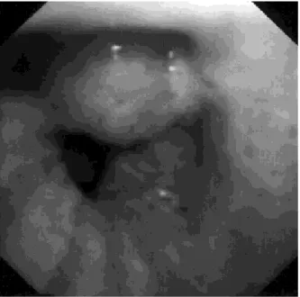
A 42-year-old man presented with a 1-year history of recurrent pneumonia in the left upper lobe. He was a current smoker (20 pack-years), and his vital signs were normal. There were decreased breath sounds over the left posterior upper portion of the thorax. Chest radiography showed linear densities and ill-defined, irregular opaci-ties in the upper lung. Computed tomographic scanning and tomographic studies showed multiple cystic spaces in the left upper lobe in which the volume was dimin-ished (Fig. 1). Flexible bronchoscopy revealed a well-circumscribed, yellow tumor localized on the upper
di-vision of the upper lobe of the left lung (Fig. 2). Numer-ous attempts at biopsy were unsuccessful because of the mobility of the lesion and the presence of a tough, fi-brous capsule. Several cytologic examinations of the sputum did not reveal any malignant cells, and culture of the sputum was negative. Preoperatively, an inflamma-tory polyp in association with chronic saccular bronchi-ectasis was suspected. A left posterolateral thoracotomy was performed. The left upper lobe bronchus was opened, and a smooth, yellow tumor was excised. Frozen sections of the tumor revealed benign fatty cells. Left upper lobectomy was performed because of destruction of the lobe resulting from recurrent pneumonia. Gross examination of the resected specimen showed a pedun-culated polypoid mass attached to the bronchial mucosa, and saccular ectatic changes of bronchi. Microscopically, the endobronchial tumor was comprised chiefly of ma-ture fatty tissue surrounded by respiratory epithelium. The submucosa contained a moderate number of chronic inflammatory cells (Fig. 3).
The patient had an uncomplicated postoperative course, was discharged 9 days after the operation, and was doing well at the 7-month follow-up.
DISCUSSION
Although lipomas are common, benign neoplasms of soft tissue, endobronchial lipoma is rare.2–4 Bronchial
Address reprint requests to Dr. I.brahim Can Ku¨rkc¸u¨og˘lu, Depart-ment of Thoracic Surgery, Medical Faculty, Atatu¨rk University, 25000 Erzurum, Turkey; e-mail: [email protected]
Journal of Bronchology
9:27–29 © 2002 Lippincott Williams & Wilkins, Inc., Philadelphia
lipomas comprise approximately 0.1% of all pulmonary tumors and approximately 13% of benign tumors.5,6The tumor is usually found in middle-age, obese men.5 Smoking may be important in the pathogenesis of this tumor.2
Endobronchial lipomas originate from fatty tissue that is normally present in the bronchial tree and consist mainly of histologically normal adult fat cells.1,3–6They
are pliable, measure 1 to 3 cm, and are often peduncu-lated. Lipomas may obstruct the bronchus partially or totally, producing varying degrees of obstructive atelec-tasis, bronchiecatelec-tasis, and inflammatory change.2–4
Presenting symptoms are frequently those of bronchial
obstruction (i.e., cough, sputum production, recurrent pneumonitis, dyspnea, and, rarely, hemoptysis).2,3,5The
symptoms depend primarily on the tumor’s location and size. Duration of symptoms varies from a few weeks to more than 20 years.2
Radiographic changes are nonspecific and are related to an intrabronchial mass. Chest radiographs may show enlargement of the hilar shadow, because endobronchial lipomas are found more commonly in the large bronchi. In addition, changes secondary to bronchial obstruction, such as incomplete aeration or atelectasis and chronic bronchiectasis, may be noted.3,5 The computed tomo-graphic findings of a homogeneous mass with fatty den-sity and no tumor contrast enhancement are considered diagnostic.3 Magnetic resonance imaging also may be useful.
The capsule often renders bronchoscopic bronchial bi-opsy specimens nondiagnostic, and bronchial washings may include some epithelial cells because of the effects of chronic irritation, leading to an erroneous diagnosis of bronchogenic carcinoma.2,3,5 An endobronchial lipoma may be indistinguishable from a bronchial adenoma be-cause of their similar appearance and their tendency to bleed during the bronchial biopsy.5
Because of their benign nature, endobronchial lipomas should be treated initially endoscopically.2,3,6With early
and complete removal, patient prognosis is excellent. There is little or no risk of malignant transformation and minimal risk of recurrence. If the tumor is destroyed, surgical resection has to be considered. Lobectomy and pneumonectomy have been performed in a majority of these cases, and only in a few instances were the masses excised by bronchotomy.2,3,5,6
In our patient, biopsy specimens were insufficient for histologic diagnosis. Diagnostic thoracotomy and left
up-FIG. 1. Computed tomography shows multiple cystic spaces in the left upper lobe, in which the volume was diminished.
FIG. 2. Bronchoscopy demonstrates the tumor localized on the upper division of the upper lobe of the left lung.
FIG. 3. Photomicrograph showing mature fat cells surrounded by respiratory epithelium. H&E, original magnification ×40 before reduction.
I˙.C. KU¨ RKC¸U¨OG˘LU ET AL. 28
per lobectomy were performed to remove the destroyed lobe.
In conclusion, endobronchial lipomas are rare, benign tumors that can cause irreversible pulmonary damage unless removed early. Accurate and early diagnosis may obviate unnecessary thoracotomy and may prevent irre-versible complications.
REFERENCES
1. Destito C, Romagnoli A, Carlucci I, et al. Endobronchial li-poma: endoscopic resection or surgical excision? Report of a
case and review of the literature [in Italian]. G Chir 1995;16: 445–7.
2. Farsad GR, Makoui C. Endobronchial lipoma. Am Surg 1981;47: 236–8.
3. Mendelsohn SL, Fagelman D, Zwanger–Mendelsohn S. Endobron-chial lipoma demonstrated by CT. Radiology 1983;148:790. 4. Dogan R, Unlu M, Gungen Y, et al. Endobronchial lipoma. Thorac
Cardiovasc Surg 1988;36:241–3.
5. Schraufnagel DE, Morin JE, Wang NS. Endobronchial lipoma.
Chest 1979;75:97–9.
6. Stey CA, Vogt P, Russi EW. Endobronchial lipomatous hamar-toma: a rare cause of bronchial occlusion. Chest 1998;113: 254–5.
ENDOBRONCHIAL LIPOMA 29