Address for correspondence: Dr. Mustafa Ozan Gürsoy, Gaziemir Devlet Hastanesi, Kardiyoloji Bölümü, 3500 İzmir-Türkiye
E-mail: [email protected] Accepted Date: 27.9.2016
©Copyright 2016 by Turkish Society of Cardiology - Available online at www.anatoljcardiol.com DOI:10.14744/AnatolJCardiol.2016.7486
Mustafa Ozan Gürsoy, Macit Kalçık
1, Mahmut Yesin
2, Süleyman Karakoyun
3,
Emrah Bayam
4, Sabahattin Gündüz
4, Mehmet Özkan
5Department of Cardiology, Gaziemir State Hospital; İzmir-Turkey, 1Department of Cardiology, Hitit University
Çorum Training and Research Hospital; Çorum-Turkey, 2Department of Cardiology, Kars Harakani State Hospital; Kars-Turkey 3Department of Cardiology, Kars Kafkas University, Faculty of Medicine; Kars-Turkey, 4Department of Cardiology,
Koşuyolu Kartal Heart Training and Research Hospital; İstanbul-Turkey, 5School of Health Sciences, Ardahan University; Ardahan-Turkey
A global perspective on mechanical prosthetic heart valve thrombosis:
Diagnostic and therapeutic challenges
Introduction
For six decades, heart valve surgery has been improving the survival and the quality of life of patients with severe valvular disease. However, it has also given rise to development of a new disease—the prosthetic heart valve disease. Although thrombus formation is less frequently observed among new-generation prosthetic valves, the hemodynamic and physical properties of mechanical valves remain thrombogenic (1). Therefore, pros-thetic valve thrombosis (PVT) is one of the major causes of pri-mary valve failure. The PVT incidence was reported to be 0.03% in bioprosthetic valves (2), 0.5%–8% in mechanical valves in the mitral and aortic positions, and as high as 20% in mechanical tricuspid valves (3). Recently, it has been reported that approxi-mately 10% of the patients with mechanical heart valves had one episode of PVT per year (4).
PVT may lead to valve dysfunction, and its onset may be acute or gradual, according to the nature of thrombi and involvement of the hinges. The most common cause of PVT is inadequate anticoagulant therapy. Unfortunately, vitamin K antagonists (VKAs) are still the only approved oral anticoagulants in patients
with heart valve prostheses. Even with the use of VKA, the risk of thromboembolism is 1%–2% per year, but the risk is consi- derably higher without or inadequate treatment with warfarin (3). There are different therapeutic modalities for PVT, including anticoagulation with heparin, thrombolytic therapy (TT) (4, 5–8), and surgery (9), which are largely influenced by the presence of valvular obstruction, valve location, and clinical features. In this review, we aimed to summarize the pathogenesis, diagnosis, and management of mechanical PVT.
Pathogenesis and clinical findings of PVT
PVT is an obstruction of a prosthesis by noninfective throm-botic material. The etiopathogenesis of PVT is based on several mechanisms. The first mechanism involves the molecular inter-action between corpuscular blood components, plasma, and the prosthetic surfaces. The initial adsorption of plasma proteins on the prosthesis is generally followed by platelet adhesion (10). The second mechanism is dependent on the effect of the transprosthetic blood flow on local thrombus formation. The tur-bulent flow may result in a blood-borne increase in shear stress and may lead to thrombosis. Furthermore, chronic hemolysis
Prosthetic valve thrombosis is one of the major causes of primary valve failure, which can be life-threatening. Multimodality imaging is neces-sary for determination of leaflet immobilization, cause of underlying pathology (thrombus versus pannus or both), and whether thrombolytic therapy attempt in the patient would be successful or surgery is needed. Current guidelines for the management of prosthetic valve thrombosis lack definitive class I recommendations due to lack of randomized controlled trials, and usually leave the choice of treatment to the clinician’s experience. In this review, we aimed to summarize the pathogenesis, diagnosis, and management of mechanical prosthetic valve thrombosis. (Anatol J Cardiol 2016; 16: 980-9)
Keywords: prosthetic heart valve, thrombosis, diagnosis, management
may occur as a result of the accelerated destruction of throm-bocytes and erythrocytes with shortened intravascular lifes-pans (11). The third mechanism is ineffective anticoagulation, which is determined by reported valve thrombosis rates for that prosthesis in relation to specific international normalized ratio levels. The other prothrombotic causes are described in Table 1 (12–17). Inherited disorders such as MTHFR A 1298 C and fibrino-gen 455G/A polymorphisms may be involved in the pathofibrino-genesis of PVT, necessitating further data from large-scale studies (15). Increased levels of specific antibodies, including anticardiolipin and anti–tissue plasminogen activator antibodies have recently been found to be associated with PVT (14, 16). Furthermore, he- parin-induced thrombocytopenia may also lead to PVT (17).
PVT usually occurs over and under the hinge points of pros-thesis and extends unidirectionally or bidirectionally over the annulus or toward the prosthetic orifice. The length and thick-ness of the thrombus are critical because these features may provide the basis for fresh thrombus attachment with an embolic risk (18).
The clinical presentation of PVT may be variable. Patients with PVT may present with symptoms such as dyspnea, dec- reased exercise capacity, palpitation, chest pain, vertigo, cerebro- vascular accident, or even flank pain (19–22). Occluder clicks are typically muffled or absent during auscultation. Also, stenotic or regurgitant murmurs may be heard. Early detection and diagno-sis may often be limited by a progressive or insidious course. The
clinical status may depend on the type of the prosthesis. Patients with bileaflet prosthetic valves might be in a good hemodynamic condition due to a well-functioning single leaflet or might be unstable due to bileaflet involvement. Echocardiographic exa- mination should be urgently performed in case of high level of clinical suspicion.
Imaging modalities
Transthoracic echocardiography (TTE) is usually the first mo-dality for detecting PVT. PVT, including large thrombotic masses, may be missed during initial TTE study, and Doppler echocar-diography is generally used for evaluation of severity of obstruc-tion (23). The principles of recording of flow velocity through prosthetic valves are similar to those used in assessing native valve stenosis (24). The use of pulsed-wave and continuous-wave Doppler as well as color Doppler are usually necessary during TTE evaluation. Doppler recordings should be performed at a sweep speed of 100 mm/s. Measurements should be taken over one to three cycles in sinus rhythm, and an average of five cycles is recommended in atrial fibrillation (23, 24). Heart rate is an important key point during TTE examination, and Doppler measurements should be performed during periods of physio- logic heart rate (65–85 bpm).
For the aortic position, the Doppler measurements needed are peak velocity, mean gradient, velocity time integral, Dopp- ler velocity index, and effective orifice area by the continuity
Table 1. The etiopathogenesis of prosthetic heart valve thrombosis
I. Molecular interaction between corpuscular Initial adsorption of plasma proteins on the prosthesis blood components and prosthetic surfaces and adhesion of the platelets (via fibrinogen, fibronectin,
von Willebrand factor, vitronectin, thrombospondin, etc.)
II. The effect of the transprosthetic blood Adenosine diphosphatase, platelet factor 4, beta-thromboglobulin flow on local thrombus formation and other proteins are released, which is associated with
the increase in blood-borne shear stress
III. Ineffective anticoagulation Subtherapeutic international normalized ratio levels IV. Other prothrombotic factors Incomplete endothelialization of the sewing ring
(the early postoperative period)
Atrial fibrillation
Left atrial enlargement
Multiple valve replacement
Ventricular dysfunction
Presence of pannus formation
Sporadic use of drugs (e.g., contraceptives)
Malignancy
Systemic diseases (i.e., systemic lupus erythematosus)
Pregnancy
Potential inherited causes (i.e., methylenetetra- hydrofolate
reductase A 1298 C, fibrinogen 455G/A polymorphisms)
Presence of specific antibodies (anticardiolipin, anti-tissue
plasminogen activator antibodies, etc.)
equation, whereas the measurements needed in the mitral and tricuspid positions are peak velocity, mean pressure gradient, velocity time integral, and pressure half-time (23). Novel Dopp- ler parameters, including ejection systolic parameters such as acceleration time (AT) and ejection time (ET), may also be very helpful during assessment of valve function and identification of prosthetic aortic valve stenosis (25). Increased transprosthetic gradients do not always indicate prosthetic dysfunction, and alternative causes, including pressure recovery in bileaflet me-chanical valves, anemia, arteriovenous fistula, and tachycardia, should also be considered. It is important to compare with base-line studies. Figure 1 shows a TTE and Doppler study in a patient with suspected mitral PVT.
Two-dimensional (2-D) transesophageal echocardiography (TEE; 2-D TEE) is limited in evaluating structural abnormalities of prosthetic valves due to attenuation and acoustic shadowing; therefore, further TEE examination is usually required (5, 6). TEE can correctly identify opening and closing angles in most
of the patients, regardless of the prosthetic type. Detailed image of the atrial side of the mitral valve prosthesis can be ob-tained because of the proximity of the esophagus to the heart and absence of interference with lungs and ribs (26). On the other hand, thrombus may not be clearly visualized by TEE in all aortic PVT cases. Acoustic shadowing from prosthetic material may often obscure the anterior part of the aortic prosthesis. This is also similar for the prosthesis on tricuspid position (24). TEE also has an indispensable value to assess thrombus size, mobil-ity, and location, which may help in treatment decisions, such as thrombolysis, anticoagulation, and surgery (5, 6). Moreover, TEE provides direct imaging of the thrombus in the body or the ap-pendage of the left atrium, which usually cannot to be detected with TTE. The presence of a left atrial thrombus is accepted as a contraindication for thrombolysis and should be ruled out by TEE before TT (5, 6).
A thrombus was defined as soft and homogeneous, with mo-bile or fixed echodensity, similar to myocardium, located at the valve occluder, hinges, and/or valve struts (5, 6). The thrombus burden usually contributes to the severity of transvalvular gra-dients. Larger thrombi are more likely to cause hemodynamic compromise and may result in thromboembolic complications. The thrombus size visualized by TEE is important in deciding on the optimal treatment strategy. PRO-TEE trial has reported that a thrombus area <0.8 cm2 confers a lower risk for embolism or
death associated with TT in left-sided obstructive PVT. There-fore, they showed that TEE could predict a low-risk group for TT (18). Figure 2a–c shows serial 2-D TEE images in a patient who received TT due to mitral PVT.
Real-time three-dimensional TEE (RT-3-D TEE) has been a milestone in the era of cardiovascular imaging of both native and prosthetic valves (19, 27). It is an excellent tool to obtain spa-tial information from cardiac structures and visualize cardiac pathologies in real-time. It has also the capacity to section the echogenic mass and visualize it from multiple angles (28).
RT-3-D TEE has especially provided a great insight into the evaluation of PVT. An en face view of the mitral valve from the left atrium can be carefully reconstructed for each patient when the mitral valve is closed because this method provides the best contrast to detect thrombus. Specific echocardiographic acqui-sition settings, including “gain,” “dynamic range,” “brightness.” and “smoothing” may be adjusted for better depiction of PVT (23). Furthermore, Vision H and Chromo map settings may be used for high-resolution color images. Linear, purple- or violet-colored echodensity on a bright cream-violet-colored base of the en-dothelialized sewing ring surrounding the prosthetic valve su-ture line or medial to it suggests thrombus (19, 23). RT-3-D TEE provides a more comprehensive delineation of PVT compared to conventional 2-D TEE, which may underestimate or even miss thrombi, particularly when it is ring-located and nonobstruc-tive “Doppler silent.” It may inform the clinician about the total thrombus burden in detail helping to organize a more strict an-ticoagulation therapy (27). Therefore, patients may benefit from
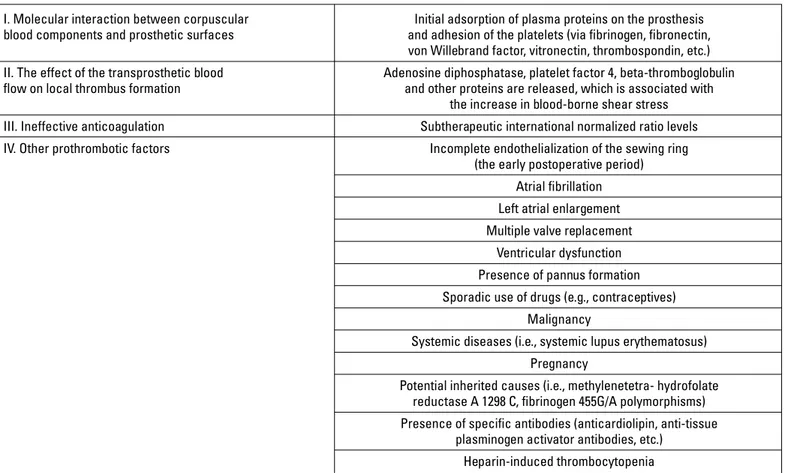
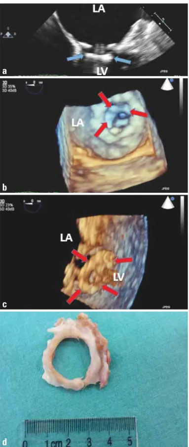
Figure 1. Two-dimensional transthoracic echocardiographic imaging of mitral prosthetic valve thrombosis (arrow) in four-chamber view (a) and increased transvalvular gradients and reduced mitral valve area, as demonstrated by Doppler imaging (b).
LA - left atrium; LV - left ventricle; RA - right atrium; RV -right ventricle
a
accurate diagnosis and correct course of therapy with the utility of RT-3-D TEE (Fig. 2a–c). This also avoids unnecessary further diagnostic workup. It must be acknowledged that RT-3-D TEE is a complementary diagnostic tool; its accuracy depends on the quality of the original 2-D images. RT 3-D TEE is time-consuming and requires added training. It has also several limitations such as reduced temporal resolution, poor visualization of anterior structures of the heart such as aortic and tricuspid valves, and poor image quality due to poor electrocardiography gating in pa-tients with arrhythmias (23). Furthermore, it has the problem of acoustic shadowing like 2-D imaging; for instance, it may be diffi-cult to visualize the pathologies (thrombus, pannus, etc.) located on the ventricular side of the mitral prosthesis (29).
Cinefluoroscopy (CF) is a low-cost, noninvasive imaging technique, which is readily available in most centers and can be performed rapidly, particularly in unstable patients, for de-tecting stuck valves (30, 31). In the case of bileaflet valves, the disks can be directly visualized, and opening and closing angles measured using a tangential view (31). Although the role of CF has declined since the introduction of TEE, it still serves as a complementary method to echocardiography in evaluation of prosthetic valve obstruction (30). It may be particularly utilized as an easily repeatable modality to follow stable patients for evaluation of valve motions during TT. CF has also limitations; it is not useful in distinguishing pannus from thrombus since neither pannus nor thrombus can be identified fluoroscopically. Therefore, TEE should be performed to confirm the findings ob-tained by CF.
Multidetector cardiac computed tomography (MDCT) is a promising technique for functional evaluation of bileaflet
me-chanical valves. Opening and closing leaflet angles can be accurately assessed. Currently, TEE is still the most reliable method in the diagnosis of PVT, but MDCT can be used as a complementary diagnostic method for a definitive diagnosis in case of clinical suspicion. It may be helpful especially in pa-tients with double left-sided mechanical valves because acous-tic shadowing can occur even during TEE study and make the interpretation difficult. It has also been proven to be a useful method for the differential diagnosis of masses amenable to TT in patients with prosthetic valve dysfunction (32) (see next sec-tion) (Fig. 3).
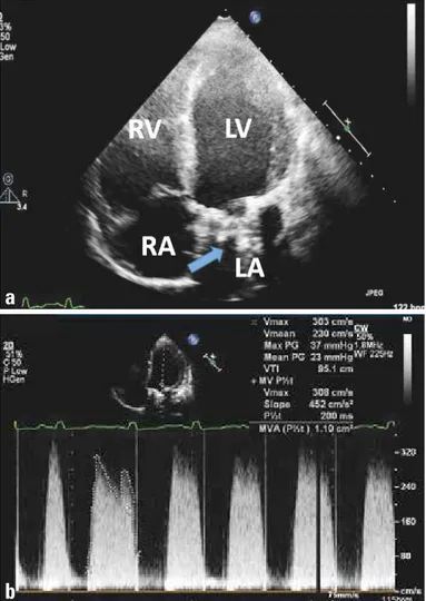
Figure 2. Two-dimensional TEE demonstrates a soft thrombotic mass (arrow) attached to the hinge of the prosthesis (a). Real-time three-dimensional transesophageal echocardiography from the left atrial side revealed thrombus (arrows) on the prosthetic mitral valve (a'). The thrombus burden was diminished (arrow) after an initial dose of TT (25 mg TPA), shown by 2-D TEE (b) and 3-D TEE (b'). After the second dose of TT, the thrombus size was completely lysed, shown by 2-D TEE (c) and 3-D TEE (c').
LA - left atrium; LAA - left atrial appendage; LV - left ventricle; TEE - transesophageal echocardiography; tPA - tissue-type plasminogen activator; TT - thrombolytic therapy
a
a'
b
b'
c
c'
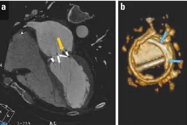
Figure 3. Multidetector computed tomography revealed a periprosthetic mass with HU: 65 (favors thrombus), which restricted the mobility of one of the leaflets (a). Volume rendering demonstration of pannus over the removed prosthetic valve (b). HU-Hounsfield unit
Differential diagnosis of PVT
The distinction between PVT and other prosthesis-related pathologies, such as pannus, vegetation, and prosthesis–patient mismatch (PPM), is important to choose the optimal treatment (23, 25, 33). Differential diagnosis based on clinical presentation may be challenging, and multimodality imaging, including echo-cardiography, CF, and MDCT is usually required. The masses re-lated to prosthetic heart valves (thrombus, pannus, and vegeta-tion) are compared in Table 2.
Pannus is an overgrowth of fibrous tissue. It is relatively more common in the aortic position. Since surgery can be avoided for some patients with mechanical valve obstruction secondary to thrombosis, but not pannus, the distinction between these two etiologies needs specific concern (23, 32). Certain findings, such as the echocardiographic homogeneity of the mass, reduced transmitral gradients, and evolution of thrombus morphology with therapeutic anticoagulation, integrated with clinical (presen- ce of inadequate anticoagulation or evidence of thromboem-bolism) observations, help distinguish thrombosis from pannus overgrowth (19, 23). Previously introduced RT-3-D TEE provides visualization of the atrial and ventricular sides of the prosthesis, improves understanding of the relation between cardiac struc-tures, and helps discriminating pannus from thrombus (34). Fur-thermore, the role of 64-slice MDCT in the differential diagnosis of thrombus versus pannus has been recently investigated by
Gündüz et al. (32). They showed that high-attenuation (HU ≥145) periprosthetic masses are resistant to TT and predict pannus whereas the low-attenuation (HU <90) periprosthetic masses are susceptible to TT and predict thrombus (Table 2). Briefly, dif-ferentiation of these two etiologies is now easier with advanced multimodality imaging. Figure 4 shows echocardiographic and postoperative view of pannus overgrowth on mitral prosthesis.
Vegetation is another entity that should be considered in dif-ferential diagnosis of PVT (Table 2). They cannot be distinguished by echocardiography alone; depiction of these sessile or pedun-culated masses with the presence of full clinical picture may lead to right diagnosis (23). Vegetation is more likely in febrile patients and in the presence of clinical signs of infective endo-carditis, perivalvular destruction, leak, or abscess formation. Re-cently, positron emission tomography–computed tomography has proven its additive role in the diagnosis of infective endocarditis in patients with negative/inconclusive echocardiography (35).
PPM is an important cause of elevated velocity and gradients across normally functioning prosthetic valves. It is present when the effective orifice area (EOA) of the inserted prosthetic valve is too small in relation to body size. It is common (20%–70% of aortic valve replacements) and has been shown to be associated with worse hemodynamic function (33). The indexed EOA ≤0.65 and ≤0.90 favors PPM in aortic and mitral prosthesis, respectively (for those with body mass index <30 kg/m2). Although EOA
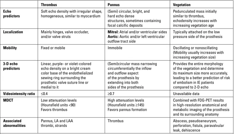
deter-Table 2. Comparison of prosthetic heart valve related-masses
Thrombus Pannus Vegetation
Echo Soft echo density with irregular shape, (Semi) circular, bright, and Pedunculated mass initially predictors homogeneous, similar to myocardium hard echo dense similar to thrombus,
structures, sometimes containing echodensity increases with
focal calcific deposits increasing vegetation age
Localization Mainly hinges, valve occluder, Mitral: Atrial and/or ventricular sides Typically attached on the low and/or valve struts Aortic: Aortic and/or left ventricular pressure side of the prosthesis
outflow tract side
Mobility Fixed or mobile Immobile Oscillating or nonoscillating
(Mobility usually increases with
increasing vegetation size)
3-D echo Linear, purple- or violet-colored (Semi)circular mass narrowing Provides the entire morphology predictors echo density on a bright cream circumferentially the inflow of the vegetation and determine color base of the endothelialized and outflow aspect its maximum size more accurately, sewing ring surrounding the of the prosthesis by leading to a better prediction of risk prosthetic valve suture line or extending into both of embolism in IE patients
medial to it sides of the prosthesis compared to 2-D echo
Videointensity ratio ≤0.4 ≥0.7 Unavailable data
MDCT Low attenuation levels High attenuation levels Combined with FDG-PET results (Hounsfield units <90) (Hounsfield units ≥145) in high-resolution anatomical and Favors thrombus Favors pannus formation metabolic imaging of the prosthesis
and its surrounding anatomy
Associated Pannus, LA and LAA Thrombus Abscess, pseudoaneurysm,
abnormalities thrombi, strands perforation, fistula, paravalvular
leak, dehiscence
2-D/3-D-dimensional; FDG-PET - 18-fluorine-fluorodesoxyglucose positron emission tomography; IE - infective endocarditis; LA - left atrium; LAA - left atrial appendage; MDCT - multide-tector-row computed tomography
mination is crucial in evaluating PPM, systolic time interval pa-rameters, AT and AT/ET could be also very helpful, especially in differentiation of PPM from prosthetic aortic valve stenosis (25).
Management of PVT
Treatment modalities for PVT include anticoagulation with heparin, TT, surgery, or even in some cases only watchful wai- ting (23).
Anticoagulation
Conclusive data is lacking regarding the effectiveness of anticoagulation in resolution of PVT. It has been previously shown that the prognosis is favorable with medical therapy by optimization of anticoagulant treatment (short-term intravenous unfractionated heparin followed by warfarin adjustment and as-pirin addition) for small asymptomatic thrombi (length <10 mm) (36). Lengyel et al. (37) demonstrated a low rate of success with heparin treatment in cases with nonobstructive PVT, but a more recent study authored by Laurent et al. (38) has reported the ef-fectiveness of prolonged heparin with oral anticoagulation in pre-venting embolic events in patients with early nonobstructive PVT of size <5 mm after mechanical prosthetic mitral valve replace-ment. Our preliminary experience shows that unfractionated heparin could be successful in 73% of the nonobstructive PVT patients who have contraindications to TT (39). In current lit-erature, the use of low-molecular-weight heparin in left-sided NOPVT is not clear yet.
TT versus surgery
Until the 1990s, the treatment of choice for mechanical valve obstruction was surgery, but over the last decade, TT has been used increasingly (40, 41). Unfortunately, randomized controlled trials to address the initial treatment strategy are lacking. The most recent European (42) and American guidelines (43) recom-mend surgery for patients with NYHA functional classes III and IV unless surgery is high risk (Class IIa). Thrombolysis is given a IIa indication in patients with right-sided valve thrombosis and a Class IIb indication in patients with a left-sided but small thrombus. The European Society of Cardiology guidelines (42) also recommend surgery for critically ill patients and restrict thrombolysis to patients with high surgical risk and/or right-si- ded valve thrombosis. On the other hand, TT is recommended as the first-line treatment for all patients with left-sided PVT by the Society for Heart Valve Disease guidelines and for patients with low thrombus burden (<0.8 cm2) (40). The diagnostic and mana-
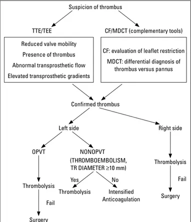
gement strategies of PVT are summarized in Figure 5.
Recently, several meta-analyses and systematic reviews have been published. Karthikeyan et al. (44) evaluated seven studies with 690 episodes of PVT (446 treated with surgery and 244 with TT) and found no significant differences in the main out-come (restoration of valve functions) or death between patients treated surgically and with TT. They stated that urgent surgery should probably be preferred over TT in experienced centers.
Figure 4. Two-dimensional transesophageal echocardiography (TEE) revealed a hyperechogenic circular mass on the left ventricular side of the prosthesis (a). Real-time three-dimensional TEE left atrial (b) and left ventricular (c) views showed circular left ventricular side pannus formation. Postoperative specimen of pannus formation (left ventricu-lar-sided) is demonstrated (d)
a
b
c
On the basis of previous data, surgical mortality may reach 69%, depending on NYHA class and need for emergency surgery (6, 9, 44-46), whereas the reported death rate is 8%–13.5% in pa-tients undergoing TT for PVT (44–46).
Recently, Castilho et al. (9) evaluated 26 studies reporting 1107 episodes of PVT treated by TT and 27 studies reporting 1132 surgeries for PVT. They reported much higher mortality rates with surgery compared with TT in the management of PVT (18.1% versus 6.6%). Nevertheless, it could be misleading to compare TT and surgery with respect to mortality rates without a head-to-head randomized trial.
Recent TT studies for PVT have shown much promise, with the results suggesting that such treatment modality might be the initial choice in these patients (6–8, 40, 47). There is no consensus regarding the optimal treatment strategy, neither the type, nor the dose or route of administration of thrombolytic agents. Due to its high fibrin specificity, recombinant tissue-type plasminogen activator (rt-PA) is widely used in the management of PVT (6–8). On the other hand, because of the relatively higher cost of tPA treatment, streptokinase is still used for TT in the developing countries. Although accelerated protocols seem attractive as they may induce more rapid lysis of the thrombus, they increase the risk of serious thromboembolism and bleeding events (4, 45).
The TROIA (Comparison of Different TRansesophageal Echocardiography Guided thrOmbolytic Regimens for prosthetIc vAlve Thrombosis) study (6), which includes the largest cohort published to date (182 consecutive patients with PVT in 220 dif-ferent episodes), evaluated a strategy of TEE-guided fibrinoly-sis with rapid infusion of streptokinase (Group 1) versus slow infusion of streptokinase (Group II) versus full-dose t-PA (100 mg) (Group 3) versus half dose (50 mg) slow infusion of t-PA (Group IV) versus low-dose (25 mg) slow infusion of t-PA (Group 5). This was a monocentric, prospective, nonrandomized study. The authors reported successful thrombolysis in 83.2% of cases without a significant difference between thrombolytic protocols (68.8%, 85.4%, 75.0%, 81.5%, and 85.5%, respectively; p = 0.46). Assessment of complication rates by groups showed a statisti-cally lower combined complication rate in low-dose slow infu-sion group. Therefore, the authors suggested that lower dose, TEE-guided, repeated, slow administration of a fibrinolytic agent could be equally efficacious with fewer complications. Although no mortality was reported in this regimen, nonfatal major complications were similar between the regimens. There-fore, in order to reduce nonfatal major complications, safety of ultra-slow TT regimen was investigated in 114 PVT patients— the PROMETEE trial (8). This study showed that, ultra-slow (25 hours) infusion of low-dose (25 mg) t-PA without bolus was as-sociated with quite low nonfatal complications and mortality for PVT patients except for those with NYHA class-IV, without compromising effectiveness.
The main limitation associated with TT of left-sided PVT in-cludes cerebral thromboembolism (48) and bleeding. In TROIA study (6), the rate of intracranial bleeding was 0.8% in PVT pa-tients undergoing low-dose slow infusion t-PA strategy. In the PROMETEE study (8), none of the patients suffered intracranial hemorrhage, and the noncerebral major bleeding rate was also quite low (1.7%). These two recent studies have shown that low-dose slow infusion of tPA can be an effective and safe therapy in the management of PVT.
While tPA is widely used as a thrombolytic agent, infusion of rt-PA may trigger the production of anti-tissue plasminogen activator (tPA) antibodies (ATA), which may interfere with the success of TT, necessitating a higher dose of rt-PA for complete success. It has also been shown that some patients with PVT have increased baseline ATA levels, which is also associated with higher risk for rethrombosis (16).
Specific population Pregnant women
PVT in pregnancy is a double jeopardizing event for both mother and fetus. The main treatment strategies include an-ticoagulation with heparin, TT, and redo surgery, each having its own pros and cons. Although no evidence-based guide-lines for pregnant patients complicated with PVT are current-ly available, recommendations of guidelines for this
compli-Figure 5. Diagnostic and therapeutic algorithm for prosthetic valve thrombosis.
CF - cinefluoroscopy; MDCT - multidetector computed tomography; NOPVT - nonobstructive prosthetic valve thrombosis; OPVT - obstructive prosthetic valve thrombosis; TEE - trans-esophageal echocardiography; TTE - transthoracic echocardiography
Reduced valve mobility Presence of thrombus Abnormal transprosthetic flow Elevated transprosthetic gradients
CF: evaluation of leaflet restriction MDCT: differential diagnosis of
thrombus versus pannus Suspicion of thrombus Confirmed thrombus Left side OPVT NONOPVT Yes No Thrombolysis Intensified Anticoagulation (THROMBOEMBOLISM, TR DIAMETER ≥10 mm) Thrombolysis Thrombolysis Surgery Surgery Fail Fail Right side TTE/TEE CF/MDCT (complementary tools)
cation are similar to the management of PVT in nonpregnant patients (42) and missing definitive class I recommendations because of lack of randomized controlled trials. Cardiac sur-gery in pregnancy is associated with very high maternal and fetal mortality (6% and 30%, respectively) and morbidity (24% and 9%, respectively) (49). Due to a recent study by Özkan et al. (7), which consisted of the largest series of pregnant patients with PVT reported to date, low-dose, slow infusion of tPA with repeated doses as needed that was guided by TEE was associated with a successful thrombolysis in all episodes, with no maternal deaths and a fetal mortality rate of 20%. The authors indicated that the incidence of mater-nal and fetal adverse events (mortality, abortion, etc.) with this protocol was lower than with surgery or medical therapy on the basis of the available published data and suggested that it should be used as first-line therapy for PVTs in preg-nant women. The high success rate of TT was attributed to the fresh and rapid development of clot in this specific patient population compared with nonpregnant patients with gradu-ally developed and organized thrombus.
Elderly
Elderly individuals are the rapidly growing segment of the population and choosing the optimal management for PVT could be challenging due to safety concerns. Surgical treatment may carry high risk especially in elderly patients with multiple comor-bidities. Furthermore, older age also poses an increased risk of hemorrhage in patients undergoing TT. In a recent single-center study, Gündüz et al. (50) has demonstrated that slow or ultra-slow infusions of low-dose thrombolytic agents (mostly t-PA) provide considerably high lysis rates with low major adverse event rates in elderly patients with PVT. Consequently, TT is also a promising therapy in elderly population.
Patients with ischemic stroke
The risk of cerebral embolism can be up to 5%–6% for left-sided PVT (40). CT should be performed in patients who admit with initial suspicion of acute ischemic stroke to exclude any in-tracranial bleeding and these patients may be considered for TT only if they are stable by neuroradiologic assessment after 3–4 weeks of anticoagulation with UFH (8). Ischemic stroke may also occur during TT and may be managed with continuing low-dose and prolonged infusion of TT in eligible patients (48).
Patients with coronary embolism
It is a potential complication of prosthetic valves and should be suspected in patients with prosthetic valves who admit with acute coronary syndrome (20). Intracoronary thrombolysis, stent implantation, and embolectomy can be performed as reperfu-sion strategies. Furthermore, intravenous TT can be successfully performed for the management of PVT and concurrent coronary embolism in eligible patients as the fresh nature of the embolic material may easily respond to TT (20).
Conclusion
Despite technological advancements, the hemodynamic and physical properties of mechanical valves remain thrombo-genic, and patients with prosthetic heart valves, therefore, are prone to developing PVT. Unfortunately, VKAs are still the only approved oral anticoagulants in patients with heart valve pros-theses and today clinicians, worldwide, are expecting for an antithrombotic agent that is at least as effective but safer and more convenient in daily clinical practice. The diagnosis of PVT and other prosthetic valve dysfunctions is now easier with the use of multimodality imaging, including RT-3-D TEE and MDCT. There is still a debate about the optimal treatment strategy for PVT. Guidelines lack definitive NYHA class I recommendations, have significant disparities, and—in most cases—leave the de-cision to the clinician’s experience. The favorable clinical out-comes of TT comparing with the surgical approach have made TT the first-line treatment in many of the developing countries. Surgical treatment could be left for patients in which TT is con-traindicated, or in those where it has already failed. Currently, the superiority of one over other remains speculative due to ab-sence of a head-to-head randomized controlled trial between TT and surgery. However, the recently initiated randomized and multicenter study (NCT02243839), which compares TT (with tPA) versus surgery for the management of patients with PVT, will provide essential data.
Conflict of interest: None declared. Peer-review: Externally peer-reviewed.
Authorship contributions: Concept – M.O.G., M.K., M.Ö., S.K.; Design – M.O.G., M.Y., E.B., M.Ö.; Supervision – M.O.G., M.K., M.Y., S.G., M.Ö.; Materials – M.K., M.Y., E.B., S.G., M.Ö.; Data collection &/or processing – M.O.G., M.K., M.Y., S.K., E.B., S.G., M.Ö.; Analysis &/or interpretation – M.O.G., M.K., M.Y., S.K., E.B., S.G., M.Ö.; Literature search – M.O.G., M.K., M.Ö.; Writing – M.O.G., M.Ö.; Critical review – M.O.G., M.K., M.Ö
References
1. Hermans H, Vanassche T, Herijgers P, Meuris B, Herregods MC, Van de Werf F, et al. Antithrombotic therapy in patients with heart valve prostheses. Cardiol Rev 2013; 21: 27-36. Crossref
2. Grunkemeier GL, Rahimtoola SH. Artificial heart valves. Annu Rev Med 1990; 41: 251-63. Crossref
3. Cannegieter SC, Rosendaal FR, Briet E. Thromboembolic and bleed-ing complications in patients with mechanical heart valve prosthe-ses. Circulation 1994; 89: 635-41. Crossref
4. Karthikeyan G, Math RS, Mathew N, Shankar B, Kalaivani M, Singh S, et al. Accelerated infusion of streptokinase for the treatment of left-sided prosthetic valve thrombosis: a randomized controlled trial. Circulation 2009; 120: 1108-14. Crossref
5. Özkan M, Kaymaz C, Kırma C, Sönmez K, Özdemir N, Balkanay M, et al. Intravenous thrombolytic treatment of mechanical prosthetic valve thrombosis: a study using serial transesophageal echocar-diography. J Am Coll Cardiol 2000; 35: 1881-9. Crossref
6. Özkan M, Gündüz S, Biteker M, Astarcıoğlu MA, Çevik C, Kaynak E, et al. Comparison of different TEE-guided thrombolytic regimens for prosthetic valve thrombosis: The TROIA Trial. JACC Cardiovasc Imaging 2013; 6:206-16. Crossref
7. Özkan M, Çakal B, Karakoyun S, Gürsoy OM, Çevik C, Kalçık M, et al. Thrombolytic therapy for the treatment of prosthetic heart valve thrombosis in pregnancy with low-dose, slow infusion of tissue-type plasminogen activator. Circulation 2013; 128: 532-40. Crossref
8. Özkan M, Gündüz S, Gürsoy OM, Karakoyun S, Astarcıoğlu MA, Kalçık M, et al. A novel strategy in the management of PROsthetic Mechanical valve Thrombosis and the prEdictors of outcomE: the Ultra-slow PROMETEE trial. Am Heart J 2015; 170: 409-18. Crossref
9. Castilho FM, De Sousa MR, Mendonça AL, Ribeiro AL, Cáceres-Lóriga FM. Thrombolytic therapy or surgery for valve prosthesis thrombosis: systematic review and meta-analysis. J Thromb Hae-most 2014; 12: 1218-28. v
10. Anderson JM, Schoen EJ. Interaction of blood with artificial sur-faces. In: Butchart E, Bodnar E, eds. Thrombosis, Embolism and Bleeding. London, ICR Publishers, 1992. p.160-71. Crossref
11. Horstkotte D, Aul C, Seipel L, Körfer R, Budde T, Schulte HD, et al. Effect of valve type and valve function on chronic intravascular hemolysis following mitral and aortic valve replacement using al-loprostheses. Z Kardiol 1983; 72: 119-31. Crossref
12. Gürsoy OM, Karakoyun S, Kalçık M, Gökdeniz T, Yesin M, Gündüz S, et al. Usefulness of novel hematologic inflammatory parameters to predict prosthetic mitral valve thrombosis. Am J Cardiol 2014; 113: 860-4. Crossref
13. Aykan AC, Gökdeniz T, Gündüz S, Astarcıoğlu MA, Gürsoy OM, Ertürk E, et al. Value of serum fibrinogen levels in the assessment of mechanical prosthetic valve thrombosis. J Heart Valve Dis 2014; 23: 222-7. Crossref
14. Aykan AÇ, Gökdeniz T, Kalçık M, Astarcıoğlu MA, Gündüz S, Kara-koyun S, et al. Role of anticardiolipin antibodies in the pathogenesis of prosthetic valve thrombosis: An observational study. Herz 2015; 40: 528-33. Crossref
15. Kalçık M, Gürsoy MO, Karakoyun S, Yesin M, Astarcıoğlu MA, Öz-kan M. Potential inherited causes of recurrent prosthetic mitral valve thrombosis in a pregnant patient suffering from recurrent miscarriage. Korean Circ J 2014; 44: 268-70. Crossref
16. Özkan M, Kalçık M, Gürsoy MO, Öcal L, Griffini S, Karakoyun S, et al. Assessment of Anti-Tissue Type Plasminogen Activator Antibodies in Patients With Prosthetic Heart Valve Thrombosis: The ATA Trial. J Cardiovasc Pharmacol Ther 2016; 21: 372-80. Crossref
17. Özkan M, Oğuz AE, Gürsoy OM, Gündüz S, Aykan CA, Astarcıoğlu MA, et al. Management of heparin-induced thrombocytopenia du- ring thrombolytic therapy for prosthetic valve thrombosis. J Heart Valve Dis 2012; 21: 636-40. Crossref
18. Tong AT, Roudaut R, Özkan M, Sagie A, Shahid MS, Pontes Júnior SC, et al; Prosthetic Valve Thrombolysis-Role of Transesophageal Echocardiography (PRO-TEE) Registry Investigators. Transesopha-geal echocardiography improves risk assessment of thrombolysis of prosthetic valve thrombosis: results of the international PRO-TEE registry. J Am Coll Cardiol 2004; 43: 77-84. Crossref
19. Özkan M, Gürsoy OM, Astarcıoğlu MA, Gündüz S, Çakal B, Kara-koyun S, et al. Real-time three-dimensional transesophageal echo-cardiography in the assessment of mechanical prosthetic mitral valve ring thrombosis. Am J Cardiol 2013; 112: 977-83. Crossref
20. Karakoyun S, Gürsoy MO, Kalçık M, Yesin M, Özkan M. A case se-ries of prosthetic heart valve thrombosis-derived coronary embo-lism. Türk Kardiyol Dern Ars 2014; 42: 467-71. Crossref
21. Gürsoy OM, Karakoyun S, Kalçık M, Özkan M. The incremental value of RT three-dimensional TEE in the evaluation of prosthetic mitral valve ring thrombosis complicated with thromboembolism. Echocardiography 2013; 30: E198-201. Crossref
22. Aykan AÇ, Gürsoy OM, Özkan M, Yıldız M, Kahveci G, Uslu Z. Suc-cessful treatment of renal artery thromboembolism with low-dose prolonged infusion of tissue-typed plasminogen activator in a pa-tient with mitral mechanical heart valve thrombosis under the gui- dance of multimodality imaging. Blood Coagul Fibrinolysis 2012; 23: 663-5. Crossref
23. Gürsoy MO, Kalçık M, Karakoyun S, Özkan M. The current status of fluoroscopy and echocardiography in the diagnosis of prosthetic valve thrombosis-a review article. Echocardiography 2015; 32: 156-64. Crossref
24. Zoghbi WA, Chambers JB, Dumesnil JG, Foster E, Gottdiener JS, Grayburn PA, et al. Recommendations for evaluation of prosthetic valves with echocardiography and Doppler ultrasound. J Am Soc Echocardiogr 2009; 22: 975-1014. Crossref
25. Ben Zekry S, Saad RM, Ozkan M, Al Shahid MS, Pepi M, Muratori M, et al. Flow acceleration time and ratio of acceleration time to ejection time for prosthetic aortic valve function. JACC Cardiovasc Imaging 2011; 4: 1161-70. Crossref
26. Muratori M, Montorsi P, Teruzzi G, Celeste F, Doria E, Alamanni F, et al. Feasibility and diagnostic accuracy of quantitative assess-ment of mechanical prostheses leaflet motion by transthoracic and transesophageal echocardiography in suspected prosthetic valve dysfunction. Am J Cardiol 2006; 97: 94-100. Crossref
27. Gürsoy OM, Özkan M. The role of real-time 3-dimensional trans-esophageal echocardiography in depiction of the concealed base of the iceberg. Anadolu Kardiyol Derg 2012; 12: E22-3. Crossref
28. Reddy VK, Faulkner M, Bandarupalli N, Nanda NC, Singh P, Dutta R, et al. Incremental value of live/real time three-dimensional trans-thoracic echocardiography in the assessment of right ventricular masses. Echocardiography 2009; 26: 598-609. Crossref
29. Sugeng L, Shernan SK, Weinert L, Shook D, Raman J, Jeevanan-dam V, et al. Real-time three-dimensional transesophageal echo-cardiography in valve disease: comparison with surgical findings and evaluation of prosthetic valves. J Am Soc Echocardiogr 2008; 21: 1347-54. Crossref
30. Kalçık M, Gürsoy OM, Astarcıoğlu MA, Özkan M. A serial fluoros-copy-guided thrombolytic therapy of a mechanical tricuspid pros-thetic valve thrombosis with low-dose and ultra-slow infusion of tissue-type plasminogen activator. Turk Kardiyol Dern Ars 2014; 42: 478-81. Crossref
31. Montorsi P, Cavoretto D, Alimento M, Muratori M, Pepi M. Prosthe- tic mitral valve thrombosis: Can fluoroscopy predict the efficacy of thrombolytic treatment? Circulation 2003; 108: II79-84. Crossref
32. Gündüz S, Özkan M, Kalçık M, Gürsoy OM, Astarcıoğlu MA, Kara-koyun S, et al. Sixty-Four–Section Cardiac Computed Tomography in Mechanical Prosthetic Heart Valve Dysfunction Thrombus or Pannus. Circ Cardiovasc Imaging 2015; 8: e003246. Crossref
33. Pibarot P, Dumesnil JG. Prosthesis-patient mismatch: definition, clinical impact, and prevention. Heart 2006; 92: 1022-9. Crossref
34. Özkan M, Gündüz S, Yıldız M, Duran NE. Diagnosis of the prosthetic heart valve pannus formation with real-time three-dimensional transoesophageal echocardiography. Eur J Echocardiogr 2010; 11: E17. Crossref
35. Saby L, Laas O, Habib G, Cammilleri S, Mancini J, Tessonnier L, et al. Positron emission tomography/computed tomography for diagnosis of prosthetic valve endocarditis: increased valvular
18F-fluorode-oxyglucose uptake as a novel major criterion. J Am Coll Cardiol 2013; 61: 2374-82. Crossref
36. Bemurat LR, Laffort PR, Deville CJ, Roques XG, Baudet EM, Rou-daut RP. Management of Nonobstructive Thrombosis of Prosthetic Mitral Valve in Asymptomatic Patients in the Early Postoperative Period: A Study in 20 Patients. Echocardiography 1999; 16: 339-46. 37. Lengyel M, Vegh G, Vandor L. Thrombolysis is superior to heparin
for non-obstructive mitral mechanical valve thrombosis. J Heart Valve Dis 1999; 8: 167-73. Crossref
38. Laurent M, Lelong B, Verhoye JP, Khattar C, de Place C, Matali P, et al. Prolonged heparin and vitamin K antagonist regimen for early non-obstructive thrombosis of mechanical mitral valve prostheses. J Heart Valve Dis 2008; 17: 533-41. Crossref
39. Yesin M, Kalçık M, Cerşit S, Gürsoy MO, Kılıçgedik A, Börekçi A, et al. Monitorization of patients with thrombotic prosthetic valves with infusion of unfractionated heparin under the guidance of se-rial transesophageal echocardiographic examinations. Anadolu Kardiyol Derg 2014; 14 (Suppl. 1): 1-165. Crossref
40. Lengyel M, Horstkotte D, Völler H, Mistiaen WP. Working Group Infection, Thrombosis, Embolism and Bleeding of the Society for Heart Valve Disease. Recommendations for the management of prosthetic valve thrombosis. J Heart Valve Dis 2005; 14: 567-75. 41. Biteker M, Altun I, Başaran O, Doğan V, Yıldırım B, Ergun G.
Treat-ment of prosthetic valve thrombosis: Current evidence and future directions. J Clin Med Res 2015; 7: 932-6. Crossref
42. Vahanian A, Alfieri O, Andreotti F, Antunes MJ, Barón-Esquivias G, Baumgartner H, et al. Guidelines on the management of valvular heart disease (version 2012). Eur Heart J 2012; 33: 2451-96. Crossref
43. Nishimura RA, Otto CM, Bonow RO, Carabello BA, Erwin JP 3rd, Guyton RA, et al. 2014 AHA/ ACC guideline for the management
of patients with valvular heart disease: a report of the American College of Cardiology/American Heart Association Task Force on Practice Guidelines. J Am Coll Cardiol 2014; 63: e57-185. Crossref
44. Karthikeyan G, Senguttuvan NB, Joseph J, Devasenapathy N, Bahl VK, Airan B. Urgent surgery compared with fibrinolytic therapy for the treatment of left-sided prosthetic heart valve thrombosis: a systematic review and meta-analysis of observational studies. Eur Heart J 2013; 34: 1557-66. Crossref
45. Bonou M, Lampropoulos K, Barbetseas J. Prosthetic heart valve obstruction: thrombolysis or surgical treatment? Eur Heart J Acute Cardiovasc Care 2012; 1: 122-7. Crossref
46. Huang G, Schaff HV, Sundt TM, Rahimtoola SH. Treatment of obs- tructive thrombosed prosthetic heart valve. J Am Coll Cardiol 2013; 62: 1731-6. Crossref
47. Caceres-Loriga FM, Perez-Lopez H, Morlans-Hernandez K, Facun-do-Sánchez H, Santos-Gracia J, Valiente-Mustelier J, et al. Throm-bolysis as first choice therapy in prosthetic heart valve thrombosis. A study of 68 patients. J Thromb Thrombolysis 2006; 21: 185-90. 48. Özkan M, Gürsoy OM, Atasoy B, Uslu Z. Management of acute
ischemic stroke occurred during thrombolytic treatment of a pa-tient with prosthetic mitral valve thrombosis: continuing throm-bolysis on top of thromthrom-bolysis. Anadolu Kardiyol Derg 2012; 12: 689-90. Crossref
49. Weiss BM, von Segesser LK, Alon E, Seifert B, Turina MI. Outcome of cardiovascular surgery and pregnancy: a systematic review of the period 1984-1996. Am J Obstet Gynecol 1998; 179: 1643-53. 50. Gündüz S, Özkan M, Yesin M, Kalçık M, Gürsoy MO, Karakoyun S,
et al. Prolonged Infusions of Low-Dose Thrombolytics in Elderly Patients With Prosthetic Heart Valve Thrombosis. Clin Appl Thromb Hemost 2015 Oct 6. Epub ahead of print.