Subclinical myocardial dysfunction in liver transplant candidates
determined by using speckle tracking imaging
Karaciğer transplantasyonu adaylarında benek takibi görüntüleme ile
belirlenen subklinik miyokart disfonksiyonu
1Department of Cardiology, Health Sciences University Dışkapı Yıldırım Beyazıt Training and Research Hospital, Ankara, Turkey 2Department of Cardiology, Başkent University Faculty of Medicine, Ankara, Turkey
3Department of Cardiology, Ministry of Health Sakarya University Training and Research Hospital, Sakarya, Turkey Saadet Demirtaş İnci, M.D.,1 Leyla Elif Sade, M.D.,2 Cihan Altın, M.D.,2 Bahar Pirat, M.D.,2
Hilal Erken Pamukcu, M.D.,1 Sabiye Yılmaz, M.D.,3 Haldun Müderrisoğlu, M.D.2
Objective: There are various cardiovascular abnormalities in end-stage liver disease (ESLD). In these patients, left ventricular (LV) systolic function is normal at rest but deteri-orates during stress. This deterioration may be due to sub-clinical myocardial dysfunction. This study evaluated global LV and right ventricular (RV) functions using 2-dimensional (2D) speckle tracking in patients with ESLD.
Methods: Forty liver transplant candidates with ESLD and 26 healthy individuals were included in the study. All of the patients underwent conventional echocardiographic mea-surements. Longitudinal, circumferential, and radial strain measurements, as well as apical and parasternal short-axis image recordings were obtained. All 2D strain measure-ments were performed with offline analysis using velocity vector imaging (VVI) software.
Results: In the apical 4- and 2-chamber measurements, the LV mean longitudinal strain was significantly lower in patients with ESLD compared with that of the control group (-16.0±3.2% versus -17.6±2.2%, -16.7±3.3% versus -18.7±2.1%; p=0.002, respectively). The LV mean circumfer-ential strain did not differ between groups. The LV mean ra-dial strain and RV longitudinal strain were significantly lower in patients with ESLD (45.4±10.7 vs. 52.7±10.8%; p=0.01 and -19.2±3.5% versus -21.5±3.6%; p=0.03, respectively).
Conclusion: Subclinical impairment of global LV and RV systolic functions were determined in liver transplantation candidates by using VVI. This deterioration was detected in longitudinal and radial deformation rather than circum-ferential deformation mechanics, which was consistent with early-stage LV myocardial dysfunction.
Amaç: Son dönem karaciğer hastalarında (SDKH) çeşitli kar-diyovasküler anormallikler olduğu bilinmektedir. Bu hastalar-da istirahatte sol ventrikül (LV) sistolik fonksiyonu normal sap-tanmasına karşın stres sırasında bozulmaktadır. Bu bozulma subklinik miyokardiyal işlev bozukluğundan kaynaklanıyor olabilir. Bu nedenle, bu çalışmada SDKH’da global LV ve sağ ventrikül (RV) fonksiyonlarını 2 boyutlu (2B) benek takibi gö-rüntüleme yöntemi ile değerlendirmesi amaçlandı.
Yöntemler: Karaciğer nakil adayı olan 40 SDKH ile 26 sağ-lıklı birey çalışmaya dahil edildi. Tüm hastaların standart ekokardiyografik ölçümleri yapıldı. Uzunlamasına, sirküm-feransiyel ve radyal strain ölçümleri için apikal ve paraster-nal kısa eksen görüntü kayıtları alındı. Tüm 2B strain öl-çümleri alınan kayıtlardan, çevrim dışı analiz ile hız vektör görüntüleme (VVI) yazılım paketi kullanılarak ölçüldü.
Bulgular: Apikal 4- ve 2- boşluk ölçümlerde, LV ortala-ma uzunlaortala-masına strain değeri, hasta grubunda kontrol grubuna göre anlamlı olarak daha düşüktü (-16.0±%3.2 ve -17.6±%2.2, p=0.03 ve -16.2±%3.3 ve -18.7±%2.1, p=0.002). LV ortalama sirkumferansiyel strain değerlerinde gruplar arasında fark saptanmazken, LV ortalama radyal strain ve RV uzunlamasına strain değerleri hasta grubun-da anlamlı derecede grubun-daha düşük saptandı (45.4±10.7 ve %52.7±10.8, p=0.01 ve -19.2±%3.5 ve -21.5±%3.6, p=0.03).
Sonuç: Karaciğer nakil adaylarında, VVI yöntemi ile global LV ve RV sistolik fonksiyonlarında subklinik bozulma tespit edildi. Bu bozulma, LV’de miyokart hasarının erken evrede olmasıyla tutarlı olan sirkumferansiyel deformasyon meka-niğinden ziyade uzunlamasına ve radyal deformasyonda saptandı.
Received:January 22, 2019 Accepted:May 31, 2019
Correspondence: Dr. Saadet Demirtaş İnci. Dışkapı Yıldırım Beyazıt Eğitim ve Araştırma Hastanesi, Kardiyoloji Kliniği, 06110 Dışkapı, Altındağ, Ankara, Turkey.
Tel: +90 312 - 596 20 00 e-mail: [email protected]
© 2019 Turkish Society of Cardiology
P
atients with end-stage liver disease (ESLD) are known to also experience cardiac abnormali-ties. ESLD causes cirrhotic cardiomyopathy (CCM), which is defined as chronic cardiac dysfunction in the absence of known heart disease in patients with cirrhosis, regardless of the etiology of the cirrhosis. [1] The specific diagnostic criteria for CCM include increased cardiac output due to peripheral vasodilata-tion, low systolic blood pressure, systolic and/or dias-tolic dysfunction, electrophysiological abnormalities, and chronotropic failure.[2] Overt heart failure is not common in CCM, though the prevalence is not known precisely since cardiac symptoms are not evident at rest. The cardiac response to stress is decreased, but stroke volume and contractile indices are typically normal or even increased at rest. However, under con-ditions of stress such as exercise, hemorrhage, or liver transplantation, cardiac function is impaired.[3] The pathogenesis includes deterioration of beta receptor functions, changes in transmembrane flow, and over-production of cardiodepressant cytokines, such as ni-tric oxide.[4,5]Speckle-tracking imaging (STI) enables quan-tification of deformation in longitudinal, radial, and circumferential directions and is more sensitive than conventional echocardiography in detecting subclini-cal ventricular dysfunction. The aim of this study was to evaluate left ventricular (LV) and right ventricular (RV) mechanical functions in liver transplant candi-dates using velocity vector imaging (VVI).
METHODS Study population
Forty liver transplant candidates with ESLD and 26 age- and gender-matched healthy subjects were stud-ied after obtaining informed consent from each pa-tient. In total, 70 subjects were initially screened for study and 4 were not be included due to inadequate echocardiographic images. Hepatic cirrhosis was di-agnosed by either liver biopsy or laboratory, clinical, and ultrasonography findings. Approval for this study was received from the Başkent University Clinical Research Ethics Committee (KA08/145).
Demographic and clinical characteristics, including age, sex, the presence of diabetes mellitus, hyperten-sion, smoking, or dyslipidemia, and the results of the Child-Turcotte-Pugh Score for cirrhosis were noted.
Patients with coronary artery disease (CAD), systolic dysfunction, di-abetes mellitus, hyper-tension, a cardiac rhythm other than sinus, valvular heart heart valve surgery, hypertrophic cardiomy-opathy, renal dysfunction, chronic obstructive pul-monary disease, hepato-pulmonary syndrome and pulmonary hypertension, sepsis, peripheral artery
arterial disease, or poor echocardiographic image qual-ity were excluded. CAD was defined as the presence of any of the following: (I) typical angina; (II) ischemic ST-T wave changes, pathological Q waves, or left bundle-branch block on ECG; (III) regional wall mo-tion abnormalities on echocardiography; (IV) ischemia or any perfusion abnormality in stress imaging; or (V) history of myocardial infarction or coronary artery stenosis ≥50% detected with coronary angiography or a history of revascularization. Patients were defined as hypertensive if they had a diastolic pressure >90 mmHg or a systolic pressure >140 mmHg or if they were being treated with antihypertensive medication. An ejection fraction (EF) of <50% was accepted as LV systolic dysfunction.
Data acquisition and analysis Echocardiography
Echocardiography was performed using an Acuson Sequoia C256 ultrasound system (Siemens Healthi-neers AG, Erlangen, Germany) equipped with a 3V2c broadband transducer. Two-dimensional (2D), M-mode, color and spectral Doppler measurements were analyzed by an experienced echocardiography spe-cialist who was blinded to the patient’s clinical data. Conventional 2D and Doppler echocardiography were performed according to the American Society of Echocardiography/European Association of Echocar-diography recommendations.[6] LV end-diastolic vol-ume, end-systolic volvol-ume, and EF were determined according to Simpson’s modified biplane model. Tissue Doppler imaging (TDI) and color-coded TDI (cTDI) were performed from the apical 4-chamber view using pulsed-wave Doppler with a 3-mm sample volume at the mitral and tricuspid annular sites. Peak
Abbreviations:
2D Two-dimensional Aa Late diastolic velocity CAD Coronary artery disease CCM Cirrhotic cardiomyopathy cTDI Color-coded TDI Ea Early diastolic color-coded
tissue velocities EF Ejection fraction ESLD End-stage liver disease LV Left ventricular MPI Myocardial performance index
RV Right ventricular Sa Peak systolic velocity STI Speckle-tracking imaging TDI Tissue Doppler imaging
systolic (Sa), early (Ea) and late (Aa) diastolic col-or-coded tissue velocities obtained from both annular sites were averaged (Fig. 1). The RV myocardial per-formance index (MPI) was calculated using the TDI of the tricuspid annulus.
Analysis of velocity vector imaging
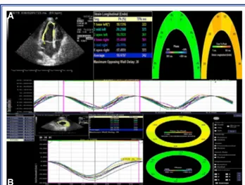
A Siemens Syngo Workplace with VVI software (Siemens Healthineers AG, Erlangen, Germany) was used to perform 2D-STI. Digital cineloops from the apical 4- and 2-chamber view and parasternal short--axis view were obtained at a frame rate of 50–90 frames/second from the peak of the R wave and stored on optical disks in the Digital Imaging and Communi-cations in Medicine format for offline analysis. All of the analyses were performed by a single operator who was blinded to all clinical data. Echocardiographic cineloops were obtained by recording 3 consecutive heart cycles. The endocardial and epicardial borders were manually traced in the end-systolic frame for each analysis, and the software tracked the margins frame by frame automatically thereafter. Careful in-spection of the tracking was conducted and manual corrections were performed if needed. In the event of unsatisfactory tracking, the segment was excluded from the analysis. Myocardial systolic strain of the LV can be assessed in 3 orthogonal directions: lon-gitudinal, circumferential, and radial strain (Fig. 2a, b). Global longitudinal and circumferential strains are expressed as negative values; a more negative value indicates good function, while a less negative value reveals impaired function. Global radial strain is ex-pressed as a positive value. The endocardial border of the RV free wall and septal wall was manually traced in the apical 4-chamber view. The RV longitudinal strain was calculated (Fig. 3).
Statistical analysis
All of the analyses were performed using SPSS Statis-tics for Windows, Version 17.0 (SPSS Inc., Chicago, IL, USA). The Shapiro-Wilk test was used to deter-mine the consistency of continuous variables with normal distribution. Homogeneity of variance was analyzed with the Levene test. Variables that had a nonhomogeneous distribution were compared using the Mann-Whitney U test. Student’s t-test was used to analyze the normally distributed variables and ho-mogeneous variables. The results were expressed as mean±SD and median value. Fisher’s exact test was
used to analyze categorical variables, which were ex-pressed as absolute values and percentages. P<0.05 was considered statistically significant.
Figure 1. Color-coded tissue Doppler imaging of the apical 4-chamber view demonstrating the systolic (Sa), early (Ea), and late (Aa) diastolic tissue velocities of mitral and tricuspit annular sites.
Figure 2. (A) Left ventricular longitudinal strain curves from 6 segments of the apical 4-chamber view are seen. The curves are negative, representing the segmental shortening during systole; (B) A dotted curve illustrates the left ventric-ular circumferential strain of the mid short-axis cut plane.
A
B
Figure 3. Regions of interest located on the corresponding right ventricular segmental longitudinal strain curves. Note that the right ventricular longitudinal strain is negative be-cause it represents shortening during systole.
end-diastolic volume and end-systolic volume were higher in the liver transplant candidates than in the controls (Table 2). Additionally, the LV mass index measurement was higher in the liver transplant can-didates than in the controls. The Ea and Ea/Aa ratio were significantly lower in the liver transplant candi-dates when compared with the control group and the E/Ea ratio, as a means of LV end-diastolic pressure, was greater in the liver transplant candidates than the controls. The Sa was significantly lower in the liver transplant candidates than in the controls at the lat-eral annulus and the septal annulus. The LV mean RESULTS
The groups were similar with regard to age and gen-der. A comparison of the clinical and biochemical pa-rameters of the study groups is presented in Table 1. The mean systolic and diastolic blood pressure was significantly lower in the liver transplant candidates than in the controls. The mean cholesterol level was lower in the liver transplant candidates (Table 1).
The 2D and Doppler echocardiographic measure-ments are presented in Table 2. There was no signif-icant difference in the EF between groups, but the
Table 2. Left ventricle 2-dimensional and Doppler echocardiographic measurements
Cirrhosis Control p
(n=40) (n=26)
Left ventricle end-diastolic diameter (cm) 4.7±0.3 4.5±0.3 0.001
Septum wall thickness (cm) 1.07±0.10 0.99±0.09 0.003
Left ventricle posterior wall thickness (cm) 1.06±0.10 0.99±0.09 0.006
Left atrial diameter (cm) 3.8±0.4 3.4±0.3 <0.001
Left ventricle end-diastolic volume (mL) 107.2±18 89.9±15.6 <0.001
Left ventricle end-systolic volume (mL) 45.5±9.3 35.1±8.3 <0.001
Left ventricle ejection fraction (%) 59.7±2.7 60.5±2.8 0.002
Early/late diastolic velocity 1.2±0.3 1.2±0.3 0.009
Mitral deceleration time (ms) 215.3±31.4 206±38.2 0.004
Isovolumic relaxation time (ms) 106.6±16.2 106.1±9.0 0.009
Left ventricle mass index (g/m2) 106.7±16.8 88.5±11.3 <0.001
Table 1. Baseline clinical charecteristics and biochemical parameters of the groups
Cirrhosis Control p
(n=40) (n=26)
Age (years) 46.2±10.1 42.2±8.6 0.001
Male/female 28/12 16/10 0.005
Body surface area (m2) 1.8±0.1 1.8±0.2 0.008
Systolic blood pressure (mmHg) 109±11 118±8 0.003
Diastolic blood pressure (mmHg) 67±7 72±8 0.001
Heart rate (beats/min) 73±11 74±8 0.007
Laboratory findings
Fasting glucose (mg/dL) 97±14 90±8 0.007
Creatinine (mg/dL) 0.7±0.2 0.8±0.2 0.006
Total cholesterol (mg/dL) 114±32 155±66 0.001
High-density lipoprotein cholesterol (mg/dL) 35±13 49±13 0.001
Low-density lipoprotein cholesterol (mg/dL) 63±20 109±20 <0.001
longitudinal strain was significantly lower in the liver transplant candidates when compared with the control group in both the apical 4- and 2-chamber views. The LV mean circumferential strain was not significantly different between the groups. However, the LV mean radial strain was significantly lower in the liver trans-plant candidates than in the controls (Table 3).
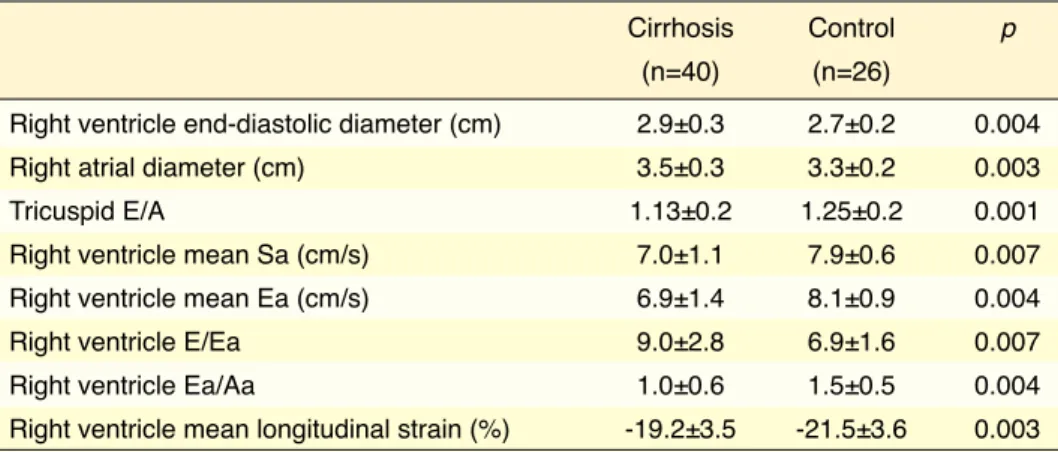
The RV diastolic diameter and right atrium diame-ter were greadiame-ter in the liver transplant candidates than in the controls (Table 4). The RV Ea velocity and Ea/ Aa ratio were significantly lower in the liver trans-plant candidates than in the controls, and the E/Ea
ratio, as a means of RV end-diastolic pressure, was higher in the liver transplant candidates compared with the controls. The RV mean Sa velocity and RV mean longitudinal strain were significantly lower in the liver transplant candidates than in the controls. Furthermore, the MPI value of the RV was greater in the liver transplant candidates than in the controls (Table 4).
DISCUSSION
STI is a newer imaging modality able to objectively quantify regional and global biventricular functions.
Table 4. Right ventricle echocardiographic measurements
Cirrhosis Control p
(n=40) (n=26)
Right ventricle end-diastolic diameter (cm) 2.9±0.3 2.7±0.2 0.004
Right atrial diameter (cm) 3.5±0.3 3.3±0.2 0.003
Tricuspid E/A 1.13±0.2 1.25±0.2 0.001
Right ventricle mean Sa (cm/s) 7.0±1.1 7.9±0.6 0.007
Right ventricle mean Ea (cm/s) 6.9±1.4 8.1±0.9 0.004
Right ventricle E/Ea 9.0±2.8 6.9±1.6 0.007
Right ventricle Ea/Aa 1.0±0.6 1.5±0.5 0.004
Right ventricle mean longitudinal strain (%) -19.2±3.5 -21.5±3.6 0.003
Aa: Late diastolic color-coded tissue velocity; A: Late diastolic velocity; Ea: Early diastolic color-coded tissue velocity; E: Early diastolic velocity; Sa: Peak systolic color-coded tissue velocity.
Table 3. Left ventricle color-coded tissue Doppler imaging and strain measurements
Cirrhosis Control p
(n=40) (n=26)
Left ventricle septal Sa (cm/s) 7.1±1.1 7.6±0.7 0.004
Left ventricle lateral Sa (cm/s) 7.1±1.5 8.1±0.7 0.008
Left ventricle mean annular Sa (cm/s) 7.1±1.0 7.8±0.5 0.004
Left ventricle septal Ea (cm/s) 6.0±2.1 7.2±1.9 0.001
Left ventricle lateral Ea (cm/s) 5.3±2.3 7.7±1.4 <0.001
Left ventricle mean annular Ea (cm/s) 5.6±2.0 7.4±1.1 <0.001
Left ventricle E/Ea 18.0±9 10.5±2 <0.001
Left ventricle Ea/Aa 0.9±0.3 1.4±0.6 <0.001
Left ventricle mean longitudinal strain (%)
Apical 4-chamber -16.0±3.2 -17.6±2.2 0.003
Apical 2-chamber -16.2±3.3 -18.7±2.1 0.002
LV mean circumferential strain (%) -23.4±3.2 -24.5±3.0 0.001
LV mean radial strain (%) 45.4±10.7 52.7±10.8 0.001
Aa: Late diastolic color-coded tissue velocity; Ea: Early diastolic color-coded tissuevelocity; Sa: Peak systolic color-coded tissue velocity.
Our study found an increase in RV and right atrium diameters in patients with ESLD compared with the controls, similar to the results of other studies mea-suring with echocardiography.[18,19] This could be explained by an increased venous return to the right heart caused by the development of portosystemic collaterals to counterbalance the increased intrahep-atic vascular resistance to portal blood flow.[10,20] In our study, liver transplant candidates had a signif-icantly higher RV MPI. MPI, which is an index of sensitive and load-independent cardiac dysfunction, shows subtle RV systolic and/or diastolic abnormal-ities.[21–23] Similarly, the diastolic and systolic indices of cTDI echocardiography revealed impairment of RV function in patients with ESLD. These findings suggest impaired diastolic and systolic RV function and increased RV filling pressure in liver transplant candidates with ESLD. Hepato-pulmonary syndrome and pulmonary hypertension may adversely affect RV myocardial function; therefore, we excluded these conditions. Additionally, the RV mean longitudinal strain was significantly lower in the liver transplant candidates. This subclinical impairment of systolic RV function in liver transplant candidates with ESLD can be detected using VVI strain imaging. Detailed evaluation of RV functions in these patients can be very important because studies have shown that RV functions are superior to LV functions in predicting poor clinical outcomes in these patients.[24]
There are several limitations to this study that should be taken into consideration. Our control group was relatively small. The analyses performed require offline processing and cannot be performed during live study. It was ideal to evaluate systolic function in patients before and after liver transplantation, but echocardiography was not performed after liver transplantation. Only 2D-echocardiographic param-eters were used to evaluate function. Novel 3D-s-train echocardiography was not used. For longitudi-nal strain, we alongitudi-nalyzed data from the apical 4- and 2-chamber views. Additional analysis of the apical long-axis view provides optimal information for lon-gitudinal strain. All of the images were analyzed by a single researcher, but intra-observer variability was not recorded at the time of the study. RV function was not evaluated using methods such as tricuspid annular plane systolic excursion, 2D fractional area change, and 3D EF measurement.
We evaluated LV and RV function in liver transplant candidates with ESLD using VVI, a 2D-strain imag-ing method based on speckle trackimag-ing.
The EF measurement was similar in the 2 groups. We determined that impairment of systolic LV func-tion in patients with ESLD could be more sensitively detected with STI than conventional echocardiogra-phy. Previous studies have indicated that STI is more sensitive than conventional echocardiography in de-tecting subclinical ventricular dysfunction in various clinical disorders.[7–10] Our results showed that this subclinical impairment was mostly related to longi-tudinal and radial rather than circumferential defor-mation mechanics, which is consistent with the early stages of myocardial damage. Global longitudinal strain is first affected because the longitudinally ori-entated subendocardial fibers are most susceptible to damage.[11] Sub-endocardial fibers generate the most significant contribution to longitudinal function. Global longitudinal strain also supports a prognostic evaluation with a good clinical value for predicting heart failure.[12] Additionally, it has been shown that global longitudinal strain is superior to LV EF and all other conventional echocardiographic parameters in predicting mortality.[13] The middle and sub-epicardial layers further contribute to thickening and to radial and circumferential function. These fibers are a key determinant of preserved global LV function.[14] This may explain why circumferential deformation was not reduced in our study. Furthermore, we found that although the EF of these patients was normal, the Sa was seen to be reduced with cTDI. This finding sup-ports the presence of subclinical myocardial dysfunc-tion. Previous studies have shown that LV diastolic dysfunction is common in patients with cirrhosis. [2,15,16] In our study, we identified diastolic dysfunction on cTDI echocardiography in patients with ESLD. We also found a greater LV mass index value, suggesting diastolic dysfunction in these patients.[17] This impair-ment cannot be detected with conventional Doppler, possibly due to hypervolemia and hyperdynamic cir-culation in these patients.
All invasive and noninvasive imaging techniques that can evaluate the structure and functions of the RV have important limitations due to the complex RV geometry. Volume calculation and estimation of EF are not ideal for the assessment of RV function. Little is known about the effects of ESLD on RV function.
cation: a report from the American Society of Echocardiogra-phy’s Guidelines and Standards Committee and the Chamber Quantification Writing Group, developed in conjunction with the European Association of Echocardiography, a branch of the European Society of Cardiology. J Am Soc Echocardiogr 2005;18:1440–63. [CrossRef]
7. Chen J, Cao T, Duan Y, Yuan L, Wang Z. Velocity vector imaging in assessing myocardial systolic function of hyper-tensive patients with left ventricular hypertrophy. Can J Car-diol 2007;23:957–61. [CrossRef]
8. Jansen C, Cox A, Schueler R, Schneider M, Lehmann J, Prak-tiknjo M, et al. Increased myocardial contractility identifies patients with decompensated cirrhosis requiring liver trans-plantation. Liver Transpl 2018;24:15–25. [CrossRef]
9. Altekin RE, Caglar B, Karakas MS, Ozel D, Deger N, Demir I. Evaluation of Subclinical Left Ventricular SystolicDysfunc-tion Using Two-Dimensional Speckle-Tracking Echocardio-graphy in Patients with Non-Alcoholic Cirrhosis. Hellenic J Cardiol 2014;55:402–10.
10. Chen Y, Chan AC, Chan CS, Chok CS, Sharr W, Fung J, et al. A detailed evaluation of cardiac function in cirrhotic patients and its alteration with or without liver transplantation Journal of Cardiol 2016;67:140–6. [CrossRef]
11. Nesbitt GC, Mankad S, Oh JK. Strain imaging in echocardio-graphy: methods and clinical applications. Int J Cardiovasc Imaging 2009;25:9–22. [CrossRef]
12. Ersbøll M, Valeur N, Mogensen UM, Andersen MJ, Møller JE, Hassager C, et al. Relationship between left ventricu-lar longitudinal deformation and clinical heart failure dur-ing admission for acute myocardial infarction: a two-di-mensional speckle-tracking study. J Am Soc Echocardiogr 2012;25:1280–9. [CrossRef]
13. Sengeløv M, Jørgensen PG, Jensen JS, Bruun NE, Olsen FJ, Fritz-Hansen T, et al. Global Longitudinal Strain Is a Supe-rior Predictor of All-Cause Mortality in Heart Failure With Reduced Ejection Fraction. JACC Cardio Vascular Imaging 2015;8:1351–59. [CrossRef]
14. Hung CL, Verma A, Uno H, Shin SH, Bourgoun M, Hassanein AH, et al. Longitudinal and circumferential strain rate, left ventricular remodeling, and prognosis after myocardial in-farction. J Am Coll Cardiol 2010;56:1812–22. [CrossRef] 15. Liu H, Gaskari SA, Lee SS. Cardiac and vascular changes
in cirrhosis: pathogenic mechanisms. World J Gastroenterol 2006;12:837–42. [CrossRef]
16. Hammami R, Boudabbous M, Jdidi J, Trabelsi F, Mroua F, Kallel R, et al. Cirrhotic cardiomyopathy: is there any correla-tion between the stage of cardiac impairment and the severity of liver disease? Libyan J Med 2017;12:1283162 [CrossRef] 17. Hinderliter AL, Light KC, Willis PW 4th. Left ventricular
mass index and diastolic filling. Relation to blood pressure and demographic variables in a healthy biracial sample. Am J Hypertens1991;4:579–85. [CrossRef]
18. Karabulut A, Iltumur K, Yalcin K, Toprak N.
Hepatopul-Conclusion
STI has gained wide clinical use in recent years and 2D-STI allows for rapid and accurate analysis of systolic ventricular function in the longitudinal, ra-dial, and circumferential directions. The technique is able to detect subtle changes in myocardial function at an early stage, even before there is a visible de-crease in conventional echocardiographic parameters. Systolic functions appeared to be normal in cirrhotic cardiomyopathy when assessed by 2D- and conven-tional Doppler echocardiography, possibly due to hypervolemia and hyperdynamic circulation in these patients at rest. This study demonstrated that liver transplant candidates with ESLD had impaired LV and RV function measured at an early stage. VVI 2D-strain imaging and cTDI are sensitive tools that can detect subclinical LV systolic and diastolic func-tion in liver transplant candidates when convenfunc-tional echocardiography measurements at rest is normal.
Ethics Committee Approval: The study protocol was
ap-proved by the Başkent University Clinical Research Ethics Committee (KA08/145).
Peer-review: Externally peer-reviewed. Conflict-of-interest: None.
Authorship contributions: Concept: B.P., S.D.İ.; Design:
L.E.S., B.P., S.D.İ.; Supervision: H.M.; Materials: S.D.İ.; Data: S.D.İ.; Analysis: L.E.S., S.D.İ.; Literature search: H.E.P., S.D.İ.; Writing: S.D.İ.; Critical revision: S.Y., C.A., L.E.S.
REFERENCES
1. Lee SS. Cardiac abnormalities in liver cirrhosis. West J Med 1989;151:530–5.
2. Gassanov N, Caglayan E, Semmo N, Massenkeil G, Er F. Cir-rhotic cardiomyopathy: a cardiologist’s perspective. World J Gastroenterol 2014;20:15492–8. [CrossRef]
3. Wong F, Girgrah N, Graba J, Allidina Y, Liu P, Blendis L. The cardiac response to exercise in cirrhosis. Gut 2001;49:268– 75. [CrossRef]
4. Moller S, Henriksen JH. Cirrhotic cardiomyopathy: a patho-physiological review of circulatory dysfunction in liver dis-ease. Heart 2002;87:9–15. [CrossRef]
5. Cahill PA, Redmond EM, Rodges R, Zhang S, Sitzmann JV. Increased endothelial nitric oxide synthase activity in the hyperemic vessels of portal hypertensive rats. J Hepatol 1996;25:370–8. [CrossRef]
6. Lang RM, Bierig M, Devereux RB, Flachskampf FA, Foster E, Pellikka PA, et al. Recommendations for chamber
quantifi-monary syndrome and right ventricular diastolic functions: an echocardiographic examination. Echocardiography 2006;23:271–8. [CrossRef]
19. Lo´pez-Candales A, Menendez FL, Shah SA, Friedrich A. Measures of right ventricular systolic function in end stage liver disease patients awaiting transplant. Int J Cardiol 2014;171:277–8. [CrossRef]
20. Zardi EM, Uwechie V, Caccavo D, Pellegrino NM, Caccia-paglia F, Di Matteo F, et al. Portosystemic shunts in a large cohort of patients with liver cirrhosis: detection rate and clin-ical relevance. J Gastroenterol 2009;44:76–83. [CrossRef] 21. Meluzín J, Spinarová L, Bakala J, Toman J, Krejcí J, Hude P,
et al. Pulsed Doppler tissue imaging of the velocity of tricus-pid annular systolic motion; a new, ratricus-pid, and non-invasive method of evaluating right ventricular systolic function. Eur Heart J 2001;22:340–8. [CrossRef]
Keywords: Echocardiography; end-stage liver disease; strain. Anahtar sözcükler: Ekokardiyografi, son dönem karaciğer hastalığı; strain.
22. Miller D, Farah MG, Liner A, Fox K, Schluchter M, Hoit BD. The relation between quantitative right ventricular ejection fraction and indices of tricuspid annular motion and myocar-dial performance. J Am Soc Echocardiogr 2004;17:443–7. 23. William V, El Kilany W. Assessment of right ventricular
func-tion by echocardiography in patients with chronic heartfail-ure. Egypt Heart J 2018;70:173–9. [CrossRef]
24. Kia L, Shah SJ, Wang E, Sharma D, Selvaraj S, Medina C et al. Role of pretransplant echocardiographic evaluation in predicting outcomes following liver transplantation. Am J Transplant 2013;13:2395–401. [CrossRef]