Global evaluation of echocardiography in
patients with COVID-19
Marc R. Dweck
1*, Anda Bularga
1, Rebecca T. Hahn
2, Rong Bing
1,
Kuan Ken Lee
1, Andrew R. Chapman
1, Audrey White
1, Giovanni Di Salvo
3,
Leyla Elif Sade
4, Keith Pearce
5, David E. Newby
1, Bogdan A. Popescu
6,
Erwan Donal
7, Bernard Cosyns
8, Thor Edvardsen
9,10, Nicholas L. Mills
1,11†, and
Kristina Haugaa
9,10†1
Centre for Cardiovascular Science, University of Edinburgh, UK;2Columbia University Irving Medical Center, NY, USA;3University Hospital Padua, Paediatric Cardiology, Padua, Italy;4
Department of Cardiology, University of Baskent, Ankara, Turkey;5
University Hospital South Manchester, Cardiology, Wythenshawe, Manchester, UK;6
Department of Cardiology, University of Medicine and Pharmacy ‘Carol Davila’-Euroecolab, Emergency Institute for Cardiovascular Diseases ‘Prof. Dr. C. C. Iliescu’, Bucharest, Romania; 7
University of Rennes, CHU Rennes, Inserm, LTSI-UMR 1099, Rennes, France;8
Centrum voor Hart en Vaatziekten, Universitair Ziekenhuis Brussel, Vrij Universiteit van Brussel, Brussels, Belgium;9Department of Cardiology, Oslo University Hospital, Rikshospitalet, Oslo, Norway;10Faculty of Medicine, University of Oslo, Oslo, Norway; and11Usher Institute, University of Edinburgh, UK
Received 22 May 2020; editorial decision 26 May 2020; accepted 2 June 2020; online publish-ahead-of-print 18 June 2020
Aims To describe the cardiac abnormalities in patients with COVID-19 and identify the characteristics of patients who
would benefit most from echocardiography.
... Methods
and results
In a prospective international survey, we captured echocardiography findings in patients with presumed or confirmed COVID-19 between 3 and 20 April 2020. Patient characteristics, indications, findings, and impact of echocardiography on management were recorded. Multivariable logistic regression identified predictors of echocardiographic abnormalities. A total of 1216 patients [62 (52–71) years, 70% male] from 69 countries across six continents were included. Overall, 667 (55%) patients had an abnormal echocardiogram. Left and right ventricular abnormalities were reported in 479 (39%) and 397 (33%) patients, respectively, with evidence of new myocardial infarction in 36 (3%), myocarditis in 35 (3%), and takotsubo cardiomyopathy in 19 (2%). Severe cardiac disease (severe ventricular dysfunction or tamponade) was observed in 182 (15%) patients. In those without pre-existing cardiac disease (n = 901), the echocardiogram was abnormal in 46%, and 13% had severe disease. Independent predictors of left and right ventricular abnormalities were distinct, including elevated natriuretic pepti-des [adjusted odds ratio (OR) 2.96, 95% confidence interval (CI) 1.75–5.05) and cardiac troponin (OR 1.69, 95% CI 1.13–2.53) for the former, and severity of COVID-19 symptoms (OR 3.19, 95% CI 1.73–6.10) for the latter. Echocardiography changed management in 33% of patients.
... Conclusion In this global survey, cardiac abnormalities were observed in half of all COVID-19 patients undergoing
echocardiog-raphy. Abnormalities were often unheralded or severe, and imaging changed management in one-third of patients.
䊏 䊏 䊏 䊏 䊏 䊏 䊏 䊏 䊏 䊏 䊏 䊏 䊏 䊏 䊏 䊏 䊏 䊏 䊏 䊏 䊏 䊏 䊏 䊏 䊏 䊏 䊏 䊏 䊏 䊏 䊏 䊏 䊏 䊏 䊏 䊏 䊏 䊏 䊏 䊏 䊏 䊏 䊏 䊏 䊏 䊏 䊏 䊏 䊏 䊏 䊏 䊏 䊏 䊏 䊏 䊏 䊏 䊏 䊏 䊏 䊏 䊏 䊏 䊏 䊏 䊏 䊏 䊏 䊏 䊏 䊏 䊏 䊏 䊏 䊏 䊏 䊏 䊏 䊏 䊏 䊏 䊏 䊏 䊏 䊏 䊏 䊏 䊏 䊏 䊏 䊏 䊏 䊏 䊏 䊏 䊏 䊏 䊏 䊏 䊏 䊏 䊏 䊏 䊏 䊏 䊏 䊏 䊏 䊏 䊏 䊏 䊏 䊏 䊏 䊏 䊏 䊏 䊏 䊏 䊏 䊏 䊏 䊏 䊏 䊏 䊏 䊏 䊏 䊏 䊏 䊏 䊏 䊏 䊏 䊏 䊏 䊏 䊏 䊏 䊏 䊏 䊏 䊏 䊏 䊏 䊏 䊏 䊏 䊏 䊏 䊏 䊏 䊏 䊏 䊏 䊏 䊏 䊏 䊏 䊏 䊏 䊏 䊏 䊏 䊏 䊏 䊏 䊏 䊏 䊏 䊏 䊏 䊏 䊏 䊏 䊏 䊏 䊏 䊏 䊏 䊏 䊏 䊏 䊏 䊏 䊏 䊏 䊏 䊏 䊏 䊏 䊏 䊏 䊏 䊏 䊏 䊏 䊏 䊏 䊏 䊏 䊏 䊏 䊏 䊏 䊏 䊏 䊏 䊏 䊏 䊏 䊏
Keywords COVID-19
•
Echocardiography* Corresponding author. BHF/University Centre for Cardiovascular Science, The University of Edinburgh, Chancellor’s Building, 49 Little France Crescent, Edinburgh EH16 4SB, UK, Email: [email protected]
†
These authors contributed equally to this work.
VCThe Author(s) 2020. Published by Oxford University Press on behalf of the European Society of Cardiology.
This is an Open Access article distributed under the terms of the Creative Commons Attribution License (http://creativecommons.org/licenses/by/4.0/), which permits unrestrict-ed reuse, distribution, and reproduction in any munrestrict-edium, providunrestrict-ed the original work is properly citunrestrict-ed.
..
..
..
..
..
..
..
..
..
..
..
..
..
..
..
..
..
..
..
..
..
..
..
..
..
..
..
..
..
..
..
..
..
..
..
..
..
..
..
..
..
..
..
..
..
..
..
..
..
..
..
..
..
..
..
..
..
..
..
..
..
..
..
..
..
..
..
..
..
..
..
..
..
..
..
..
..
..
..
..
..
..
..
..
..
..
.
Introduction
Coronavirus disease 2019 (COVID-19) has emerged as a major cause of morbidity and mortality that is placing unprecedented
pres-sure on healthcare services across the world.1,2Whilst the severe
acute respiratory syndrome coronavirus 2 (SARS-CoV-2)
respon-sible for COVID-19 predominantly affects the respiratory tract,3
patients with cardiovascular risk factors or established disease4and
those with elevated cardiac biomarkers appear to be more
suscep-tible and to have a worse prognosis.5,6The mechanisms underlying
these initial observations remain unclear.
Early case reports suggest that COVID-19 can cause a wide range
of cardiac conditions that include acute myocardial infarction,7
myo-carditis,8and takotsubo cardiomyopathy.9Acute left and right
ven-tricular failure may be a direct consequence of cardiac pathology, with the latter also arising secondary to elevations in right ventricular
afterload due to pulmonary embolism or pneumonia.10Virus
par-ticles have been observed in the myocardium and vascular
endothe-lium in patients with COVID-19 and cardiogenic shock.11,12
However, the incidence of these cardiac complications and the sub-sequent implications for treatment and resource allocation are un-known. Consequently, there is an urgent need to better understand the interactions between COVID-19 and the heart.
Echocardiography is well placed to help further this understanding, being inexpensive, portable, and widely accessible. However, a large systematic evaluation of echocardiography in all patients with COVID-19 would be highly challenging due to the logistical consider-ations of testing, consumption of personal protective equipment (PPE), and the risk of further viral transmission. We therefore con-ducted a global survey to capture the findings of echocardiography performed on clinical grounds in patients with confirmed or a high probability of COVID-19. We aim to improve our understanding of the cardiac manifestations of COVID-19, and to provide insights into the characteristics of patients who would benefit most from echocardiography.
Methods
This global online survey of echocardiography in patients with COVID-19 was designed by the European Association of Cardiovascular Imaging (EACVI), with input from external international experts.13 Any trans-thoracic echocardiogram that was performed on a patient with con-firmed, or a high probability of, COVID-19 in the hospital setting was eligible for inclusion. Patients were imaged as part of routine care, and non-identifiable patient data were captured. As such, this audit did not re-quire individual patient consent and this approach was approved by the European Society of Cardiology and by local research ethics committees. An online format was developed (https://www.surveymonkey.com/r/ 2FBFFQD) that allows rapid completion of 11 questions on a smartphone by sonographers or clinicians immediately after completion of the echo-cardiogram. For most questions, the operator selects from several pre-specified answers, with the option to select multiple answers and to pro-vide free-text comments (Supplementary material online, Appendix). First, several baseline characteristics are recorded: age, sex, comorbidities, symptom severity, COVID-19 status, presence of pneumonia, and the lo-cation in hospital where imaging was performed. Secondly, the indilo-cation for imaging is recorded: suspected left heart failure, suspected right heart failure, chest pain with ST-segment elevation on the electrocardiogram,
cardiac biomarker elevation [troponin or brain natriuretic peptide (BNP)], ventricular arrhythmia, suspected tamponade, or cardiogenic shock. Thirdly, echocardiographic findings are captured for left ventricu-lar abnormalities (normal, mild, moderate, or severe systolic dysfunction, dilatation, evidence of new myocardial infarction, myocarditis, or takot-subo cardiomyopathy), right ventricular abnormalities (normal, mild or moderate, or severe systolic dysfunction, dilatation, D-shaped left ven-tricle, or elevated pulmonary artery pressure), or cardiac tamponade. The survey then captures whether the echocardiogram changed patient management.
This prospective survey (www.escardio.org/eacvi/surveys) was distrib-uted to the EACVI network and its wider membership,14,15to a pre-established European Society of Cardiology database of cardiologists with an interest in cardiac imaging, and to the presidents and chairpersons of national societies and working groups in imaging across the world. It was also distributed widely on social media platforms.
Statistical analysis
Survey entries were excluded if there were incompatible, incom-plete, or conflicting data, or if there were no echocardiographic find-ings recorded. In this analysis, missing values were not imputed. Age was reported as median with an interquartile interval, and categorical variables were reported as frequencies (%). Between-group
compari-sons were performed using the v2test or an independent samples
t-test. In the primary analysis, patients with either confirmed or prob-able COVID-19 were included. A sensitivity analysis was performed restricted to those with confirmed COVID-19. A critical care setting was defined as intensive care, high dependency, or coronary care units, the emergency department, or the cardiac catheterization la-boratory. A normal echocardiogram was defined as normal left and right ventricular function, with no other reported abnormalities. To evaluate associations between clinical variables and cardiac abnor-malities that were more likely to be due to COVID-19, an analysis was performed in patients without pre-existing cardiac disease, after excluding those with previous ischaemic heart disease, heart failure, or valvular heart disease. Univariable and multivariable logistic regres-sion models were constructed separately with an abnormal left ven-tricle (any degree of left ventricular dysfunction or dilatation, myocardial infarction, myocarditis, or takotsubo cardiomyopathy) or abnormal right ventricle (any right ventricular dysfunction or dilata-tion, a D-shaped left ventricle, or pulmonary hypertension) as the de-pendent variables. Covariates included age, gender, scan location, symptom severity, hypertension and diabetes mellitus, and the indica-tion for echocardiography. Analysis was performed using R version 3.5.0 (R Foundation for Statistical Computing, Vienna, Austria).
Results
The survey was launched on 3 April 2020. The results reported here comprise data from the first 17 days of the survey, with the last date of collection on 20 April 2020. Data from 1272 patients undergoing echocardiography were collected from 69 countries across six
conti-nents where COVID-19 has been reported (Figure1). Data were
available for analysis in 1216 (96%) patients [62 (52–71) years, 70% male], of whom 813 (73%) had confirmed COVID-19, and 298 (27%)
had a high probability at the time of scanning (Table1).
..
..
..
..
..
..
..
..
..
..
..
..
..
..
..
..
..
..
..
..
..
Overall, 60% of scans were performed in a critical care setting (54% intensive care, 2% high dependency unit and coronary care unit, 5% emergency room, and 1% cardiac catheter laboratory), with the remainder performed in general medicine, cardiology, respiratory,
and dedicated COVID-19 wards (Table1). Correspondingly, 54% of
patients had severe symptoms and 19% had evidence of pneumonia. Pre-existing cardiac disease was reported in 26% of patients due to a combination of ischaemic heart disease (14%), heart failure (9%), or valvular heart disease (7%). Hypertension (37%) and diabetes mellitus (19%) were also common. The most common indications for echo-cardiography were suspected left-sided heart failure (40%), elevated cardiac biomarkers (26%), and right-sided heart failure (20%). Chest pain with ST-segment elevation on the electrocardiogram (9%), cir-culatory shock (8%), ventricular arrhythmia (3%), and suspected
cardiac tamponade (2%) were less frequent, as were other indica-tions, such as suspected pulmonary embolism (5%), endocarditis (6%), and myocarditis (1%).
Echocardiographic findings
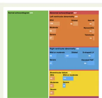
Compared with patients with a normal echocardiogram (n = 549, 45%), patients with an abnormal scan (n = 667, 55%) were older and had a higher prevalence of pre-existing ischaemic heart disease, heart failure, or valvular heart disease, but a similar prevalence of hypertension or diabetes mellitus. The proportion
of males was similar in both groups (Table 1; Figure 2, Central
Illustration).
Figure 1Prevalence of COVID-19 and countries contributing to the global online survey of echocardiography. (A) The prevalence of COVID-19 on 20 April for the 185 countries for which data were available through the Johns Hopkins Center for Systems Science and Engineering COVID-19 dashboard.25(B) The location and number of scans reported in the global online survey of echocardiography during a 17-day period from 3 to 20 April 2020.
..
..
..
..
..
..
..
..
..
..
..
..
Left ventricular abnormalities were reported in 479 (39%) patients, with echocardiographic evidence of new myocardial infarc-tion in 36 (3%), myocarditis in 35 (3%), and takotsubo cardiomyop-athy in 19 (2%). Left ventricular impairment was classified as mild, moderate, or severe in 17, 12, and 9% of patients, respectively. Right ventricular abnormalities were reported in 397 (33%) patients, with mild or moderate right ventricular impairment in 19% and severe im-pairment in 6%. Right ventricular dilatation (15%), elevated
pulmonary artery pressures (8%), and a D-shaped left ventricle (4%) were reported less frequently. Cardiac tamponade and endocarditis were reported in 11 (1%) and 14 (1%) patients, respectively. Severe cardiac disease, defined as severe left or right ventricular dysfunction or cardiac tamponade, was reported in 1 in 7 patients (n = 182, 15%; Supplementary material online,Table S1).
Abnormalities on the echocardiogram were more common in those where the indication for imaging was chest pain with
ST-...
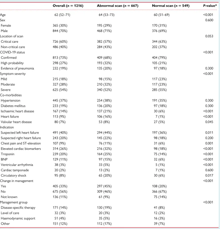
Table 1 Patient characteristics and indications for echocardiography
Overall (n 5 1216) Abnormal scan (n 5 667) Normal scan (n 5 549) P-value*
Age 62 (52–71) 64 (53–73) 60 (51–69) <0.001 Sex 0.600 Female 365 (30%) 195 (29%) 170 (31%) Male 844 (70%) 468 (71%) 376 (69%) Location of scan 0.053 Critical care 726 (60%) 382 (57%) 344 (63%) Non-critical care 486 (40%) 284 (43%) 202 (37%) COVID-19 status <0.001 Confirmed 813 (73%) 409 (68%) 404 (79%) High probability 298 (27%) 193 (32%) 105 (21%) Evidence of pneumonia 232 (19%) 135 (20%) 97 (18%) 0.300 Symptom severity <0.001 Mild 215 (18%) 98 (15%) 117 (23%) Moderate 327 (28%) 210 (32%) 117 (23%) Severe 625 (54%) 340 (52%) 285 (55%) Co-morbidities Hypertension 445 (37%) 254 (38%) 191 (35%) 0.300 Diabetes mellitus 233 (19%) 136 (20%) 97 (18%) 0.300
Ischaemic heart disease 167 (14%) 137 (21%) 30 (6%) <0.001
Heart failure 113 (9%) 106 (16%) 7 (1%) <0.001
Valvular heart disease 80 (7%) 53 (8%) 27 (5%) 0.045
Indication
Suspected left heart failure 491 (40%) 294 (44%) 197 (36%) 0.011 Suspected right heart failure 243 (20%) 145 (22%) 98 (18%) 0.200 Chest pain and ST-elevation 107 (9%) 76 (11%) 31 (6%) 0.001 Elevated cardiac biomarkers 314 (26%) 216 (32%) 98 (18%) <0.001
Troponin 239 (20%) 164 (25%) 75 (14%) <0.001 BNP 129 (11%) 97 (15%) 32 (6%) <0.001 Ventricular arrhythmia 38 (3%) 33 (5%) 5 (1%) <0.001 Cardiac tamponade 20 (2%) 13 (2%) 7 (1%) 0.600 Circulatory shock 95 (8%) 65 (20%) 30 (6%) 0.017 Change in management <0.001 Yes 405 (33%) 297 (45%) 108 (20%) No 675 (56%) 309 (46%) 366 (67%) Not known 136 (11%) 61 (9%) 75 (14%) Management group <0.001 Disease-specific therapy 171 (14%) 130 (19%) 41 (8%) Level of care 32 (3%) 20 (3%) 12 (2%) Haemodynamic support 51 (4%) 35 (5%) 16 (3%) Other 151 (12%) 112 (17%) 39 (7%)
Median (interquartile range), number (%). Abbreviations: BNP, brain B-type natriuretic peptide; COVID-19, coronavirus disease 2019. *Between-group comparisons are v2
test or independent samples t-tests
Missing values in the overall population: age = 18; sex = 7; location of scan = 4; COVID-19 status = 8; symptom severity = 49; indication = 9.
..
..
..
..
..
..
..
..
..
..
..
..
..
..
..
..
..
..
..
..
..
..
..
..
..
..
..
..
..
..
..
..
..
..
..
..
..
..
..
..
..
..
..
..
..
..
..
..
..
..
..
..
..
..
..
..
..
..
..
..
..
..
..
..
..
..
..
..
..
..
..
..
..
..
..
..
..
..
..
..
..
..
..
..
..
..
.
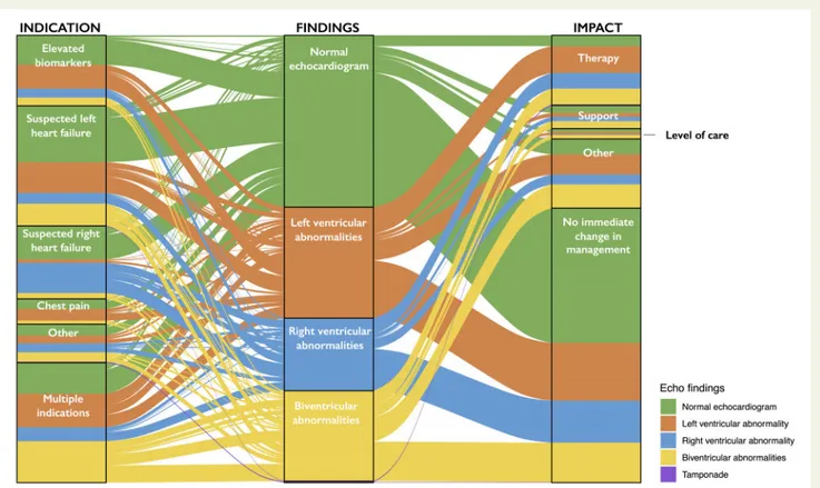
segment elevation (71%), elevated biomarkers (69%), suspected left ventricular failure (60%), suspected right ventricular failure (60%), or
where multiple indications were present (72%) (Table2; Figure3). In a
sensitivity analysis restricted to the 813 patients with confirmed COVID-19, the proportion with an abnormal echocardiogram was similar to the overall population at 50% (409/813), and 1 in 7 patients had severe cardiac disease (n = 119, 15%).
Patients without pre-existing
cardiac disease
After excluding 315 patients with pre-existing ischaemic heart dis-ease, heart failure, or valvular heart disdis-ease, 901 patients were
identi-fied [60 (50–69) years, 68% male] (Supplementary material online,
Table S2). These patients were more likely to have a normal echocar-diogram (54%, 488/901) than those with pre-existing heart disease (19%, 61/315; P < 0.001). Suspected left ventricular failure was the most common indication for scanning (35%), followed by suspected right heart failure (23%) and elevated cardiac biomarker concentra-tion (23%). A quarter of patients had an abnormal left ventricle and a third had an abnormal right ventricle. On both univariable and multi-variable analysis, the covariates and indications for echocardiography
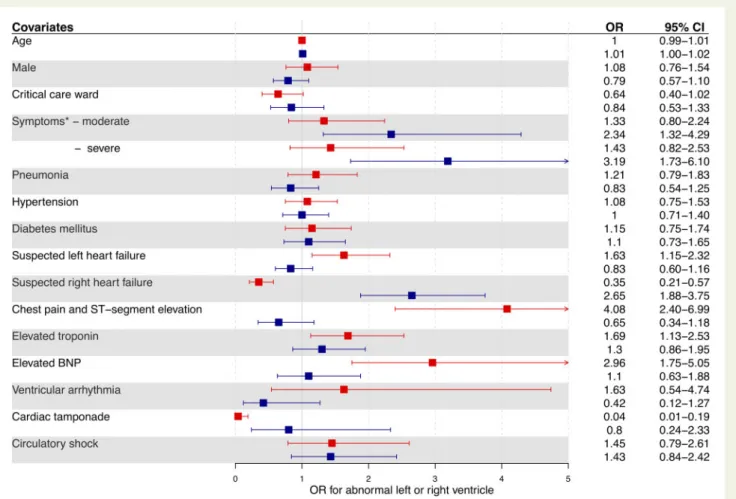
that predicted an abnormal left and right ventricle differed (Figure4;
Supplementary material online, Tables S3 and S4). The independent
predictors of an abnormal left ventricle were suspected left heart fail-ure [odds ratio (OR) 1.63, 95% confidence interval (CI) 1.15–2.32], chest pain with ST-segment elevation (OR 4.08, 95% CI 2.40–6.99), troponin elevation (OR 1.69, 95% CI 1.13–2.53), and BNP elevation (OR 2.96, 95% CI 1.75–5.05). In contrast, the independent predictors of an abnormal right ventricle were suspected right heart failure (OR 2.65, 95% CI 1.88–3.75) and moderate (OR 2.34, 95% CI 1.32–4.29) or severe COVID-19 symptoms (OR 3.19, 95% CI 1.73–6.10). Overall, 1 in 8 of these patients without pre-existing cardiac disease (13%) had severe cardiac disease identified on echocardiography.
Changes in management
In 405 (33%) patients, an immediate change in management due to
the echocardiogram was reported (Table 1). In the remaining 811
(67%) patients, no change in management was reported in 675 (56%), or it was not clear to the echocardiographer whether there had been a change in management in 136 (11%), as the individuals performing the scan were not directly involved in guiding the patients’ care. An immediate change in management occurred more often in those patients with an abnormal compared with a normal
echocar-diogram (45% vs. 20%, P < 0.001, Figure3) and similarly in those with
severe disease compared with those with no severe disease identified
on echocardiogram (59% vs. 29%, P < 0.001;Supplementary material
online,Table S1). Specific changes in management reported in the free-text comments were collated into four groups. In patients in whom a change in management was reported, the echocardiogram led to changes in disease-specific therapy in 42% (171/405), such as initiating therapy for heart failure, acute coronary syndrome, tampon-ade, or pulmonary embolism, and commencing antimicrobial therapy for endocarditis. The echocardiogram also facilitated decisions regarding changes in the level of patient care in 8% (32/405), and guided titration of haemodynamic support in 13% (51/405). In the remaining 37% (151/405) where management changed, the change was not described. Changes in management were reported in a higher proportion of patients with pre-existing cardiac disease com-pared with those without (38% vs. 32%, P = 0.005), and in those with elevated cardiac biomarkers compared with the remaining popula-tion (39% vs. 31%, P < 0.001).
Discussion
We report findings from the first international survey of echocardiog-raphy in patients with confirmed or suspected COVID-19. Data from 1216 patients scanned in 69 countries across six continents demon-strated left or right ventricular abnormalities in half of all patients with COVID-19 undergoing echocardiography, and that these abnor-malities were severe in 1 in 7 patients. The majority had non-specific patterns of ventricular dysfunction, although new myocardial infarc-tion, myocarditis, and takotsubo cardiomyopathy were observed in a minority of patients. Echocardiography was reported to directly change patient management in a third of cases including alterations to disease-specific management, haemodynamic support, and the level of care received by the patients.
The simple online format of this survey allowed rapid capture of the echocardiographic findings from a large number of patients with
Figure 2Central Illustration. Mosaic plot illustrating the findings on echocardiography in patients with COVID-19.
Mosaic plot illustrating the distribution of normal and abnormal echocar-diogram findings in patients with suspected or confirmed COVID-19 infec-tion. Box size is proportional to the number of patients per category. Left ventricular abnormalities are shown in orange and right ventricular abnormalities are shown in blue, both in the independent boxes and in the biventricular failure box. Survey respondents could enter data for multiple categories of left or right ventricular abnormality, therefore sub-categories are not mutually exclusive. Eleven patients had evidence of cardiac tamponade which was an isolated finding in three patients, illus-trated in purple. LV, left ventricle; MI, myocardial infarction; PAP, pulmon-ary arterial pressure.
..
..
..
..
..
..
..
..
..
..
..
..
..
..
..
..
..
..
..
..
..
..
..
..
..
..
..
..
..
..
..
..
..
..
..
..
.
COVID-19 during the pandemic’s peak. This was facilitated by our ability to disseminate and to publicize the survey via social media and through an established global network of imaging specialists. This for-mat allowed us to keep pace with the rapid spread of COVID-19 around the world. Most scans were performed in the current epi-centres of the outbreak: the UK, Italy, Spain, France, and the USA. While undoubtedly a global survey, our data remain representative of the current geographical distribution of the virus.
Whilst our previous understanding of how COVID-19 affects the
heart was limited to case reports and case series,7–9consistent
epi-demiological data have demonstrated that patients with established cardiovascular disease, risk factors, or elevated cardiac biomarkers have an increased susceptibility to infection and an increased risk of
severe disease and death.3–6Severe cardiac disease was observed in
1 in 7 patients across the whole cohort and in 1 in 8 patients without pre-existing cardiac disease. This proportion rose to 1 in 5 when the indication for imaging included raised cardiac biomarkers. The pro-portion of abnormal echocardiograms and those demonstrating se-vere cardiac disease were similar after excluding patients with previously established cardiac disease (heart failure, valve disease, or ischaemic heart disease), suggesting that in this population the cardiac abnormalities relate to COVID-19 infection.
The pattern of cardiac injury observed in our survey appears to be consistent with the cardiovascular involvement observed in patients
with other severe viral respiratory infections.16–19Right ventricular
abnormalities were observed in a quarter of patients and were more common in patients with more severe symptoms of COVID-19. These are likely to reflect severe respiratory disease, including the viral pneumonia itself, as well as both clinical and subclinical
pulmon-ary thrombo-embolism.20 Left ventricular abnormalities were
pre-sent in a third of patients and were predominantly non-specific in nature. Further research is required to define the mechanism of this dysfunction as only occasionally were echocardiographic patterns consistent with myocardial infarction, myocarditis, or takotsubo car-diomyopathy. The latter conditions are often difficult to recognize during an isolated echocardiogram, particularly when performed in a critical care setting, and, as such, their true prevalence may have been underestimated.
In a third of patients who underwent echocardiography on clinical indication, imaging was reported to result in an immediate change in patient management. This included changes in disease-specific thera-pies, such as pericardiocentesis or therapy for heart failure, pulmon-ary embolism, or acute coronpulmon-ary syndromes. It also contributed to decisions regarding the level of patient care, such as the admission of patients to critical care, and the need for titration of haemodynamic support. In practice, this proportion may have been underestimated as echocardiographers may not have fully appreciated the conse-quences of their scan at the time of imaging. In addition, a majority of
Figure 3Alluvial plot illustrating the indications for echocardiography and impact on patient management grouped according to the scan findings. Colours represent findings reported on echocardiography. All patients for whom complete data were available for indication, findings, and change in manage-ment were included in this plot [89% (1080/1216)]. The elevated biomarker indication subgroup includes both indication for raised troponin and indication for ele-vated BNP. Changes in management (impact) include those where changes in disease-specific therapy, titration of haemodynamic support, or changes in the level of patient care were described.
...
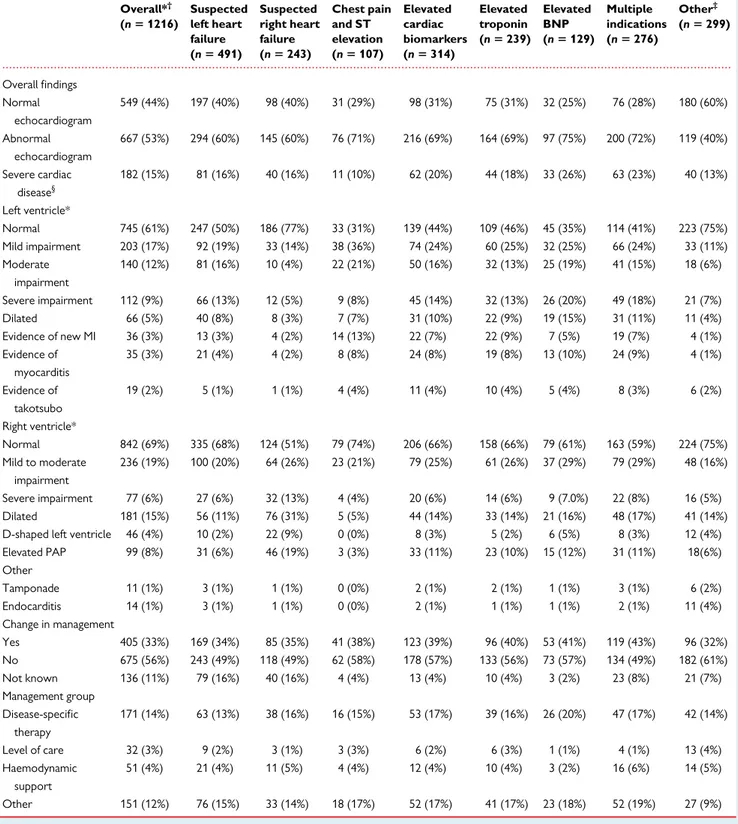
Table 2 Echocardiographic findings stratified by indication
Overall*† (n 5 1216) Suspected left heart failure (n 5 491) Suspected right heart failure (n 5 243) Chest pain and ST elevation (n 5 107) Elevated cardiac biomarkers (n 5 314) Elevated troponin (n 5 239) Elevated BNP (n 5 129) Multiple indications (n 5 276) Other‡ (n 5 299) Overall findings Normal echocardiogram 549 (44%) 197 (40%) 98 (40%) 31 (29%) 98 (31%) 75 (31%) 32 (25%) 76 (28%) 180 (60%) Abnormal echocardiogram 667 (53%) 294 (60%) 145 (60%) 76 (71%) 216 (69%) 164 (69%) 97 (75%) 200 (72%) 119 (40%) Severe cardiac disease§ 182 (15%) 81 (16%) 40 (16%) 11 (10%) 62 (20%) 44 (18%) 33 (26%) 63 (23%) 40 (13%) Left ventricle* Normal 745 (61%) 247 (50%) 186 (77%) 33 (31%) 139 (44%) 109 (46%) 45 (35%) 114 (41%) 223 (75%) Mild impairment 203 (17%) 92 (19%) 33 (14%) 38 (36%) 74 (24%) 60 (25%) 32 (25%) 66 (24%) 33 (11%) Moderate impairment 140 (12%) 81 (16%) 10 (4%) 22 (21%) 50 (16%) 32 (13%) 25 (19%) 41 (15%) 18 (6%) Severe impairment 112 (9%) 66 (13%) 12 (5%) 9 (8%) 45 (14%) 32 (13%) 26 (20%) 49 (18%) 21 (7%) Dilated 66 (5%) 40 (8%) 8 (3%) 7 (7%) 31 (10%) 22 (9%) 19 (15%) 31 (11%) 11 (4%) Evidence of new MI 36 (3%) 13 (3%) 4 (2%) 14 (13%) 22 (7%) 22 (9%) 7 (5%) 19 (7%) 4 (1%) Evidence of myocarditis 35 (3%) 21 (4%) 4 (2%) 8 (8%) 24 (8%) 19 (8%) 13 (10%) 24 (9%) 4 (1%) Evidence of takotsubo 19 (2%) 5 (1%) 1 (1%) 4 (4%) 11 (4%) 10 (4%) 5 (4%) 8 (3%) 6 (2%) Right ventricle* Normal 842 (69%) 335 (68%) 124 (51%) 79 (74%) 206 (66%) 158 (66%) 79 (61%) 163 (59%) 224 (75%) Mild to moderate impairment 236 (19%) 100 (20%) 64 (26%) 23 (21%) 79 (25%) 61 (26%) 37 (29%) 79 (29%) 48 (16%) Severe impairment 77 (6%) 27 (6%) 32 (13%) 4 (4%) 20 (6%) 14 (6%) 9 (7.0%) 22 (8%) 16 (5%) Dilated 181 (15%) 56 (11%) 76 (31%) 5 (5%) 44 (14%) 33 (14%) 21 (16%) 48 (17%) 41 (14%) D-shaped left ventricle 46 (4%) 10 (2%) 22 (9%) 0 (0%) 8 (3%) 5 (2%) 6 (5%) 8 (3%) 12 (4%) Elevated PAP 99 (8%) 31 (6%) 46 (19%) 3 (3%) 33 (11%) 23 (10%) 15 (12%) 31 (11%) 18(6%) Other Tamponade 11 (1%) 3 (1%) 1 (1%) 0 (0%) 2 (1%) 2 (1%) 1 (1%) 3 (1%) 6 (2%) Endocarditis 14 (1%) 3 (1%) 1 (1%) 0 (0%) 2 (1%) 1 (1%) 1 (1%) 2 (1%) 11 (4%) Change in management Yes 405 (33%) 169 (34%) 85 (35%) 41 (38%) 123 (39%) 96 (40%) 53 (41%) 119 (43%) 96 (32%) No 675 (56%) 243 (49%) 118 (49%) 62 (58%) 178 (57%) 133 (56%) 73 (57%) 134 (49%) 182 (61%) Not known 136 (11%) 79 (16%) 40 (16%) 4 (4%) 13 (4%) 10 (4%) 3 (2%) 23 (8%) 21 (7%) Management group Disease-specific therapy 171 (14%) 63 (13%) 38 (16%) 16 (15%) 53 (17%) 39 (16%) 26 (20%) 47 (17%) 42 (14%) Level of care 32 (3%) 9 (2%) 3 (1%) 3 (3%) 6 (2%) 6 (3%) 1 (1%) 4 (1%) 13 (4%) Haemodynamic support 51 (4%) 21 (4%) 11 (5%) 4 (4%) 12 (4%) 10 (4%) 3 (2%) 16 (6%) 14 (5%) Other 151 (12%) 76 (15%) 33 (14%) 18 (17%) 52 (17%) 41 (17%) 23 (18%) 52 (19%) 27 (9%)
Values are number (%). BNP, brain natriuretic peptide; PAP, pulmonary artery pressure; LV, left ventricle; MI, myocardial infarction; RV, right ventricle. *Groups are not mutually exclusive as patients may have more than one indication for echocardiography or abnormality.
†
Nine patients included in the analysis had missing indications. ‡
The other group includes patients with indication of ventricular arrhythmia, tamponade, circulatory shock, and a combination of free-text indications such as suspected endo-carditis, or pulmonary embolus.
§
Severe cardiac disease is defined as severe left ventricular or right ventricular dysfunction or cardiac tamponade.
..
..
..
..
..
..
..
..
..
..
..
..
..
..
..
..
..
..
..
..
..
..
..
..
..
..
..
..
..
..
..
.
patients had echocardiography performed in an intensive care unit. In this setting, optimization of management may have been previously instituted or changes in management limited by severe respiratory or haemodynamic compromise. Few previous studies have reported the impact of echocardiography on changes in management, and
none has been performed in a critical care setting.21To put our
find-ings into context, Bethge et al. report in an outpatient setting that whilst 22% of patients had abnormal findings, management changed
in only 3% of patients.22Finally, we suggest that information
support-ing the continuation of a management strategy may be as clinically relevant as information that leads to the initiation of an alternative strategy.
The complex logistics involved in performing echocardiography in patients with COVID-19 and the risk of virus transmission
necessi-tates robust selection of patients for imaging.23Our data do not imply
that all patients with COVID-19 require an echocardiogram. Indeed, patients undergoing echocardiography here had clearly defined clinic-al indications. Our data suggest that cardiac biomarkers may help im-prove the selection of patients for imaging, with elevated BNP and cardiac troponin concentrations independent predictors of left and right ventricular abnormalities, respectively. Building on this study,
there is now a need for future imaging and biomarker studies to sys-tematically investigate the cardiovascular manifestations of COVID-19, and to establish their true prevalence. The CAPACITY-COVID European Registry aims to determine the role of cardiovascular dis-ease in the COVID-19 pandemic through standardized large-scale
data collection.24Imaging with echocardiography and cardiovascular
magnetic resonance following recovery from COVID-19 will be more readily achievable and will be well placed to define any residual cardiac damage caused by the condition. Similarly, studies investigat-ing whether cardiac biomarkers can better direct clinical imaginvestigat-ing and improve patient outcomes would be welcome.
Our study suffers from the usual limitations associated with an ob-servational survey. Whilst by design we sought to conduct a rapid survey capturing key echocardiographic findings during the pandem-ic’s peak, this limited the amount and granularity of the data we could capture. We are reliant on operator-reported findings, as is common in clinical practice, and acknowledge that definitive assessment and core lab verification of cardiac function with echocardiography in crit-ically ill patients is challenging. A proportion of the data was collected from free text-fields, and as such may be biased and represent an underestimate of these findings or clinical variables. Additionally, this
Figure 4Predictors of an abnormal left (red) and right (blue) ventricle on echocardiography in patients with COVID-19 without pre-existing car-diac disease.
Two multivariable logistic regression models examined the associations of clinical covariates with abnormal left ventricular or abnormal right ventricular findings on echocardiography. Categorical covariate data comprised only those answers that were pre-defined in survey questions and were selected a priori based on clinical relevance. *Those with mild symptoms were the referent group for symptom severity. BNP, brain type natriuretic peptide.
..
..
..
..
..
..
..
..
..
..
..
..
..
..
..
..
..
..
..
..
..
..
..
..
..
..
..
..
..
..
..
..
..
..
..
..
..
..
..
..
..
..
..
..
..
..
..
..
..
..
..
..
..
..
..
..
..
..
..
..
..
..
..
..
..
..
..
..
..
..
..
..
..
..
..
..
..
..
..
..
..
..
..
..
..
survey is subject to substantial case selection bias. For example, we do not know the prevalence of abnormalities in those who did not under-go scanning. In view of the complex logistics around scanning, echocar-diography was probably limited to those with clear clinical indications or those with increased disease severity. Furthermore, the use of echo-cardiography has probably decreased in the current pandemic due to concerns over viral transmission, and this may further contribute to the selection of patients for scanning. We did not capture patient out-comes, but many of the relevant outcomes have yet to occur. Finally, there were relatively few data from certain countries, including China. As the survey continues, we will seek to better target and gather more information from these countries, with further reports to follow.
In this global survey, cardiac abnormalities were observed in half of all COVID-19 patients undergoing echocardiography. Abnormalities were often unheralded or severe, and imaging changed management in one-third of patients.
Supplementary material
Supplementary material is available at European Heart Journal – Cardiovascular Imaging online.
Acknowledgements
We would like to acknowledge all of the sonographers and clinicians world-wide who contributed data to this survey under challenging conditions to further our collective knowledge. We also wish to rec-ognize the significant help and support provided by the EACVI staff in conducting this survey, in particular Oceane Marie, as well as national
echocardiographic societies including the British Society of
Echocardiography. We acknowledge Professor Ewen Harrison and the DataSurg collaborative who inspired the mosaic plot.
Funding
This work was supported by a British Heart Foundation (BHF) Research Excellence Award to the University of Edinburgh (RE/18/5/34216). M.R.D., D.E.N., and N.L.M. are supported by the BHF through the award of an Intermediate Clinical Research Fellowship (FS/14/78/31020), Chair (CH/09/002), and Senior Clinical Research Fellowship (FS/16/14/32023), respectively. D.E.N. is the recipient of a Wellcome Trust Senior Investigator Award (WT103782AIA). M.R.D. is the recipient of the Sir Jules Thorn Award for Biomedical Science (15/JTA). A.R.C. is supported by a Starter Grant for Clinical Lecturers from the Academy of Medical Sciences, which is supported by the Wellcome Trust, the Medical Research Council, the British Heart Foundation, Versus Arthritis, Diabetes UK, and the British Thoracic Society [SGL021\1075].
Conflict of interest: N.L.M. has received honoraria and consultancy from Abbott Diagnostics, Roche Diagnostics Siemens Healthineers, and LumiraDx. E.D. has received material facilities from General Electric Healthcare and an educational grant from Bristol-Myer-Squibb. All other authors have no conflicts to declare.
Data availability: Data will be made available upon request to the cor-responding author.
References
1. Guan WJ, Ni ZY, Hu Y, Liang WH, Ou CQ, He JX, Liu L, Shan H, Lei CL, Hui DSC, Du B, Li LJ, Zeng G, Yuen KY, Chen RC, Tang CL, Wang T, Chen PY,
Xiang J, Li SY, Wang JL, Liang ZJ, Peng YX, Wei L, Liu Y, Hu YH, Peng P, Wang JM, Liu JY, Chen Z, Li G, Zheng ZJ, Qiu SQ, Luo J, Ye CJ, Zhu SY, Zhong NS. Clinical characteristics of coronavirus disease 2019 in China. N Engl J Med 2020; 382:1708–1720.
2. Gates B. Responding to Covid-19 – a once-in-a-century pandemic? N Engl J Med 2020;382:1677–1679.
3. Huang C, Wang Y, Li X, Ren L, Zhao J, Hu Y, Zhang L, Fan G, Xu J, Gu X, Cheng Z, Yu T, Xia J, Wei Y, Wu W, Xie X, Yin W, Li H, Liu M, Xiao Y, Gao H, Guo L, Xie J, Wang G, Jiang R, Gao Z, Jin Q, Wang J, Cao B. Clinical features of patients infected with 2019 novel coronavirus in Wuhan, China. Lancet 2020;395: 497–506.
4. Li B, Yang J, Zhao F, Zhi L, Wang X, Liu L, Bi Z, Zhao Y. Prevalence and impact of cardiovascular metabolic disease on COVID-19 in China. Clin Res Cardiol 2020; 109:531–538.
5. Zhou F, Yu T, Du R, Fan G, Liu Y, Liu Z, Xiang J, Wang Y, Song B, Gu X, Guan L, Wei Y, Li H, Wu X, Xu J, Tu S, Zhang Y, Chen H, Cao B. Clinical course and risk factor for mortality of adult inpatients with COVID-19 in Wuhan, China: a retro-spective cohort study. Lancet 2020;395:1054–1062.
6. Shi S, Qin M, Shen B, Cai Y, Liu T, Yang F, Gong W, Liu X, Liang J, Zhao Q, Huang H, Yang B, Huang C. Association of cardiac injury with mortality in hospi-talized patients with COVID-19 in Wuhan, China. JAMA Cardiol 2020:doi: 10.1001/jamacardio.2020.0950.
7. Bangalore S, Sharma MHA, Slotwiner A, Yatskar L, Harari R, Shah B, Ibrahim H, Friedman GH, Thompson C, Alviar CL, Chadow HL, Fishman GI, Reynolds HR, Keller N, Hochman JS. ST-segment elevation in patients with Covid-19 – a case series. N Engl J Med 2020;doi: 10.1056/NEJMc2009020.
8. Hu H, Ma F, Wei X, Fang Y. Coronavirus fulminant myocarditis saved with
gluco-corticoid and human immunoglobulin. Eur Heart J 2020;doi:
10.1093/eurheartj/ehaa190.
9. Meyer P, Degrauwe S, Delden CV, Ghadri JR, Templin C. Typical takotsubo syn-drome triggered by SARS-CoV-2 infection. Eur Heart J 2020;41:1860.
10. Chapman AR, Bularga A, Mills NL. High-sensitivity cardiac troponin can be an ally in the fight against COVID-19. Circulation 2020;141:1733–1735.
11. Tavazzi G, Pellegrini C, Maurelli M, Belliato M, Sciutti F, Bottazzi A, Sepe PA, Resasco T, Camporotondo R, Bruno R, Baldanti F, Paolucci S, Pelenghi S, Iotti GA, Mojoli F, Arbustini E. Myocardial localization of coronavirus in COVID-19 cardiogenic shock. Eur J Heart Fail 2020;22:911–915.
12.. Varga S, Flammer, AJ, Steiger P, Haberecker M, Andermatt R, Zinkernagel, AS, Mehra MR, Schuepbach RA, Ruschitzka F, Moch, H.. Endothelial cell infection and endotheliitis in COVID-19. Lancet 2020;395:1417–1418.
13. Haugaa KH, Marsan NA, Cameli M, D’Andrea A, Dweck MR, Carvalho RF, Holte E, Manka R, Michalski B, Podlesnikar T, Popescu BA, Schulz-Menger J, Sitges M, Stankovic I, Maurer G, Edvardsen T. Criteria for surveys: from the European Association of Cardiovascular Imaging Scientific Initiatives Committee. Eur Heart J Cardiovasc Imaging 2019;20:963–936.
14. Cameli M, Marsan NA, D’Andrea A, Dweck MR, Fontes-Carvalho R, Manka R, Michalski B, Podlesnikar T, Sitges M, Popescu BA, Edvardsen T, Fox KF, Haugaa KH. EACVI survey on multimodality training in ESC countries. Eur Heart J Cardiovasc Imaging 2019;20:1332–1336.
15. Michalski B, Dweck MR, Marsan NA, Cameli M, D’Andrea A, Carvalho RF, Holte E, Robert TP, Haugaa KH. The evaluation of aortic stenosis, how the new guide-lines are implemented across Europe: a survey by EACVI. Eur Heart J Cardiovasc Imaging 2020;21:357–362.
16. Madjid M, Miller CC, Zarubaev VV, Marinich IG, Kiselev OI, Lobzin YV, Filippov AE, Casscells SW 3rd. Influenza epidemics and acute respiratory disease activity are associated with a surge in autopsy-confirmed coronary heart disease death: results from 8 years of autopsies in 34 892 subjects. Eur Heart J 2007;28: 1205–1210.
17. Kwong JC, Schwartz KL, Campitelli MA, Chung H, Crowcroft NS, Karnauchow T, Katz K, Ko DT, McGeer AJ, McNally D, Richardson DC, Rosella LC, Simor A, Smieja M, Zahariadis G, Gubbay JB. Acute myocardial infarction after laboratory-confirmed influenza infection. N Engl J Med 2018;378:345–353.
18. Vardeny O, Solomon SD. Influenza vaccination: a one-shot deal to reduce car-diovascular events. Eur Heart J 2017;38:334–337.
19. Madjid M, Connolly AT, Nabutovsky Y, Safavi-Naeini P, Razavi M, Miller CC. Effect of high influenza activity on risk of ventricular arrhythmias requiring ther-apy in patients with implantable cardiac defibrillators and cardiac resynchroniza-tion therapy defibrillators. Am J Cardiol 2019;124:44–50.
20. Cui S, Chen S, Li X, Liu S, Wang F. Prevalence of venous thromboembolism in patients with severe novel coronavirus pneumonia. J Thromb Haemost 2020;doi: 10.1111/jth.14830.
21. Singh K, Mayo P. Critical care echocardiography and outcomes in the critically ill. Curr Opin Crit Care 2018;24:316–321.
22. Bethge A, Penciu O, Baksh S, Parve S, Lobraico J, Keller AM. Appropriateness versus value: echocardiography in primary care. Clin Cardiol 2017;40:1212–1217.
..
..
..
..
..
..
..
..
23. Skulstad H, Cosyns B, Popescu BA, Galderisi M, Salvo GD, Donal E, Petersen S, Gimelli A, Haugaa KH, Muraru D, Almeida AG, Schulz-Menger J, Dweck MR, Pontone G, Sade LE, Gerber B, Maurovich-Horvat P, Bharucha T, Cameli M, Magne J, Westwood M, Maurer G, Edvardsen T. COVID-19 pandemic and car-diac imaging: EACVI recommendations on precautions, indications, prioritization, and protection for patients and healthcare personnel. Eur Heart J Cardiovasc Imaging 2020;21:592–598.
24. Linschoten M, Asselbergs FW on behalf of CAPACITY-COVID collaborative consortium. CAPACITY-COVID: a European registry to determine the role of cardiovascular disease in the COVID-19 pandemic. Eur Heart J 2020;41: 1795–796.
25. Dong E, Du H, Gardner L. An interactive web-based dashboard to track COVID-19 in real time. Lancet Infect Dis 2020;20:533–534.
IMAGE FOCUS
doi:10.1093/ehjci/jeaa067Online publish-ahead-of-print 25 April 2020
...
A young man with a ST-elevation myocardial infarction
Jesse R. Kimman 1*Wouter J. van Leeuwen2, andFelix Zijlstra1
1
Department of Cardiology, Erasmus Medical Center, Doctor Molewaterplein 40, 3015 GD Rotterdam, The Netherlands; and2
Department of Cardiothoracic Surgery, Erasmus Medical Center, Doctor Molewaterplein 40, 3015 GD Rotterdam, The Netherlands
* Corresponding author. Tel: 131 107034280. E-mail: [email protected]
A 34-year-old man was seen at the emergency department with acute retrosternal chest pain. His medical history reported an asymptomatic bicuspid aortic valve. The electrocardiogram revealed signs of left ventricular hypertrophy and convex ST-segment elevations in leads V1– V4 without clear reciprocal
depressions (Panel A). The
patient underwent a coronary
angiography which showed
non-atherosclerotic coronary
arteries with a small calcic embolus in the distal left ante-rior descending artery (arrow in Panel B; circled in zoomed-in image). Because the pain was disappeared spontaneously and the ST segments were normal-ized, no intervention was
per-formed. The invasive mean
gradient of the aortic valve was
49 mmHg. Laboratory tests
revealed a rise and fall of cardiac enzymes with a maximum detected creatine kinase myocardial band level of 219 lg/L. Transthoracic echo-cardiogram showed an extensively calcified aortic valve with severe aortic stenosis, a dilated ascending aorta (maximum diameter 46 mm) and akinesia of the left ventricular apex (Panel C). The patient underwent an urgent surgical mechanical aortic valve and Bentall aortic arc replacement (Panel D). He recovered well and was discharged 6 days later. In conclusion, we report a rare case of a patient presenting with an anterior ST-elevation myocardial infarction as a first symptom of a severe aortic stenosis.
VCThe Author(s) 2020. Published by Oxford University Press on behalf of the European Society of Cardiology.
This is an Open Access article distributed under the terms of the Creative Commons Attribution Non-Commercial License (http://creativecommons.org/licenses/by-nc/4. 0/), which permits non-commercial re-use, distribution, and reproduction in any medium, provided the original work is properly cited. For commercial re-use, please con-tact [email protected]