Original Article
Frequency of subclinical peripheral neuropathy in cases of untreated
brucellosis
Hilal Sanivar1, Hatice Köse Özlece1, Nergiz Hüseyinoglu1, Emsal Aydin2, Faik Ilik3 1 Department of Neurology, Medical Faculty, Kafkas University, Kars, Turkey
2 Department of Infectious Disease, Medical Faculty, Kafkas University, Kars, Turkey 3 Department of Neurology, Medical Faculty, Baskent University, Konya, Turkey
Abstract
Introduction: Brucellosis is a common zoonotic disease in some areas of the world. It may affect several organs and is known to involve the nervous system in 2.7–17.8% of affected patients. During the progression of brucellosis, peripheral neuropathies (PNs) have been reported. However, there are few studies investigating the presence of subclinical neuropathy in asymptomatic patients. In our study, we aimed to evaluate the presence of peripheral neuropathy using electrophysiological methods in newly-diagnosed untreated brucellosis patients.
Methodology: The study included a control group of 60 healthy volunteers and 60 untreated brucellosis patients with a positive result of 1/160 or above on a brucella tube agglutination test. The patient and control groups were evaluated by electrophysiological methods.
Results: In the patient group, all investigated motor nerves had slower average motor conduction speeds, reduced compound muscle action potential (CMAP) amplitudes and delayed F response and terminal latency compared to the control group. The sural nerve sensory conduction speed was slower and the sensory nerve action potential (SNAP) was found to be reduced.
Conclusion: Among the 60 patients with acute brucellosis, 18% had sensorimotor peripheral neuropathy of widespread axonal character. Brucellosis can have many effects in the nervous system, including clinical or subclinical peripheral neuropathy in the peripheral nervous system. Brucellosis should be considered for differential diagnosis of patients with unexplained neurological and clinically relevant electrophysiological findings, especially in regions with endemic brucellosis.
Key words:brucellosis; peripheral neuropathy; electrophysiological evaluation. J Infect Dev Ctries 2017; 11(10):753-758. doi:10.3855/jidc.8056
(Received 29 December 2015 – Accepted 09 August 2016)
Copyright © 2017 Sanivar et al. This is an open-access article distributed under the Creative Commons Attribution License, which permits unrestricted use, distribution, and reproduction in any medium, provided the original work is properly cited.
Introduction
Brucellosis is a widespread zoonosis globally. In Turkey, especially in Eastern and Southeastern Anatolia, brucellosis is frequently encountered as a subacute and chronic infection [1]. It is transmitted by direct or indirect contact with infected animals. The mortality of the disease is low; however, as morbidity is high, early diagnosis and treatment is important [2,3]. Brucellosis is often seen in regions where animal husbandry is common. Due to the wide clinical spectrum of brucellosis in humans and the heterogeneity of this clinical spectrum, brucellosis should be excluded from differential diagnosis where risk factors are present [4].
Brucellosis affects many organs and systems and may present different clinical findings in the central and peripheral nervous systems. Studies have demonstrated that 4–13% of patients have neurological involvement [5,6]. Neurological involvement has been reported in both the acute or chronic period of brucellosis. Central
nervous system involvements of meningitis, meningoencephalitis, myelitis, arachnoiditis, brain abscess and epidural abscess may be presented [5-7]. Peripheral nervous system involvements of polyradiculoneuritis, cranial nerve neuropathies and peripheral neuropathies have been reported. However, only a single study in the literature could be found which evaluated the presence of subclinical neuropathy in patients asymptomatic for peripheral neuropathies [8]. As a result, in Turkey, where brucellosis is endemic, we aimed to evaluate the presence of subclinical peripheral neuropathies using electrophysiology in newly-diagnosed untreated brucellosis patients with no clinical or examination findings to indicate peripheral neuropathy.
Methodology
Clinical and demographic characteristics
This study included a control group of 60 healthy volunteers and 60 patients with a clinical diagnosis of
brucellosis and from laboratory investigations within the Neurology Clinic and the Infectious Disease Clinic of Kafkas University Faculty of Medicine in Eastern Anatolia from January 2013 to January 2014. The study was completed via a case-control approach and this study obtained the relevant local hospital ethical committee permission (09.01.2013/02/09). All the patients gave their written informed consent for participation in the study.
Diagnosis of brucellosis was made after a positive (> 1/160 or a 4-fold or greater rise within three weeks) standard tube agglutination test (STAT) [1,9] in the presence of clinical signs and findings. No patient had findings of central nervous system involvement, so cerebrospinal fluid investigation was not performed.
Detailed neurological examinations were performed for all participants and tests undertaken including full blood counts, formula leukocytes, erythrocyte sedimentation rates, C-reactive proteins, full urine analysis and biochemical tests (glucose, HbA1c, SGOT, SGPT, bilirubin, alkali phosphatase, urea, gamma glutamyl transferase, creatinine, vitamin B12 and thyroid function tests).
All patients were chosen because they were newly diagnosed, had not yet received medical treatment for brucellosis, had fully normal neurological examination results and had no potential diseases that may cause peripheral neuropathy.
As a control group, we included age- and gender-matched healthy volunteer subjects with no history of brucellosis or any other neurological diseases. The tube agglutination testing for brucella, as well as all other blood tests performed in the patient group, were negative or within an expected range for all healthy participants.
The patient and control groups were evaluated by electrophysiological methods. Motor conduction studies were performed on the unilateral median, ulnar and bilateral tibial and peroneal nerves. Sensorial conduction studies were performed for the unilateral median, ulnar and bilateral sural nerves. F latency was also performed for each nerve.
Nerve conduction was completed as described by Preston and Shapiro [10]. During nerve transmission measurements using a, (Nihon Kohden,Co., Ltd, Tokyo,Japan) EMG-EP electromyography device the room temperature was 22 Cº, with body temperature kept above 36 Cº. All study participants had their skin cleaned with alcohol before the tests to reduce skin resistance. Filters set the motor transmission to 10 Hz– 10 kHz, sensory transmission to 20 Hz–2 kHz and F latency to 100 Hz–10 kHz. Sensitivity and sweep speed
for motor transmissions were 1 mV, 5 ms/div; for sensory transmission they were 20 μV, 2 ms/div; and for F latency they were 200 μV, 100 ms/div.
Measurements were calculated as the distance between the first negative peak and positive peak of CMAP amplitude and the distance between negative-positive peaks of SNAP amplitude. Motor nerve transmission speed was obtained by dividing the distance between the two stimulation points by the time of transmission between the two stimulation points. The sensory nerve transmission speed was obtained by dividing the distance between the cathode of the stimulator electrode and the recording electrode by the latency for the same segment.
Polyneuropathy diagnosis and typing were completed taking account of the criteria below.
- On nerve conduction studies, if the disease of the nerve measured progresses with segmental demyelinization, motor conduction speed and sensory conduction speed fall below 40% of normal and distal latency lengthens. CMAP flattens and loses amplitude. This slowing, the degree of which changes from nerve to nerve, is widespread in all peripheral nerves, mainly in the legs combined to the arms, with distal slower compared to proximal. Additionally there is clear temporal dispersion of CMAP and SNAP.
- If the disease progresses with axonal degeneration, the slowing of motor and sensory conduction are less, generally a slowing of less than 30% of normal is observed or may be below the lower limit of normal. However, there is a clear reduction in CMAP and SNAP [11].
In our laboratory, reference intervals are used, as described by Preston [10].
Statistical method
The data obtained in this study were analyzed using the SPSS 18.0 (Statistical Package for Social Sciences v.18, Chicago, USA) program. Descriptive statistics was undertaken including the mean, standard deviation, minimum and maximum values, count and percentages. The normal distribution and pairwise comparison were completed with the t test, three-way comparisons were completed using variance analysis for statistics of the conductance measurement solutions. The Mann-Whitney U test and the Kruskall-Wallis test were used for non-normal data. For data with normal distribution, the Shapiro-Wilk test was used. Significance was classed as significant where p < 0.05.
Results
The average age of participants in the patient group was 37.90 ± 13.64 years, varying from 18 to 69 years, while the average age in the control group was 46.22 ± 8.98 years varying from 19 to 62 years. When evaluated for gender, 55% of participants in the patient group were male and 45% were female, while in the control group 51.6% were female and 48.3% were male. No
statistically significant association was found in terms of age and gender of patients in comparison to the controls.
The electrophysiological diagnostic criteria for PN found that of 60 untreated brucellosis patients, 11 (18%) were found to have electrophysiological findings in accordance with sensorimotor axonal peripheral
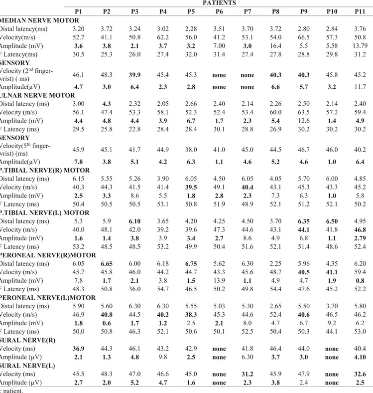
Table 1.The result of the nerve conductance studies of brucellosis patients with peripheral neuropathy. PATIENTS
P1 P2 P3 P4 P5 P6 P7 P8 P9 P10 P11 MEDIAN NERVE MOTOR
Distal latency(ms) 3.20 3.72 3.24 3.02 2.28 3.51 3.70 3.72 2.80 2.84 3.76 Velocity(m/s) 52.7 41.1 50.8 62.2 56.0 41.2 53.1 54.0 66.5 57.3 50.8 Amplitude (mV) 3.6 3.8 2.1 3.7 3.2 7.00 3.0 16.4 5.5 5.58 13.79 F Latency(ms) 30.5 25.3 26.0 27.4 32.0 31.4 27.4 27.8 28.8 29.8 31.2 SENSORY Velocity (2nd
finger-wrist) ( ms) 46.1 48.3 39.9 45.4 45.3 none none 40.3 40.3 45.8 45.2
Amplitude(µV) 4.7 3.0 6.4 2.3 2.8 none none 6.6 5.7 3.2 11.7
ULNAR NERVE MOTOR
Distal latency (ms) 3.00 4.3 2.32 2.05 2.66 2.40 2.14 2.26 2.50 2.14 2.40 Velocity (m/s) 56.1 47.4 53.3 58.1 52.3 52.4 53.4 60.0 63.5 57.2 59.4 Amplitude (mV) 4.4 4.8 4.4 3.9 6.7 1.7 2.3 5.4 12.6 1.4 4.9 F Latency (ms) 29.5 25.8 22.8 28.4 28.4 30.1 28.8 26.9 30.2 30.2 30.2 SENSORY Velocity(5th finger-wrist) (ms) 45.9 45.1 41.7 44.9 38.0 41.0 45.0 44.5 46.7 46.0 40.2 Amplitude(µV) 7.8 3.8 5.1 4.2 6.3 1.1 4.6 5.2 4.6 1.0 6.4
P.TIBIAL NERVE(R) MOTOR
Distal latency (ms) 6.15 5.55 5.26 3.90 6.05 4.50 6.05 4.05 5.70 6.00 4.85
Velocity (m/s) 40.3 44.3 41.5 41.4 39.5 49.1 40.4 43.1 45.3 43.3 45.2
Amplitude (mV) 2.5 3.3 8.6 5.5 1.8 2.8 2.3 7.3 6.3 1.0 5.8
F Latency (ms) 50.4 50.5 50.5 53.1 50.8 51.9 48.9 52.1 51.2 52.1 50.2
P.TIBIAL NERVE(L) MOTOR
Distal latency (ms) 5.3 5.9 6.10 3.65 4.20 4.25 4.50 3.70 6.35 6.50 4.95 Velocity (m/s) 40.0 48.1 42.0 39.2 39.6 47.3 44.6 43.1 44.1 41.8 46.8 Amplitude (mV) 1.6 1.4 3.8 3.9 3.4 2.7 8.6 4.9 6.8 1.1 2.79 F Latency (ms) 53.2 48.5 48.5 53.2 49.9 50.4 51.6 52.1 51.4 48.6 52.4 PERONEAL NERVE(R)MOTOR Distal latency (ms) 6.05 6.65 6.00 6.18 6.75 5.62 6.30 2.25 5.96 4.35 6.20 Velocity (m/s) 45.7 45.8 46.0 44.2 44.7 43.3 45.6 48.7 40.5 41.1 59.4 Amplitude (mV) 7.8 1.7 2.1 3.8 1.5 13.9 1.1 4.9 4.7 1.9 0.8 F Latency (ms) 48.3 50.8 36.0 54.7 46.5 50.2 49.8 54.4 47.6 45.2 52.2 PERONEAL NERVE(L)MOTOR Distal latency (ms) 5.90 5.60 6.30 6.30 5.55 5.03 5.30 2.65 5.50 3.70 5.80 Velocity (m/s) 46.9 40.8 44.5 40.2 38.3 45.3 44.6 52.4 40.6 46.5 46.2 Amplitude (mV) 1.8 0.6 1.7 1.2 2.5 2.1 8.0 4.7 6.7 9.2 6.2 F Latency (ms) 50.0 50.8 46.3 52.1 50.6 50.1 52.5 50.4 50.3 44.1 53.0 SURAL NERVE(R)
Velocity (ms) 36.9 44.3 46.1 43.2 42.9 none 41.8 46.4 44.0 none 40.4
Amplitude (µV) 2.1 1.3 4.8 9.8 2.5 none 6.30 3.7 3.0 none 4.10
SURAL NERVE(L)
Velocity (ms) 45.5 48.3 47.0 46.6 45.0 none 31.2 45.9 47.9 none 32.6
Amplitude (µV) 2.7 2.0 5.2 4.7 1.6 none 2.3 3.8 2.4 none 2.5
neuropathy with more definite involvement of motor fibres (Table 1).
Of patients found to have PN, 6 (54.5%) were male and 5 (45.5%) were female. The average age of these patients was 46.27 ± 14.85 years (range 22–69 years). In the brucellosis patient group, the average age of those without peripheral neuropathy was 33.57 ± 12.35 years. No statistically significant association was found in terms of gender of untreated brucellosis patients with or without peripheral neuropathy (p > 0.05) whereas the difference in average age was statistically significant. The average age of individuals with peripheral neuropathy was higher (p < 0.05).
Comparing the untreated brucellosis patients with the control group, the nerve transmission speeds of the motor median, ulnar, bilateral peroneal and tibial in the patient group were lower than for the control group and were statistically significant (p ˂ 0.05). Additionally, the terminal latencies of the median and ulnar nerves were lengthened compared to controls and there were
statistically significant (p ˂ 0.05). Also, bilateral peroneal and tibial nerve terminal latencies were lengthened compared to the control group but no statistically significant association was found (p > 0.05).
For CMAP amplitudes, the median, ulnar and bilateral peroneal were lower than the control group and were statistically significant (p ˂ 0,05).
For sensory transmission studies, while speeds for the median, ulnar and bilateral sural were slower in the patient group, there was a fall in CSAP for the median, ulnar and bilateral sural.
Similarly in the patient group, F latency lengthened for median, ulnar, bilateral peroneal and tibial nerves. The results of the nerve conductance studies of patient and control groups are shown in Table 2.
Discussion
There are very few prospective studies researching peripheral nervous system involvement of brucella. The
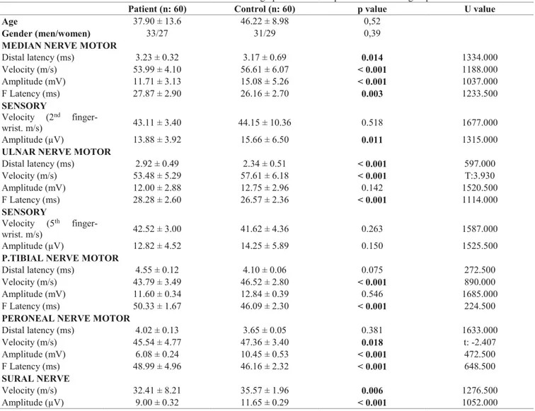
Table 2. The results of the nerve conductance studies and sociodemographic features of patient and control groups.
Patient (n: 60) Control (n: 60) p value U value
Age 37.90 ± 13.6 46.22 ± 8.98 0,52
Gender (men/women) 33/27 31/29 0,39
MEDIAN NERVE MOTOR
Distal latency (ms) 3.23 ± 0.32 3.17 ± 0.69 0.014 1334.000 Velocity (m/s) 53.99 ± 4.10 56.61 ± 6.07 ˂ 0.001 1188.000 Amplitude (mV) 11.71 ± 3.13 15.08 ± 5.26 ˂ 0.001 1037.000 F Latency (ms) 27.87 ± 2.90 26.16 ± 2.70 0.003 1233.500 SENSORY Velocity (2nd finger-wrist. m/s) 43.11 ± 3.40 44.15 ± 10.36 0.518 1677.000 Amplitude (µV) 13.88 ± 3.92 15.66 ± 6.50 0.011 1315.000
ULNAR NERVE MOTOR
Distal latency (ms) 2.92 ± 0.49 2.34 ± 0.51 ˂ 0.001 597.000 Velocity (m/s) 53.48 ± 5.29 57.61 ± 6.18 ˂ 0.001 T:3.930 Amplitude (mV) 12.00 ± 2.88 12.75 ± 2.96 0.142 1520.500 F Latency (ms) 28.28 ± 2.60 26.57 ± 2.36 ˂ 0.001 1114.000 SENSORY Velocity (5th finger-wrist. m/s) 42.52 ± 3.00 41.62 ± 4.36 0.263 1587.000 Amplitude (µV) 12.82 ± 4.52 14.25 ± 5.89 0.150 1525.500
P.TIBIAL NERVE MOTOR
Distal latency (ms) 4.55 ± 0.12 4.10 ± 0.06 0.075 272.500
Velocity (m/s) 43.79 ± 3.49 46.52 ± 2.80 ˂ 0.001 890.000
Amplitude (mV) 11.60 ± 0.34 12.84 ± 0.39 0.546 1685.000
F Latency (ms) 50.33 ± 1.67 46.09 ± 2.30 ˂ 0.001 224.500
PERONEAL NERVE MOTOR
Distal latency (ms) 4.02 ± 0.13 3.65 ± 0.05 0.381 1633.000 Velocity (m/s) 45.54 ± 4.77 47.36 ± 3.40 0.018 t: -2.407 Amplitude (mV) 6.08 ± 0.24 10.45 ± 0.53 ˂ 0.001 472.500 F Latency (ms) 48.99 ± 4.96 46.16 ± 2.32 ˂ 0.001 648.500 SURAL NERVE Velocity (m/s) 32.41 ± 8.21 35.57 ± 1.96 0.006 1276.500 Amplitude (µV) 9.00 ± 0.32 11.65 ± 0.29 ˂ 0.001 1052.000
majority of information is from case reports. One study by Kutlu et al. in 2009 involved 38 brucellosis patients and another study by Benbir et al. in 2013 involved 57 brucellosis and 42 control patients [8,12].
The Kutlu et al. study investigated the nerve conductance of brucellosis patients before and after treatment by electrophysiological approaches by examining the motor and sensory transmittance and F response of median, ulnar, tibial, peroneal and sural nerves using standardized methods and found sensorimotor PNs in 35% of the patients [8].
In 2013 Benbir et al. researched the presence of PNs in brucellosis patients, and similar to our study, found sensorial axonal PNP in 19% of the patients with slight transmission abnormality in motor and sensory branches [12].
In addition to these clinical studies, Kaya et al. presented a brucellosis patient with macular rash and peripheral neuropathy. Electrophysiological investigation of the patient indicated findings suggesting a moderate stocking-glove patterns demyelinizing sensory peripheral neuropathy.[13]. Aygul et al. presented a case with Guillain-Barré syndrome (GBS) linked to Brucella spp. infection and found nerve transmission blocks, slowing of nerve transmission speeds, lengthening of distal latency and inability to obtain F response on the electrophysiological examination of the patient [14]. Shoja et al. presented a nerve conductance study of a brucellosis case with renal failure and peripheral neuropathy and found symmetric, distal, axonal-type sensorimotor peripheral neuropathy especially in the lower extremities [15]. Kang et al. in an electrophysiological study of a brucellosis patient described findings suggesting demyelinizing PNs such as lengthened distal latency, reduced nerve transmission speed, fall in CMAP amplitudes, delayed F response in upper extremities and no F response for lower extremities and monitored the patient for a diagnosis of chronic inflammatory demyelinizing PN linked to brucellosis [16].
In our study, the nerve conductance was completed unilaterally for upper extremities (on the right) and bilaterally for lower extremities. Motor transmission speeds slowed in the median and ulnar nerves of the upper extremity and terminal latency and delays in F latencies lengthened for these nerves. While slowing was found only for transmission speeds of motor nerves in patients in our study, Kutlu et al. found slowing of transmission speeds for both motor and sensory branches of median and ulnar nerves in the brucellosis group [8].
In the whole brucellosis group, the average age of those with PN was higher than those without PN (p = 0.004). Previously Kutlu et al. and Benbir et al. described similar studies and found that the patients with PN in the brucellosis group had a higher average age than the average of the whole brucellosis group and stated that the age factor may be a reason for identification of PN [8,12]. This fits in with our study where the average age of PN patients was higher, leading to the consideration that age may create a susceptibility for PN. However, the average age of the control group without PN (46.22±8.98) is similar to the average age of the group with PN (46.27 ± 14.85), leading to the consideration that it may not be appropriate to link peripheral neuropathy to the age factor alone. In the control group, no patient was found to have PN.
While the pathogenesis of peripheral nervous system involvement of brucellosis is not fully understood, it has been shown that the characteristics of
Brucella spp. bacteria within cells (phagocytes) may
damage peripheral nerve cells [5]. Damage to nerves during the direct invasion of the Brucella spp. organism or production of endotoxins in the acute period may be the mechanism for axonal-type peripheral neuropathy in peripheral nerves. Immune-modulated mechanisms may cause demyelinizing-type peripheral neuropathy especially in the chronic period [5,15,17]. Inflammation of proximal nerve roots and spinal cord compression due to granuloma or abscess may be shown as another cause of peripheral neuropathy [3]. A study investigating a brucellosis patient with GBS suggested that antibodies against gangliosides were responsible for the pathogenesis of peripheral nerve involvement. This study found that GM1 ganglioside antibodies were produced against molecules similar to gangliosides expressed on the surface of B.melitensis and caused symptoms similar to weakness in extremities and ataxia in rats [18]. As a result, it was proposed that polyradiculopathies related to brucellosis may cause nerve damage due to the antibody response to similarities between GM1 ganglioside molecules in peripheral nerves and Brucella spp.
lipooligosaccharides [16,18]. Studies indicate that systemic treatment of brucellosis has caused regression of peripheral neuropathy linked to Brucella spp. infection [8,12]. This shows that though the mechanism of peripheral neuropathy linked to Brucella spp. is not fully understood, it is reversible via appropriate treatment [8].
In this study, nerve conduction tests in our untreated brucellosis patient group identified abnormal findings
in comparison to the control group and according to PN diagnostic criteria, 18% had axonal PN identified. This abnormality may be linked to Brucella spp. infection or may be related to other factors. Even with the careful selection of the patient and control group, and with the exclusion of other known factors that cause PN, abnormalities on nerve conduction studies may be caused by other unforeseen factors (e.g., genetic factors, nutritional habits, medications used, etc.).
Conclusions
Our electrophysiological investigation discovered subclinical, widespread axonal-type PN in patients who received a diagnosis of brucellosis, had not started medical treatment, had no specific complaints of peripheral nervous system involvement and had no neurological exam findings. With many different involvements of central and peripheral nervous systems, brucellosis may be encountered as a cause of clinical or subclinical PN; furthermore, PN may regress with appropriate brucellosis treatment. As a result, in regions where brucellosis is endemic, a differential diagnosis should always be considered for PN.
References
1. Guven T, Ugurlu K, Ergonul O, Celikbas AK, Gok SE, Comoglu S, Baykam N, Dokuzoguz B (2013) Neurobrucellosis: clinical and diagnostic features. Clin Infect Dis 56: 1407-1412.
2. Kim EJ, Lee SJ, Ahn EY, Ryu DG, Choi YH, Kim TH (2015) Relapsed brucellosis presenting as neurobrucellosis with cerebral vasculitis in a patient previously diagnosed with brucellar spondylitis: a case report. Infect Chemother. Dec 47: 268-271.
3. Young EJ (2005) Brucella species. In: Mandell GL, Bennett JE, Dolin R, editors. Principles and practice of infectious diseases. 6th edition Philadelphia: Churchill Livingstone p 2669-2674.
4. Young EJ, Borchert M, Kretzer FL, Musher DM (1985) Phagocytosis and killing of brucella by human polymorphonuclear leukcocytes. J Infect Dis 151: 682. 5. Al Deeb SM, Yaqub BA, Sharif HS, Phadge JG (1989)
Neurobrucellosis: clinical characteristics, diagnosis and outcome. Neurology 39: 498-501.
6. Yetkin MA, Bulut C, Erdinç FŞ, Oral B, Tulek N (2006) Evaluation of the clinical presantation in neurobrucellosis. İnt J Infect Dis 10: 446-452.
7. Gotuzo E, Carillo C, Guerra J, Liosa L (1986) An evaluation of diagnostic methods for brusellosis - the value of bone marrow culture. J Infect Dis 153: 122.
8. Kutlu G, Ertem GT, Coskun O, Ergun U, Gomceli YB, Tulek N (2009). Brucella: A cause of peripheral neuropathy. Eur Neurol 61: 33-38.
9. Karsen H, Koruk ST, Duygu F, Yapici K, Kati M (2012) Review of 17 cases of neurobrucellosis: clinical manifestations, diagnosis, and management. Arch Iran Med 15: 491 – 494.
10. Presto DC, Shapiro BE (2005) Electromyography and neuromuscular disorders: clinical-electrophysiologic correlations. In Presto DC editor. Appendix; Nerve Conduction Studies: Normal Adult Values. Philadelphia: Pennsylvania.Elsevier Press. 663-666.
11. Ertekin C (2006) Central and peripheral electromyography. In Ertekin C editor. Conduction Characteristics In Pathological Signals. Izmir: Meta Press. 140-146.
12. Benbir G, Tursun I, Akkoyunlu Y (2013) An electrophysiological study of peripheral polyneuropathy in brucellosis. Arch Iran Med 16: 446-448.
13. Kaya S, Kostakoğlu U (2009) A brucellosis case with macular rash and peripheral neuropathy. Mikrobiyol Bul 43: 147-151. 14. Aygul R, Deniz O, Guzelcik M, Kotan D (2010) Guillain-Barre
syndrome during active brusellosis. Eurasian J Med 42: 157-159.
15. Shoja MM, Khosroshahi HT, Tubbs RS, Varshochi M, Fervenza FC (2008). Brucellosis mimicking vasculitis in a patient with renal failure and peripheral neuropathy. Am J Med Sci 336: 285-287.
16. Kang BH, Lim YM, Kim KK (2012). Brucellosis manifesting as chronic inflammatory demyelinating polyneuropathy. Can J Neurol Sci 39: 536-538.
17. Shakir RA, Al Din ASN, Araj GF, Lulu AR, Mousa AR, Saadah MA (1987) Clinical categories of neurobrucellosis: A case report on 19 cases. Brain 110: 213-223.
18. Watanabe K, Kim S, Nishiguchi M, Suzuki H, Watara M (2005) Brucella melitensis infection associated with Guillian-Barré syndrome through molecular mimicry of host structures. FEMS Immunol Med Microbiol 45: 121-127.
Corresponding author
Hatice Kose Ozlece MD,
Department of Neurology, Kafkas University, Turan Celik street, 180, 36001, Kars, Turkey.
Phone: +9005317901791 Fax: +90 (474) 225 11 61 e-mail: haticekse@hotmail.com