Kocatürk et al. J Brain Disord 2018, 2(1):44-46 Volume 2 | Issue 1
Copyright: © 2018 Kocatürk M, et al. This is an open-access article distributed under the terms of the Creative Commons Attribution License, which permits unrestricted use, distribution, and reproduction in any medium, provided the original author and source are credited.
Case Presentation Open Access
SCHOLARLY
PAGES
Journal of Brain Disorders
• Page 44 •
*Corresponding author: Özcan Kocatürk, MD, Department of Neurology, Harran University Şanlıurfa, Osman bey Cam-pus, Mardin Yolu, Şanlıurfa, Turkey, Tel: +905074191621, E-mail: [email protected]
Received: November 01, 2017; Accepted: January 08, 2018; Published online: January 10, 2018
Citation: Kocatürk M, Aksoy M, Kocatürk Ö (2018) Do Not Overlook Zona Herpes Ophthalmicus in a Patient Presenting with Pupil-Sparing Oculomotor Nerve Palsy. J Brain Disord 2(1):44-46
Do Not Overlook Zona Herpes Ophthalmicus in a Patient
Presenting with Pupil-Sparing Oculomotor Nerve Palsy
Mehtap Kocatürk
1, Mustafa Aksoy
2and Özcan Kocatürk
1*1Department of Neurology, Harran University Şanlıurfa, Turkey 2Department of Dermatology, Harran University Şanlıurfa, Turkey
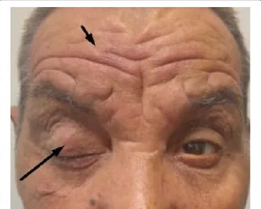
infraduction and adduction movements of eye ball. The pupils was equally reactive to light and isocoric. There were some squamatous and hyperemic cutaneous lesion along ophthalmic division of right trigeminal nerve, whi-ch is consistent with healing herpes zoster lesion (Figure 1). Visual acurity of both eye and rest of the examination were normal.
Herpes Zoster (HZ), relatively common syndrome, is the result of reactivation of Varicella-Zoster Virus (VZV) that lying in the ganglion after a prior bout of varicella. Reactivation is usually caused by a decline in the specific cell-mediated immunity to VZV with aging, immunosup-pression, trauma, and psychological stress [1].
In the HZ, various neurologic complications are seen. Those are postherpetic neuralgia (chronic pain), VZV vas-culopathy, meningoencephalitis, meningoradiculitis, cere-bellitis, myelopathy, and involvement of cranial nerves [2]. When HZ is involved in the ophthalmic branch of the tri-geminal nerve, it is termed as Herpes Zoster Ophthalmicus (HZO). HZO accounts for 10-15% of all herpes zoster cases. Half of the HZO cases involve the ocular component like keratit, cataracts, glaucoma, uveitis [3].
Oculomotor Nerve Palsy (ONP) could result from le-sions anywhere along its path between the oculomotor nucleus and extraocular muscles. Differential diagnosis of ONP can be made according to presence of pupil invol-ment, Pupil sparing oculomotor nerve palsy typically result from ischemic cranial neuropathy, often associated with vascular risk factors, which improves (and usually fully re-solves) within 3 months.
We present herein a patient exhibiting oculomotor nerve palsy associated with herpes zoster ophthalmicus.
Presentation
A 70-year-old man presented to the neurology outpa-tient clinic with acute onset of painful ptosis of his right eye. Past medical history revealed hypertension, hyperli-pidemia and cerebrovascular disease. Family, social and surgical history was not significant. His blood pressure, pulse, respiration and body temperature were within normal limits. At first physical examination of patient showed complete ptosis and absence of supraduction,
Figure 1: Patient photography that taken at first presentation shown that complete third nerve palsy with ptosis and healing ulcer of Herpes Ophthalmicus.
• Page 45 •
Citation: Kocatürk M, Aksoy M, Kocatürk Ö (2018) Do Not Overlook Zona Herpes Ophthalmicus in a Patient Presenting with Pupil-Sparing Oculomotor Nerve Palsy. J Brain Disord 2(1):44-46
SCHOLARLY PAGES
Kocatürk et al. J Brain Disord 2018, 2(1):44-46
A
B
C
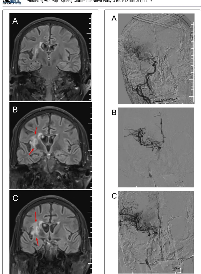
Figure 2: Brain Magnetic Resonance Imaging (MRI) shown right peri-insular old infarct (red arrow).
A
B
C
Figure 3: Digital subtraction angiography (DSA) of brain vessel shown that there is no intracranial aneurism.
• Page 46 •
Citation: Kocatürk M, Aksoy M, Kocatürk Ö (2018) Do Not Overlook Zona Herpes Ophthalmicus in a Patient Presenting with Pupil-Sparing Oculomotor Nerve Palsy. J Brain Disord 2(1):44-46
SCHOLARLY PAGES
Kocatürk et al. J Brain Disord 2018, 2(1):44-46
Brain Magnetic Resonance Imaging (MRI) revealed right peri-insular old infarct (Figure 2). There was no pathologic signal that considered as oculomotor nerve palsy. Digital subtraction angiography of brain vessel that was done 3 months ago due to acute stroke treat-ment was re-evaluated and revealed no brain aneurism (Figure 3). Blood biochemistry, hemogram, sedimantas-yon was normal.
Ten days ago, the patient presented to the dermato-logy clinic with sharp pain and sudden onset vesicular cutaneous eruption at right periorbital region and was diagnosed as herpes zoster ophthalmicus. Oral valacyc-lovir treatment initiated within 24 hours of symptoms and continued for 10 days. Ophthalmalogic examination was within normal limits.
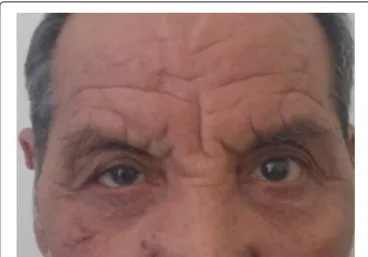
Third nerve palsy associated with HZO was conside-red and deflazacort 60 mg/day was given and tapeconside-red. At three weeks later, patient symptom improve and at the 2 months, there was no residual symptoms (Figure 4).
Discussion
HZ is a localized disease characterized by unilateral radicular pain with vesicular eruption and nearly 1 in 3 people will develop it during their lifetime [1]. After thoracic dermatomes, cranial nerves are commonly affe-cted by herpes zoster. Beside the sensory nerves, motor neurons very rarely could be affected by HZ. HZO occur between 10%-25% of all cases of herpes zoster and half of these cases affect the eye [3,4]. Ocular signs include conjunctivitis, keratitis, episcleritis, scleritis, uveitis, se-condary glaucoma, cataract, and retinal necrosis [4].
Ophthalmoplegia is seen in 5%-31% of patients with HZO. The oculomotor nerve is the most common site amongst them and the trochlear nerve is the least. Complete ophthalmoplegia is seen very rarely. The
extra-ocular muscle palsies usually are seen 2-4 weeks after the vesicular eruption, but sometimes occurs simultaneously with the eruption or more than 1 month later [3]. In our patient, there was painful-pupil sparing complete oculo-motor nerve palsy that appeared 10 days after the rush.
The pathogenesis of oculomotor nerve palsy is cont-roversial and several mechanisms have been suggested. Some of them are following; direct cytopathic effect of virus, immune response of the central nervous system to the virus, occlusive vasculitis induced by the virus and another latent neuropathic virus activation within the brain [5]. Given that the patient has vascular risk factors and pace of symptom recovery, occlusive vasculitis and microinfarction of oculomotor nerve could be respon-sible in our patient.
Treatment of herpes zoster ophthalmicus require sys-temic antiviral drugs. It is most effective if it is started within 72 hours of symptom onset. Administration of antiviral medications reduce viral spreading and ocular complications. The use of systemic steroid along with an-tiviral treatment is recommended because it may be effe-ctive to prevent occlusive vasculitis and reduce duration of acute pain [1]. This patient received valacyclovir for 10 days before the our presentation. After oculomotor palsy related to HZO was confirmed, we had started oral steroid for 3 weeks. It was shown that duration of ptosis and diplopia related to third nerve was from 2 months to 23 months. In our patient, pupil sparing oculomotor nerve palsy has improved and show complete recovery within the 2 months.
It is important to consider zona zoster as a reversib-le cause of oculomor nerve plasy. Therefore physicians should examine the periorbital skin of patients and look for any healing lesion.
References
1. Vrcek I, Choudhury E, Durairaj V (2017) Herpes zoster oph-thalmicus: A review for the internist. Am J Med 130: 21-26. 2. Nagel MA, Gilden D (2013) Complications of varicella zos-ter virus reactivation. Curr Treat Options Neurol 15: 439-453.
3. Shin HM, Lew H, Yun YS (2005) A case of complete oph-thalmoplegia in Herpes Zoster Ophthalmicus. Kor J Opht-halmol 19: 302-304.
4. Kemal Balci, Ufuk Utku, Bahar Özbek (2008) Altinci kranial sinir paralizisine neden olan bir herpes zoster oftalmikus olgusu/herpes zoster ophthalmicus with sixth cranial nerve palsy : A case report. Turk J Neurol 14: 350-352.
5. Yıldız ÖK, Seğmen H, Bolayır E, et al. (1998) Journal of neurological sciences (Turkish). Aegean Neurological So-ciety 26: 500-504.
Figure 4: Patient photography that taken 3 month later show that resolution of pitosis and Herpes Ophthalmicus lesion.