Oral and Maxillofacial Surgery Cases 6 (2020) 100196
Available online 16 October 2020
2214-5419/© 2020 The Authors. Published by Elsevier Inc. This is an open access article under the CC BY-NC-ND license (http://creativecommons.org/licenses/by-nc-nd/4.0/).
Very rare intraoral appearance of ossifying fibro myxoid tumor: A
case report
Ümit Ertas
a, ¨Omer Gunhan
b, Yunus Emre AS¸ÇI
c,*aAtatürk University Faculty of Dentistry Head of Oral and Maxillofacial Surgery Department, Department of Oral and Maxillofacial Surgery, Faculty
of Dentistry, Ataturk University, 25240, Erzurum, Turkey
bDepartment of Pathology, TOBB ETU School of Medicine, Ankara, Turkey
cDepartment of Oral and Maxillofacial Surgery, Faculty of Dentistry, Ataturk University, 25240, Erzurum, Turkey
A R T I C L E I N F O Keywords:
Ossifying fibro myxoid tumor Mezenchymal neoplasm Subcutaneous
A B S T R A C T
Ossifying fibro myxoid tumor (OFMT) is a rare soft tissue neoplasm and may present diagnostic difficulty. Only a few cases of oral cavity are described. A 49 –year-old male presented with a mass in the vestibular region of the mandible middle line, complaining of pain for the last three months. The mass contains spindle and polygonal mesenchymal cells on the myxoid ground in histochemical examinations. The tumor had a partial fibrous capsule surrounded by a non- metaplastic bone trabeculae. Histopathological examination was typical for OFMT. Although most cases of OFMT follow a benign course, recurrence and metastasis have been reported. Therefore, this type of tumor should be followed carefully.
1. Introduction
Ossifying fibro myxoid tumor (OFMT), first described by Enzinger et al., in 1989, is a rare mesenchymal neoplasm that typically arises in the extremities. These types of tumors often appear as a painless, well-defined subcutaneous mass usually attached to the nerve, muscle, or cartilaginous. In more than 70% of cases, extremities are involved; less frequently involved areas include the head and neck, and the mediastinum [1]. These tumors are uncommon in the oral cavity; however, a few cases have been reported [2–4]. OFMTs particularly affect adults [2]. OFMTs are usually follow a benign course and are generally treated with complete excision [5]. Recurrence in the head and neck area is reported 21% of the time and metastases are quite rare [1,6]. OFMTs are rare soft tissue neoplasms of uncertain etiology, characterized by the proliferation of round to spindle-shaped cells arranged in cords and embedded in a fibro myxoid matrix [7]. OFMTs are multilobular and appear to be an incomplete metaplastic bone shell [3,8]. After the charac-terization of this tumor by Enzinger et al., about 220 cases have been published, mostly seen in the extremities and found in subcu-taneous tissues [8,9]. The trunk, retroperitoneum, mediastinum, and to a lesser extent, the head and neck region may also be affected. It is very rare to see OFMTs only in the oral cavity [8–10]. Most cases of OFMT exhibit a mostly symptom-free biological behavior. Local recurrence, which is treated with local curettage, has been reported at a rate of 20–27%. In rare cases, it may show an aggressive biological behavior. The presence of these cases has been associated with the development of metastasis [10,11].
The present article reports an OFMT case in the intraoral region of a 49-year-old male patient and includes the histogenesis, histopathological features, biological behavior, and surgical treatment management of the OFMT.
* Corresponding author. Tel./Fax: 90 442 231 2734.
E-mail addresses: [email protected] (Ü. Ertas), [email protected] (¨O. Gunhan), [email protected] (Y.E. AS¸ÇI).
Contents lists available at ScienceDirect
Oral and Maxillofacial Surgery Cases
journal homepage: www.oralandmaxillofacialsurgerycases.comhttps://doi.org/10.1016/j.omsc.2020.100196
2.1. Histopathological and molecular findings
In histological examination of the OFMT, a proliferation of round to spindle-shaped cells embedded in the myxoid matrix is observed in the sockets and cords. An incomplete mineralized bone trabecular crust is observed just below a fibrous pseudo capsule [5].
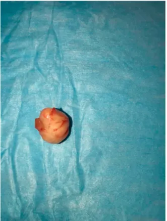
In a macroscopic examination, the OFMT measured 2.5cm in diameter, and appeared as a round yellowish mass circumscribed with a lobulated rubbery cut surface (Fig. 1).
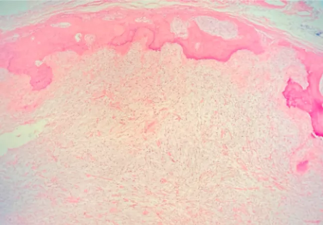
In histological sections the tumor; an unencapsulated, well-circumscribed fibro myxoid tumor was seen. It was incompletely covered with a lamellated shell of bone (Fig. 2).
The tumor is composed of chondroid-like lobules of the spindle or polygonal fibroblastic cells in a myxohyaline background (Fig. 3). Neoplastic cells have bland nuclei with indistinct nucleoli and eosinophilic cytoplasm. Mitotic figures were rare (<2/50hpf). Myxoid stroma contained cellular strands, hyaline bundles, mucinous microcysts (Fig. 4). There was neither invasion outside the main tumor nor necrosis vascular permeation. Immunohistochemically, the tumor shows diffuse S-100 positivity (Fig. 5). Ki-67 activity reached 10% in the hot spots. The histopathologic appearance is accepted as a typical type of ossifying fibro myxoid tumor.
In the sections in histopathological examination, a tumoral lesion with a fibrous capsule in some periphery and a bone-shaped bone in some areas is observed. The tumor consisted of spindle and polygonal mesenchymal cells in most areas on a myxoid surface. Myxoid change is evident locally on the ground and includes cystic areas. Fig. 6The tumor also contains focal nodularity surrounded by collagen bands and cellular areas consisting of spindle cells. Mitotic activity and necrosis have not been observed. Infiltration is not present in the surrounding tissue.
In controlled immunohistochemical examinations, widespread staining of neoplastic cells is seen with the S100 antibody. Morphological findings showed that the diagnosis is compatible with ossifying fibro myxoid tumor.
3. Discussion
OFMTs are often seen in adult individuals and appear to be located in the subcutaneous tissues of extremities [2]. Men are typically affected more than women, with the gender ratio ranging from 1.8:1 to 1.5:1. Although there are reports of OFMTs in newborns, most cases are diagnosed in adults at a mean age of 50 years [2–15]. In this case report, the patient was a 49-year-old male Fig. 7with the lesion presenting in an intraoral region. Only few cases have been reported in the literature in which the masses were located in oral
areas including buccal mucosa, gingiva, and lips [2–15]. Most often the tumor appears as a painless, well-defined subcutaneous mass usually attached to the nerve, muscle, or cartilaginous [1,13–15]. In the presented case, the mass was seen anterior to the mentum region, which is not often seen with OMFT.
Previous studies have suggested that loss of S100 protein may be associated with atypia and malignant transformation [17–21]. In this case report, staining with the S100 antibody was found positive. In this case, mitotic activity was not detected so that there may be a relationship between the S100 antibody and malignant transformation and atypia.
Differential diagnosis:
✓ Peripheral bone construction is not observed in the schwannoma.
✓ Low-grade fibro myxoid sarcoma is large, deep tissue and more pleomorphic.
Fig. 2. The mass uncovered as a result of the incision.
✓ Malignant peripheral schwannoma is more aggressive, NF-1 relation is not observed in ossified fibro myxoid tumor [ [22–25]]. In this case report, differential diagnosis was made with diseases such as myxoid chondrosarcoma, low-grade fibro myxoid sarcoma and schwannoma. Low-grade sarcoma contains atypical spindle cells with myxoid and collagenous matrix and can be misinterpreted as a malignant OFMT with immunohistological features such as S100 protein deficiency. However, low-grade sarcoma contains hyaline
Fig. 4. Tumor covered with a peripheral ossifying layer with an inner myxohyaline component.
Fig. 5. Lobulated chondroid-like with intervening spindle cell bundles.
rosettes which aids in distinguishing it.
The lesion may be present from 4 to 30 years before treatment is required due to its slow and painless progression [16]. In this patient, progress occurred faster than suggested by the literature. The patient had no pain initially, but this advanced quickly over a period as short as three months, being noticed in activities such as chewing or speaking on the phone. The lesion is difficult to spot, as most OFMTs occur in deep soft tissues. But in this particular case, the patient noticed at a very early stage due to swelling.
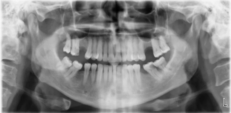
Radiographically, it may appear as multiple focal irregular calcifications surrounded by an incomplete calcification ring inside the lesion. There are similar clinical findings in this case. Although rare, OFMTs may infiltrate the bone or periosteal reactions may be observed. In this case, although different irregularities of the underlying alveolar bone were described, a significant periosteal reaction and bone infiltration were observed during the operation [7,22].
OFMTs generally follow a benign clinical prognosis as a course. Despite this, three forms of OFMT have been defined. These are: typical, atypical and malignant forms. Looking at the OFMT classification of Folpe and Weiss, the lesion presented in this case can be described as typical [26]. In previous research, the recurrence and metastasis rates of typical OFMTs vary between 5 and 17%. In this case, it was suggested that the tumor be considered an intermediate malignancy lesion [5]. In the case report, recurrence was not observed in the follow-up, which is compatible with the literature. However, recurrence can be observed up to 10 years after treatment [16]. Therefore, long-term follow-up is required.
4. Conclusion
The significance of this case report lies in the rarity of ossifying fibro myxoid tumors in the oral cavity. Here, an ossifying fibro myxoid tumor is presented for the first time in the mandibula middle line, which is an extremely rare location given that the tumor was thought to occur exclusively in the extremities. With this case report, ossified fibro myxoid tumors should be considered among the differential diagnoses for mandibular swellings with cystic lesions that are well-defined radiographically.
Declaration of competing interest
The authors declare that they have no known competing financial interests or personal relationships that could have appeared to influerıce the work reported in this paper.
References
[1] Weiss SW. Soft tissue tumors of intermediate malignancy of uncertain type. Enzinger and Weiss’s soft tissue tumors 2008:1093–160.
[2] Mollaoglu N, Tokman B, Kahraman S, Cetiner S, Yucetas S, Uluoglu O. An unusual presentation of ossifying fibromyxoid tumor of the mandible: a case report. J Clin Pediatr Dent 2007:136–8.
[3] Sharif MA, Mushtaq S, Mamoon N, Khadim MT. Ossifying fibromyxoid tumor of oral cavity. J Coll Physicians Surg Pak 2008:181–2.
[4] Nonaka CF, Pacheco DF, Nunes RP, Freitas Rde A, Miguel MC. Ossifying fibromyxoid tumor in the mandibular gingiva: case report and review of the literature. J Periodontol 2009:687–92.
[5] Folpe AL, Weiss SW. Ossifying fibromyxoid tumor of soft parts: a clinicopathologic study of 70 cases with emphasis on atypical and malignant variants. Am J Surg Pathol 2003:421–31.
[6] Kondylidou-Sidira A, Kyrgidis A, Antoniades H, Antoniades K. Ossifying fibromyxoid tumor of head and neck region: case report and systematic review of literature. J Oral Maxillofac Surg 2011:1355–60.
[7] Rubin BP, Stenman G. Ossifying fibromyxoid tumor. In: Fletcher CDM, Unni KK, Mertens F, editors. World health organization classification of tumors, pathology and genetics: tumors of soft tissue and bone. Lyon, France: IARC Press; 2002. p. 196–7.
[8] Al-Mazrou KA, Mansoor A, Payne M, Richardson MA. Ossifying fibromyxoid tumor of the ethmoid sinus in a newborn: report of a case and literature review. Int J
Pediatr Otorhinolaryngol 2004;68:225–30.
[9] Miettinen M, Finnell V, Fetsch JF. Ossifying fibromyxoid tumor of soft parts – a clinicopathologic and immunohistochemical study of 104 cases with long-term follow-up and a critical review of the literature. Am J Surg Pathol 2008;32:996–1005.
analysis. Histopathology 1993:101–12.
[21]S¸ıraneci P, Tekkes¸in Ms. Ossifying fibromyxoid tumor; an unusual tumor in oral cavity: case report. Turkiye Klinikleri J Case Rep 2016:73–6.
[22]Williams SB, Ellis GL, Meis JM, Heffner DK. Ossifying fibromyxoid tumour (of soft parts) of the head and neck: a clinicopathological and immunohistochemical study of nine cases. J Laryngol Otol 1993:75–80.
[23]Miettinen M, Finnell V, Fetsch JF. Ossifying fibromyxoid tumor of soft parts–a clinicopathologic and immunohistochemical study of 104 cases with long-term follow-up and a critical review of the literature. Am J Surg Pathol 2008:996–1005.
[24]Graham RP, Dry S, Li X, Binder S, Bahrami A, Raimondi SC, et al. Ossifying fibromyxoid tumor of soft parts: a clinicopathologic, proteomic, and genomic study. Am J Surg Pathol 2011:1615–25.
[25]Demiryont M. Uncommon soft tissue tumors with controversial histogenesis. Turk J Pathol 2007:181–6.