İZMİR KATİP ÇELEBİ UNIVERSITY « GRADUATE SCHOOL OF SCIENCE AND ENGINEERING
M.Sc. THESIS
SEPTEMBER 2016
EVALUATION OF EFFECTIVENESS OF COLD ATMOSPHERIC PLASMA TREATMENT METHODS FOR PREVENTION OF
VENTILATOR-ASSOCIATED PNEUMONIA
Thesis Advisor: Asst. Prof. Dr. Utku Kürşat ERCAN FATMA İBİŞ
Department of Biomedical Technologies Biomedical Technologies Programme
Anabilim Dalı : Herhangi Mühendislik, Bilim Programı : Herhangi Program
SEPTEMBER 2016
İZMİR KATİP ÇELEBİ UNIVERSITY « GRADUATE SCHOOL OF SCIENCE AND ENGINEERING
EVALUATION OF EFFECTIVENESS OF COLD ATMOSPHERIC PLASMA TREATMENT METHODS FOR PREVENTION OF
VENTILATOR-ASSOCIATED PNEUMONIA
M.Sc. THESIS FATMA İBİŞ
Y140101005
Department of Biomedical Technologies Biomedical Technologies Programme
Anabilim Dalı : Herhangi Mühendislik, Bilim Programı : Herhangi Program
EYLÜL 2016
İZMİR KATİP ÇELEBİ ÜNİVERSİTESİ « FEN BİLİMLERİ ENSTİTÜSÜ
VENTİLATÖR İLİŞKİLİ PNÖMONİNİN ÖNLENMESİ AMAÇLI ATMOSFERİK SOĞUK PLAZMA MUAMELE YÖNTEMLERİNİN
ETKİNLİĞİNİN DEĞERLENDİRİLMESİ
YÜKSEK LİSANS TEZİ FATMA İBİŞ
Y140101005
Biyomedikal Teknolojileri Anabilim Dalı Biyomedikal Teknolojileri Programı
Anabilim Dalı : Herhangi Mühendislik, Bilim Programı : Herhangi Program
v
Thesis Advisor : Asst. Prof. Dr. Utku Kürşat ERCAN ... İzmir Katip Çelebi University
Jury Members : Prof. Dr. Bahattin TANYOLAÇ ... Ege University
Asst. Prof. Dr. Ozan Karaman ... İzmir Katip Çelebi University
Prof. Dr. Aydın AKAN ... İzmir Katip Çelebi University
Asst. Prof. Dr. Nermin TOPALOĞLU... İzmir Katip Çelebi University
Fatma İbiş, an M.Sc. student of IKCU Graduate School of Science and Technology student ID Y140101005, successfully defended the thesis entitled “EVALUATION OF EFFECTIVENESS OF COLD ATMOSPHERIC PLASMA TREATMENT METHODS FOR PREVENTION VENTILATOR-ASSOCIATED PNEUMONIA”, which she prepared after fulfilling the
requirements specified in the associated legislations, before the jury whose signatures are below.
Date of Submission : 19 September 2016 Date of Defense : 26 September 2016
vii
ix FOREWORD
I would especially like to thank my advisor, Asst.Prof.Dr. Utku Kürşat Ercan, for his healthy degree of optimisms when experiments disappointed and all seemed lost. I would also like to thank him for the warm and friendly atmosphere he garnered in his group; it encouraged sharing of ideas, insightful discussions and a productive work environment.
Next, I would like to thank TÜBİTAK 2211 Graduate Scholarship Programme (TÜBİTAK 2211 Yurt İçi Lisansüstü Burs Programı) for support and aid for my thesis project.
We would like to acknowledge The Scientific and Technology Research Council of Turkey for financial support of this study under grant 114E163.
Additionally, I would like to thank Res.Asst.Eyyüp Yalçın for helping and patient for the viewing of AFM in our university.
Without great friends and family, this endeavor would have concluded before it began. I would like to thank them for believing in me, encouraging me to continue going, and providing distractions from work when they were needed.
I want to thank my parents. It is with their help for all my life that I became who I am today. Thanks for always being there for me, believing in me, and motivating me to set out on my own path. I cannot begin to describe how lucky I feel for having them as my parents. All opportunities and accomplishments I owe to them.
xi TABLE OF CONTENTS
... Page
FOREWORD ... ix
TABLE OF CONTENTS ... xi
LIST OF FIGURES ... xvii
INTRODUCTION ... 1
1. 1.1 Hospital –acquired infections ... 1
1.1.1 Urinary Tract Infections ... 2
1.1.2 Surgical Site Infections ... 2
1.1.3 Ventilator Associated Pneumonia ... 3
1.2 Microorganisms ... 5
1.2.1 Antibiotic-resistant microorganisms ... 5
1.2.2 Fungi ... 6
1.3 Prevention of Infection ... 6
1.3.1 Antiseptics and Antisepsis ... 6
1.3.2 Disinfection ... 7
1.3.3 Sterilization ... 8
1.4 Plasma ... 10
1.4.1 Dielectric Barrier Discharge (DBD) Plasma ... 12
1.4.2 Plasma Chemistry ... 13
1.4.3 Plasma Medicine ... 14
1.4.3.1 Plasma Assisted Blood Coagulation ... 15
1.4.3.2 Plasma Applications for Cancer Theraphy ... 15
1.4.3.3 Dental Applications of Cold Atmospheric Plasma ... 16
1.4.3.4 Plasma Applications in Dermatology and Wound Healing ... 16
1.4.3.5 Cold Atmospheric Plasma - Cell Interactions ... 17
1.4.3.6 Plasma sterilization ... 17
1.5 Nebulization ... 18
1.6 Aim of Study ... 18
MATERIALS AND METHODS ... 20
2. 2.1 Culvitation of Microorganisms ... 20
2.2 Preperation of Frozen Stock Cultures ... 20
2.3 Preperation of N-Acetyl Cysteine (NAC) Solution ... 21
2.4 Designing and Manufacturing of Electrode and Fluid Holder ... 21
2.4.1 Dielectric Barrier Discharge (DBD) Electrode ... 21
2.4.2 Plasma Jet Electrode ... 22
2.4.3 Fluid Holder ... 23
2.5 Optimization of Plasma Parameters for Appropriate Antimicrobial Effects ... 23
2.6 Quantifications Methods for Antimicrobial Effect of Plasma Treatment ... 24
2.6.1 XTT Assay ... 24
2.6.2 Safranine Assay ... 25
2.7 DBD Plasma Treatment of Liquids ... 25
2.8 Antimicrobial Activity of Nebulized Plasma Treated Liquids ... 26
xii
2.10 Optimization of Drip Flow Reactor (DFR) for Formation of Biofilm ... 28
2.11 Tests for Prevention of Formation of Biofilm ... 29
2.12 Antibiofilm Effect of Plasma Treatment ... 31
2.12.1 Antibiofilm Effect of Plasma Nebulized Treated Liquid ... 31
2.12.2 Antibiofilm Effect Air Plasma Jet ... 32
2.13 Microscopic Characterization of Biofilm on Endotracheal Tube Surfaces ... 32
RESULT ... 34
3. 3.1 Optimization of Plasma Parameters for Appropriate Antimicrobial Effects ... 34
3.2 Evaluation of the Antimicrobial Activity of Plasma Jet Application ... 36
3.3 Biofilm Inactivation Tests with Liquid mediated Plasma Treatment ... 36
3.4 Optimization of Drip Flow Reactor for Formation of Biofilm ... 39
3.5 Tests for Prevention of Formation of Biofilm ... 39
3.6 Biofilm Inactivation Efficacy of Nebulized Plasma Treated Liquids ... 42
3.7 Biofilm Characterization on Endotracheal Tubes Surfaces ... 50
DISCUSSION ... 56
4. CONCLUSION ... 60
5. CURRICULUM VITAE ... 67
xiii ABBREVIATIONS
HAI : Hospital acquired infection HCAI : Healthcare-associated infection ICU : Intensive care unit
MRSA : Methicillin-resistant Staphylococcus aureus VAP : Ventilator associated pneumonia
UTI : Urinary tract infection SSI : Surgical site infection CFU : Colony forming unit
VRE : Vancomycin resistant Enterococcus UV : Ultraviolet
EO : Ethylene oxide DC : Direct current AC : Alternate current
DBD : Dielectric barrier discharge
FE-DBD : Floating electrode dielectric barrier discharge ROS : Reactive oxygen species
RNS : Reactive nitrogen species APC : Argon plasma coagulation RBC : Red blood cells
PIM : Powder injection molding NAC : N-Acetyl Cysteine
ATCC : American Type Culture Collection TSB : Trypticase soy broth
TSA : Trypticase soy agar
YPD : Yeast extract-peptone-dextrose
XTT : (2,3-Bis(2-methoxy-4-nitro-5-sulfophenyl)-2H-tetrazolium-5-carboxanilide)
PBS : Phosphate buffered saline rpm : Revolution per minute DFR : Drip flow reactor
SEM : Scanning Electron Microscopy AFM : Atomic Force Microscopy HDMS : Hexamethyldisilazane
xv LIST OF TABLES
Page Table 3.1: Summary of results of antimicrobial effects that were obtained to
determine optimum plasma jet parameters. ... 36
xvii LIST OF FIGURES
Page
Figure 1.1: SSI classification depend on deepness infection ... 3
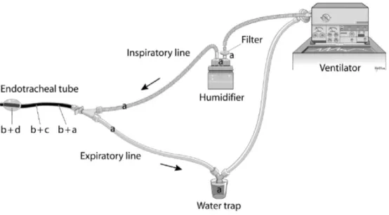
Figure 1.2: Endoracteal tube and its connections ... 4
Figure 1.3: The patient is connected to ventilator ... 5
Figure 1.4: Resistance of microorganisms against disinfectants. (Prions has resistance to disinfectants because of this it is not above) ... 7
Figure 1.5: Cleaning, Disinfection and Sterilization Methods [34]. ... 9
Figure 1.6: Floating Electrode Dielectric Barrier Discharge (FE-DBD) for direct plasma treatment [39]. ... 11
Figure 1.7: Plasma Jet for indirect plasma treatment [40]. ... 12
Figure 1.8: DBD Schematic (A) [41]. The treatment view of hand made DBD in our laboratory (B). ... 12
Figure 1.9: Effect of plasma and plasma species on cells ... 14
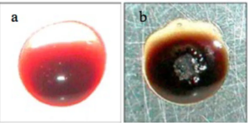
Figure 1.10: Plasma assisted blood coagulation: (a) Untreated blood, (b) Blood drop after 15 seconds of FE-DBD plasma treatment ... 15
Figure 1.11: Plasma interaction with living tissue ... 17
Figure 2.1: Prepared stock cultures for using in experiments agar (A) and glycerol (B) ... 21
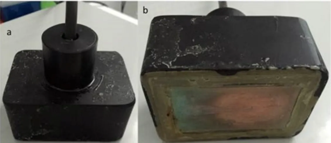
Figure 2.2: In front of custom made copper electrode (A) and the below of electrode (B) the glass on the electrode surface as a dielectric material and copper ... 22
Figure 2.3: In the system for the plasma treatment, power supply, gas tube and electrode were seen (A). The supply connections which were high ... 22
Figure 2.4: The custom made quartz fluid holder ... 23
Figure 2.5: Chemical reaxion of XTT [52]. ... 24
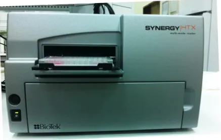
Figure 2.6: Samples were read via Multi-mode reader ... 25
Figure 2.7: Direct plasma treatment of suspended organisms culture with 1 mm discharge gap. ... 26
Figure 2.8: The application of plasma treated liquid on the suspense organisms .... 27
Figure 2.9: Plasma jet plasma (He/O2) treatment of suspended organisms culture. . 28
Figure 2.10: The overview of a drip flow reactor system established for biofilm formation in endotracheal tubes (A), peristaltic pump (B), growing ... 29
Figure 2.11: The inner surface of the endotracheal tube designed to be treated with plasma DBD electrode (A): schematic, (B): side and (C): in front of it. ... 30
Figure 2.12: Air plasma jet treatment on the endotracheal tube ... 32
Figure 3.1: Antimicrobial effect of direct plasma treatment on P. aureginosa (A) A.baumannii (B) and, C.albicans (C) depending on changing frequency. ... 35
Figure 3.2: The antimicrobial effect of the liqud that was treated with plasma during ... 38 Figure 3.3: The amount of biofilm in the drip flow reactor depend maturing time. 39
xviii
Figure 3.4: The blocking effect of biofilm formation depends on plasma pre- ... 40
Figure 3.5 : XTT Results following plasma pre-treatment ... 41
Figure 3.6: Results of Safranine Test after Plasma Pre-treatment ... 42
Figure 3.7: Antimicrobial Activity Accord Colony Counting Results of Nebulisation Plasma treated (A) DIW, (B) PBS, and (C) NAC Solutions on the Tested ... 43
Figure 3.8: XTT Results of Nebulisation Plasma treated (A) DIW, (B) PBS, and (C) NAC Solutions on the Tested Organisms ... 45
Figure 3.9: Safranine Tests Results of Nebulisation Plasma treated (A) DIW, (B) PBS, and (C) NAC Solutions on the Tested Organisms ... 47
Figure 3.10: Antimicrobial activity of air plasma jet application on biofilm inner the ... 48
Figure 3.11: XTT tests results after the air plasma jet treatment on biofilm was formed on the endotracheal tubes ... 49
Figure 3.12: Safranine tests results after the air plasma jet treatment on biofilm was ... 50
Figure 3.13: Scanning Electron Microscopy was used to take views of untreated .. 51
Figure 3.14: Scanning Electron Microscopy was used to take views of nebulization ... 52
Figure 3.15: Nebulization of plasma treated NAC on endotracheal tube ... 52
Figure 3.16: Representative images of morphological modifications on ... 53
Figure 3.17: Atomic-force microscopy (AFM) was used to take views of non- ... 54
xix
EVALUATION OF EFFECTIVENESS OF COLD ATMOSPHERIC PLASMA TREATMENT METHODS FOR PREVENTION
VENTILATOR-ASSOCIATED PNEUMONIA SUMMARY
Hospital-acquired infections (HAI) (nosocomial infection) are serious health problems that occur when person acquires infection during stay in a hospital for causes other reasons than that infection. Colonization of microorganisms pave the way for deaths, in detail, Ventilator Associated Pneumonia (VAP) is remarkable in terms of mortality accounted for 15% of nosocomial infections. Ventilator associated pneumonia (VAP) is common a disease, especially among patients that are on connected ventilators in intensive care units. It is defined as the pneumonia, in which occurs in at least 48 hours after mechanical ventilation, is provided via endotracheal tubes or tracheostomy. This infection occurs because of assault of microorganisms that settle in endotracheal tubes in respiratory tract and then in lung parenchyma tissue. The most common settling organisms in respiratory tract are S.aureus, P. aureginosa, K. pneumonia, E.coli and Acinetobacter, Candida, Enterobacter species. Due to multi drug resistant microorganisms, the current treatments are inadequate and the mortality rate increases. Therefore, plasma can play significant role for the management of ventilator-associated pneumonia. Plasma is ionized gas and defined as the fourth state of matter. The treatments of plasma in health care can ensure quick and pretty effect to protect from resistant microorganism to antibiotics to patients. The purpose of present study is to develop a method of plasma treatment which can be utilized for the control and prevention of ventilatör associated pneumonia. For this concept, we have designed different systems and determined optimum parameters to prevent and eradicate biofilms of Staphylococcus aureus, Acinotabacter baumannii, Pseudomonas aureginosa, and Candida Albicans in endotracheal tubes. Our results indicate that, various plasma treatment modalities are capable of prevention and eradication of biofilms grown in endotracheal tubes. Therefore, cold atmospheric pressure plasma treatment methods could be considered as for the management of ventilator associated pneumonia.
xxi
VENTİLATÖR İLİŞKİLİ PNÖMONİNİN ÖNLENMESİ AMAÇLI ATMOSFERİK SOĞUK PLAZMA MUAMELE YÖNTEMLERİNİN
ETKİNLİĞİNİN DEĞERLENDİRİLMESİ ÖZET
Hastane kökenli enfeksiyonlar (hastane enfeksiyonları) kişinin bu enfeksiyon dışında bir sebepten hastanede yatışı sırasında meydana gelen ciddi bir sağlık problemidir. Mikroorganizmaların kolonizasyonu ölüme yol açıyor, detaylandırırsak, Ventilatör İlişkili Pnömoni (VİP) hastane enfeksiyonlarının %15’ini oluşturup ölüm oranı bakımından dikkat çekmektedir. VİP yoğun bakım ünitelerinde ventilatöre bağlı hastalarda görülen yaygın bir hastalıktır. Bu hastalıkta endotrakeal tüpler veya trakeostomi aracılığıyla mekanik ventilasyonun sağlanmasıyla 48 saat içinde pnömoni gelişir. Öncelikle solunum yollarına ardından akciğer parankima dokusuna yerleşen mikroorganizmaların saldırıları nedeniyle bu enfeksiyon meydana gelir. Solunum yollarına en sık olarak yerleşen bakteriler S.aureus, P. aureginosa, K. pneumonia, E.coli, Acinetobacter, Candida, ve Enterobacter türleridir.
Antibiyotiklere dirençli organizmalar nedeniyle mevcut tedaviler yetersiz kalmakta ve ölüm oranı artmaktadır. Bu hastalığı çözmek için plazma önemli bir rol oynayabilir. Plazma iyonize gaz olup maddenin dördüncü halidir. Plazma tıp alanında sıcak olmayan plazma olarak adlandırılır ve tıbbi plazma yeni ve gelecek vaat eden bir alandır. Tedavi alanındaki plazma uygulamaları hastaları antibiyotiğe dirençli organizmalardan korumak için hızlı ve güzel etki sağlayabilir. Araştırmanın amacı endotrakeal tüpte oluşan biyofilmi önleyici ve biyofilmi inactive edici plazma uygulamasını geliştirmektir. Biz bu amaç için farlı sistemler tasarladık ve endotrakeal tüpte oluşan Staphylococcus aureus, Acinotabacter baumannii, Pseudomonas aeruginosa ve Candida albicans biyofilmini önlemek ve ortadan kaldırmak için uygun plazma parametrelerine karar verdik. Sonuçlarımız çeşitli plazma uygulama yöntemlerinin endotrakeal tüp içinde oluşan biyofilmin önlenmesinde ve yok edilmesinde yeterli olduğunu gösteriyor. Bu nedenle, atmosferik basınçlı soğuk plazma uygulama yöntemlerinin ventilator ilişkili pnömoninin tedavisinin yönetiminde dikkate alınabilir.
1 INTRODUCTION
1.
1.1 Hospital –acquired infections
Infection is a process where an infectious factor can develop and increase number in the tissue of a macro-organism [1]. Hospital-acquired infection (HAI) (nosocomial infection) is a serious health problem that occurs when person acquires infection during stay in a hospital for a cause other than that infection. The patient with HAI does not have this infection or the infection is not in incubation period of HAI before hospitalization. Nosocomial infections can be seen after discharge of patient and it threates not only patient but also health care staff and the public.[2, 3] Under favor of close adaptation to human system and environmental conditions, organisms that are bacteria, viruses and fungi can cause HAIs [1]. HAIs are the main reasons of deaths and rise morbidity of hospitalized patients. Therefore, HAI is an important issue for patients, healthcare professionals and public health. Longer stay of infected patients in hospital is a significant contributor to healthcare cost [2, 4]. In the United States, almost 4.5 million patients are affected by HAI that causes almost 100,000 mortality/year. Morever it causes high expense that is nearly from 28 billion dollars to 336 billion dollars per year in excess hospital cost [2, 5-7]. According to The World Health Organization 1.7 million patients are influenced by HAI and annually 37,000 deaths are reported due to HAI in Europe [7]. Hospital acquired infection rate had increased from 0.72% to 0.98% in a perod of 1970-2000s due to increased number of hospitalized patients and older age of patients [8]. Eventhough the efforts for prevention of HAI and progress in public health, nosocomial infections continue to develop in patients who stay in hospital and health care professionals [4]. According to publication of the World Health Organisation, the most common HAIs are surgical site, urinary tract and respiratory tract infactions. [2, 4]. Development of HAI depends on various factors such as properties of microorganisms, immune system of patient, intrinsic virulence, environment that includes using some materials from hospital and quantity of infective materials [2, 4, 9]. According to National Healthcare Safety Center, during 2009-2010, about 20% of nosocimal infections are
2
caused by of multidrug resistant organisms (MDROs) [5, 10]. Antibiotic resistances in microorganisms are led by excessive and unnecessary use of antibiotics. Once an organism acquires resistance to a specific antibiotic, then either antibiotic will remain inefficient or higher dose will be needed to eradicate the infection. Methicillin resistant Staphylococcus aureus (MRSA) (8.5%), Vancomycin resistant Enterococci (VRE) (3%), Escherichia coli (2%), Resistant Enterobacteriaceae, Multi–drug resistance Acinetobacter spp., Pseudomonas aeruginosa, Klebsiella pneumoniae (ESBL), Clostridium difficile associated diarrhea (CDAD) and Candida albicans are common MDR organisms responsible for nosocomial infection. They are capable to form biofilms that are sources of contamination and infection in hospitals. Therefore, they are responsible for hospital-acquired penumonias, wound infections, catheter related bacteremia that is seen frequently in hospitals [4, 5, 10, 11].
1.1.1 Urinary Tract Infections
Bacteria are the main reason for causing of Urinary Tract Infection (UTIs) that contain E. coli, which is responsible of 80-90% of these diseases, and S. saprophyticus. According to reports, UTI accounts for one-third of HAI. Normally, any organisms do not stay in urine. If the bacteria enter urethra, it causes infections that are treated easily in most situations. However, a lower urinary tract infection because of infection may spread in ureters and kidneys. The spreading of bacteria in kidney causes permanent kidney damage. [2, 4, 5, 12]. Urinary tract infections contain; urethritis, cystitis, ureteritis and, pyelonephritis. The prevelange UTIs occur in women and affect bladder and urethra. Cystitis, in other words, infection of the bladder is usually brought on E. coli that found in the gastrointestinal tract. Urethritis, put it differently, infection of the urethra can take place if gastrointestinal bacteria expand from anus to urethra [4, 9, 13, 14].
1.1.2 Surgical Site Infections
Surgical site infections (SSIs) may present on surgical wounds that is over or under aponeurosis. SSI is defined as any infection on the site of surgery that develops in 30 days after surgery and increases hospitalization up to 7 to10 days. Depending on the typem of surgery, SSI can be detected in few days to weeks. Patient’s age, gender, type of surgical wound, treatment time, anesthesia types are important factors for the development of SSIs. [1, 9, 15] Contamination of operation room, using non-sterile
3
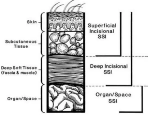
equipment, inadequate preparation of patient before operation are main causes for the development of SSIs [4]. SSI can be divided into two types, which are endogenous and exogenous. While endogenous is related to patient floras that are skin, mucous membrane and gastrointestinal tract, exogenous are related to aseptic techniques, insufficient hand cleaning, flora of surgical staff, contaminated equipment, unhygienic environment and ventilation. Aerobic and anerobic microorganisms such as Staphylococcus aureus, Escherichia coli, Candida albicans, Staphylococcus epidermis, and Enterococcus species from patients’ samples and MDR organisms such as Methicillin-resistant Staphylococcus (MRSA) and Vancomycin resistant Enterococcus (VRE) are common organisms that cause SSIs. Surgical site infections can be categorized as finding etiher incisional organ or space. Also incisional SSIs are separated related to involving just skin and subcutaneous tissue. Moreover, it seen that at Figure 1.1, deep incisional SSI for more deeply soft tissues of the incision and organ or space SSIs contain organ or space in body [1, 9, 15].
Figure 1.1: SSI classification depend on deepness infection 1.1.3 Ventilator Associated Pneumonia
Ventilator associated pneumonia (VAP) is prevalent disease especially among patients that are connected to ventilators in intensive care units. It is defined as the pneumonia that develops in at least 48 hours after mechanical ventilation is provided via endotracheal tubes or tracheostomy. VAP significantly increases mortality rate in
4
patients who are treated with invasive mechanical ventilation that is need for intubation in intensive care [16].
Figure 1.2: Endoracteal tube and its connections
In the process of mechanical ventilation, an endotracheal tube is inserted into trache, which will direct the air pumped by mechanical ventilator towards lung. During insertion of endotracheal tube and ventilation, the respiratory tract can be damaged. Moreover, the tube is not biocompatible, and serves as a perfect surface for biofilm formation. Also, mechanically ventilated patients are seriously ill and have weaker imnmune system, which makes them more susceptible to VAP. There are significant mortality in VAP cases accompanied with other diseases. This infection occurs because of assault of microorganisms that settle in endotracheal tubes in respiratory tract and then lung parenchyma tissue. Colonization of microorganisms pave the way for deaths, in detail, VAP includes 15% of HAIs, and these are the most prevalent reasons of deaths because of nosocomial infections having > 75% of mortality rate.
Most common settling organisms in respiratory tract are S.aureus, P. aureginosa, K. pneumonia, E.coli and Acinetobacter, Candida, Enterobacter species [4, 9, 17].
5
Figure 1.3: The patient is connected to ventilator
Proper disinfection and careful tubing, and using humidifiers should done to decrease contamination [4]. These methods are insufficient to eradicate organisms that cause ventilator-associated pneumonia.
1.2 Microorganisms
Microorganisms were presented before plants and animals seemed billions years ago in the world. Microorganisms are smallest form of living organisms. Bacteria are classified as gram stain gram (+) and gram (-). Gram negative bacteria has thiner peptidoglycan than gram positive bacteria [18, 19]. Later gram staining, gram (+) bacteria look like purple-violet and gram (-) bacteria seem pink. The same method is red stain safranin, the cells types can be recognized [19, 20]. Gram-negative bacteria examples are Acinetobacter, Escherichia Coli, Pseudomonas, Enterobacter, Salmonella, Helicobacter etc. [21]. The examples of gram-positive bacteria are Staphylococci (Staphylococcus aureus), Streptococci, Enterococci etc. [22]. According to size, the distinguishing of bacteria is not possible. However they can separate according to shapes that are coccus (Staphylococcus aureus), bacillus (Escherichia coli), and spirillum (Treponema pallidum) [19, 20]
1.2.1 Antibiotic-resistant microorganisms
There are serious threats in hospital environment related to microorganisms. For instance methicillin-resistant S.aureus in other words multi resistant strains of S.aureus can transport on the health staff’s hands [4]. Multi drug resistant organisms (MDROs) cause common health problems in the world seriously [23]. In the United States, annually 2-million hospital infections occur and drug resistance bacteria cause more than half of nosocomial infections. It plays an important role about morbidity
6
and mortality of patients and economic cost. Numbers that are between $4 billion-$30 billion can explain the cost. These microorganisms are not only seen in the patients that are stayed in hospital or discharged from hospital but also seen among patients that are taken some treatments such as indwelling catheters, surgical wounds, and dialysis [24]. Frequently, the arrangement of using antibiotics with limited dose causes insufficient to eliminate MDR pathogens. Because of this reason after effective infection threapy, the patients body are likely to be colonized with MDR pathogens [25]. Multi drug resistant bacteria are such as Methicillin resistant Staphylococcus aureus, Vancomycin-resistant Enterococcus, Mycobacterium tuberculosis, and Klebsiellapneumoniae. In order to prevent antibiotic resistance, unnecessary antibiotics should not be used, efficient hygiene must be provided and the infection control procedure must be followed [26].
1.2.2 Fungi
Fungi may present as a single cell or complex multicellular form. They live on the solid or in liquid. They act as a responsible duty of carbon cycles. However, they induce diseases in human such as skin disease that can be ringworm. The fungi are divided three main types that are multicellular filamentous moulds, macroscopic filamentous and single celled microscopic yeast. Yeast is almost as small as red blood cell nearly. Some yeast play important roles in necessary reactions. However, some of yeasts such as Candida Albicans induce infection that are related immune system [27, 28] .
1.3 Prevention of Infection
Common methods are used for infection prevention are listed and briefly explained below:
1.3.1 Antiseptics and Antisepsis
Antiseptics are defined as biocidal compunds that harm and hinder the development of microorganisms in or on living structures such as tissue, skin. The skin, the largest organ, constitutes nearly one sixth of human body weight, has enormous of function that are temperature regulation, playing role as a barrier, preventing water loss. So living tissues and skin are more susceptible than hard tissue, it is important to use
7
right and limited antisepsics, due to their toxicity. There are many formulations and preparations such as surgical scrubs, mouthwashes, antimicrobial hand washes that are utilized as antiseptics because of being biocidal. They provide antisepsis conditions that represent inhibitiation microorganisms mentioned before; it provides decrease or blocks the hazard of infection. Antisepsis treatments are such as periodic skin hygiene, skin medication before surgical response or wound infection and mucosal membranes treatment. The most common used skin antiseptics and washes are biocides [1, 29, 30].
1.3.2 Disinfection
Disinfection is defined as the eradication of microorganisms on living and nonliving environment. Chemical and physical disinfection are two major types of disinfection methods and these methods may not completely eliminate all types of microorganisms. In physical disinfection, physical energy is used for eradication of microorganisms. In chemical disinfection, a chemical substance can kill the living microorganisms via denaturation of the major proteins, enzyme, and genetic material. In the physical disinfection applications, the ratio of killing organisms relies on the dose of physical energy and application time. In chemical disinfection, efficacy relys on killing organisms related to toxicity, quantity of disinfectant and application time. These methods are used to against microbial contamination but are not completely effective to all types of organisms [1, 29, 31]
Figure 1.4: Resistance of microorganisms against disinfectants. (Prions has resistance to disinfectants because of this it is not above)
8
Chemical disinfectants have various chemical groups such as alcohol, aldehyde, clorine, alkylamine, iodine, phenol, and guanidine, alkaline, peroxide, inorganic, and organic acids that show antimicrobial efficay. They have different classes related to resistance of microorganisms against disinfectants.
1.3.3 Sterilization
Sterility infers disappearance of living organisms in any form. Sterilization is the process to make sterile tools or anything through killing all forms of living organisms. In the medical field, in order to obtain surgical asepsis, sterilization technic is enhanced for eradicating potencial infectious element in environment without harming the sterilized tools. This technique is used for obtaining complete disinfection on hazardous tools used for operation on the patient’s body. Morever it is used for preventive procedure related to infectious diseases [1, 32, 33]. Invasive applications require contact within patients’s tissue or mucous membranes and medical tools. This contact could cause infection due of entrance of microorganisms. Any error related to disinfection and sterilization has risk for patients. The strelization procedure is adjusted according to characterization of surgical items, treatments and patients tissue and mucous membrane [32]. Sterilization techniques are separated two major units as physical and chemical sterilization. In physical sterilization, the process includes using of moist heat (steam), dry heat, and radiation. The steam procedure is a common and safe method depends on existence of steam under pressure. This sterilization process is utilized in controlled vessels such as autoclaves, steam sterilizers, or pressure vessels. Even if this method is reliable, it is not appropriate for temperature or pressure delicate devices or products. Dry-heat or hot air sterilization involves incineration of waste materials. The dry heat sterilization are utilized sterilization ovens and incineration whose temperatures are ranging from 800 to 1,300 0C. The general dry air sterilization is usually applied at 160 0C for 120 minutes, at 170 0C for 60 minutes, at 180 0C for 30 minutes, and at 190 0C for 6 minutes. Even though dry heat sterilization is cheaper and easier than steam process, many temperature sensitive materials or liquid can not be sterilized with this technique. The last common method is the high-energy ionizing radiation method that utilizes X rays and electron beams. Ionizing radiation sterilization is used for sterilization of medical devices, food, wastewater, liquid, solid materials, and pharmaceutical products. Radiation sterilization has limited applicability because
9
of cost and safety concern. These methods for sterilization and disinfections are seen that in Figure 1.5. There are some technics that are used for special applications, for instance; plasma, pulsed light and supercritical fluids [1, 29, 34].
Moreover, there is a new method for sterilization from United Kingdom. Superoxide water is the mixture of oxidizing species. This product is under trademark
STERILOX®. The major products are hypochlorous acid in 144 mg/L of concentration
and chlorine radicals. The process takes place at pH 5.0- 6.5 and the potential of oxidation-reduction is higher than 950 mV. During this process, a saline solution is passed over titanium-covered electrodes at a current of about 6 to 9 Amps. Studies have shown that this process effectively causes 5-log reduction of Mycobacterium tuberculosis, M.chelonae, HIV, MRSA, E.coli, C.albicans, P.aureginosa in less than 2 minutes. However, antimicrobial efficacy of this process decreases with the existence of organic material and further studies are needed [29, 35, 36].
Figure 1.5: Cleaning, Disinfection and Sterilization Methods [34].
However, these technics are not enough to eradicate some types of organisms on the surfaces or living tissues.
10 1.4 Plasma
Plasma that is fourth state of matter is an ionized gas and first defined by Irving Langmuir in 1928. He thought that this matter resembles blood plasma because of multicomponent structure and it has free electrons, ionized gas, and reactive species. Plasma constitutes more than 99% of the universe and aurora borealis, lightning sun are examples of natural plasmas. Additionally, plasma can be generated artificially. Plasma is an ionized gas. Ionization is defined as a process that an atom or molecule gains or looses at least one electron. In consequence of ionization the molecule or atom turns into positive or negative form depending on the gain or lost of electron respectively. When the temperature rises, molecules gains energy, it occurs transform plasma as a fourth state after solid, liquid, gas phases. During generation of plasma, electrons take energy much faster than heavier particles with the effect of electrical field. Electrons and ions cause plasma electrically conducting and responsive [37]. According to thermal characteristics, plasma could be generated in various ways. Plasma is categorized as non-thermal, cold or non-equilibrium and thermal, hot or equilibrium plasma according to temperature of free electrons and gas. The electric field helps to generate plasma and electrons gain energy quicker than havier particles. In non-thermal plasma or nonequilibrium plasma, cooling of ions and neutral molecules are more efficient than energy transference from electrons, the gas remains at low temperature. As for thermal plasma, energy flux transfers from electrons to heavy particles balance and from heavy particles to environment. The temperature of heavy particles and electrons become nearly equal. The plasma temperature is substantial for treating temperature-sensing material [9, 37, 38]. According to treatment method, cold atmospheric plasma can be divided two types as direct and indirect plasma treatment. For direct plasma treatment, the material that is treated such as tissue, skin, cell serve as an electrode, and the discharge is generated between high voltage electrode and the treated material. The example of direct plasma is in Figure 1.6.
11
Figure 1.6: Floating Electrode Dielectric Barrier Discharge (FE-DBD) for direct plasma treatment [39].
In indirect plasma treatment, the plasma discharge is generated in a region that is not in contact with the treated material and the plasma products are carried with a gas flow. Plasma needles or torches are examples of plasma devices that are utilized in indirect plasma treatment. In addition to direct and indirect plasma treatment also, fluid mediated plasma treatment has been defined. In fluid mediated plasma treatment, firstly, a fluid is treated with direct plasma treatment, which leads chemical modifications in the fluid, and then the treat fluid is transferred to a substance such as bacterial cell. In fluid mediated plasma treatment of fluid is considered as direct and transferring fluid to a substance is considered as indirect plasma treatment. Eventhough it is defined as fluid mediated plasma treatment various materials such as alginate gels metals polymers might be treated in the same way in order to obtained varios plasma mediated effects [38].
12
Figure 1.7: Plasma Jet for indirect plasma treatment [40]. 1.4.1 Dielectric Barrier Discharge (DBD) Plasma
Dielectric Barrier Discharge (DBD) was first defined by Siemens in late 1800s for ozone production. DBD set up consists of one high voltage and one grounding electrode. In the DBD set up at least one of these electrodes should be covered with a dielectric material such as glass, ceramic, quartz. Presence of dielectric material is used to avoid arc formation and limits and it distributes the current over a large area. DBD generates localised shock waves and noise. DBD is largely implemented for ozone product in UV-source. DBD plasma electrode could be set in plane and cylindrical configurations. DBD is one of the most used plasma for medical application due to its flexibility.
A B
Figure 1.8: DBD Schematic (A) [41]. The treatment view of hand made DBD in our laboratory (B).
13 1.4.2 Plasma Chemistry
While the electric field is implemented to gas that is in the environment, electrons are accumulated to get higher energy. These electrons collide with gas atoms and molecules. It causes to generation of positively charged ions because of electron lost from ambient gas. The positive ions are essential components related to plasma-chemical processes. Their exothermic reactions do not have any activation energy. It makes their contribution important in plentiful plasma-chemical process. Morever, for high chemical activity, atom and molecule ions are able to have substantial kinetic energy that decides their additive in reactions. Chemical species formed by plasma treatment. Active plasma is a complex system that is ready to reaction because of high concentrations of charged particles that are electrons and ions, active atoms, radicals and UV-photons. During the plasma generatingh, chemical species act part on the biological influence of cold atmospheric plasma. During formation of plasma reactive oxygen species (ROS), such as hydrogen peroxide (H2O2), ozone
(O3), hydroxyl radical (OH), singlet oxygen, superoxide (O2−), and reactive nitrogen
species (RNS) such as nitric oxide (NO), nitrite (NO2), nitrate (NO3). While
generating plasma, there is energy transfer that is from electrons to ambient gas as exit and ionization. These processes cause formation of radicals that are H·, OH· and O·. For example, the reactions for obtaining hydrogen peroxide and oxygen molecule can be explained below;
H2O H++ OH
-e -+ H
2O H· + OH·
H· + O2 + M HO2· + M OH· + H2O2 HO2 ·+ H2O
OH· + O3 HO2· + O2 OH·+ OH· H2O2 HO2·+ HO2· H2O2 + O2 [9, 37].
14 1.4.3 Plasma Medicine
Plasma medicine is new and up-and-coming area. Thermal plasmas can be used for cauterization, tissue removal, cutting tissue, sterilization of medical tools in health areas. In addition, treatments of plasma health care can ensure quick and pretty influential according to protecting resistant microorganism to antibiotics, and hospital infections. It can be supplied the entering of reactive species from plasma to cell wall and cytoplasm, result of this stage cells affect [9, 38, 42]. Plasma medicine is come across some healthy treatments that are used for improving of wound healing and treatment of skin, cardiovascular, and digestive system. During these treatments, some reactions take place; these are shown in Figure 1.9. Morever, dental treatment is applied by plasma nowadays. Plasma holds promise for cure of cancer. The bleeding controlling can be ensured by plasma applications. As well as treatment, plasma can improve biomaterial quality and it can provide tissue analysis, diagnosing of diseases. Cold atmospheric plasma is also purposive creating new drugs in pharmacological area [38, 43].
15 1.4.3.1 Plasma Assisted Blood Coagulation
Blood coagulation is a critical subject regard to wound healing in the medical area. It is a natural response of the human body to provide homeostasis, because of vessel damage. Friedman et al. confirmed to expedite blood coagulation with floating electrode-dielectric barrier discharge plasma (FE-DBD) by activating some of coagulation proteins and by rising Ca2+ ion concentration. These proteins provide rapid fibrinogen gathering.
Figure 1.10: Plasma assisted blood coagulation: (a) Untreated blood, (b) Blood drop after 15 seconds of FE-DBD plasma treatment
The tests are demonstrated that the cold plasma treatment can trigger natural coagulation processes in Figure 1.10. The plasma can change the ionic strength of the solution and pH [9, 38].
1.4.3.2 Plasma Applications for Cancer Theraphy
Cancer is called as a disease which in some cells have rised metabolism and avoid the processes of natural regulated growth and death for different causes. They can pile up in the body than other normal cells. According to current studies, the non-thermal plasma treatment could play a role in cancer therapy. Fridman et al, 2013, have researched that melanoma cells go through necrosis at specific plasma treatment doses that were 15 s at 1.4 W/cm2 and apoptosis at lower plasma doses 5 s at 0.8 W/cm2. Additionally, Vandamme et al. have reported antitumor activity of cold atmospheric DBD plasma on U87MG malignant glioma and HCT-116 colorectal carcinoma. According to results, accumulation of cancer cells in S phase, which is indicator of decayed of cancer cell proliferation and generation of DNA damage guided to apoptosis and multiphase cell cycle arrested due by the reason of plasma generation of ROS. Kim et al. have submitted a new way to cancer treatment by
non-16
thermal plasma treatment. They have demonstrated that cultured TC-1 lung carcinoma cells are more vulnerable to non-thermal Helium microplasma jet treatment than cultured fibroblasts and undergo apoptosis. According to forementioned information, non-thermal plasma treatment could be a new method for cancer therapy [9, 38].
1.4.3.3 Dental Applications of Cold Atmospheric Plasma
There are three major applications in dentistry. First one is related to killing of bacteria is subjected teeth surfaces and inner tooth root canals. Also, it provides some anti-inflammatory activity. Secondly, the plasma treatment enhances adhesion and incorporation of materials like included in tooth fillings. The last one is whitening of the tooth enamel [9, 38]. There were two practices about plasma and dentistry in our plasma medicine laboratory. Firstly the using atmospheric low temperature plasma activated hydrogen peroxide and deionized water on dental bleaching. Also, the second one was to evaluate the bleaching efficacy and colour stability of non-equilibrium plasma source activated agents . For this purpose two hundred bovine enamel-dentin samples were used by treating 40% hydrogen peroxide gel and plasma treatments during specific time. After these experiments, we obtained that plasma treatment could provide more effective and significant bleaching safely.
1.4.3.4 Plasma Applications in Dermatology and Wound Healing
Non-thermal plasma could be promoted wound healing by decreasing the microbial colonization and direct influence on skin cells. For this context, argon plasma jet (MicroPlaSter®) was used as an indirect treatment for wound healing firstly. The plasma treatment is efficient to give acceleration wound healing by decreasing number of bacteria such as Klebsiella oxytoca and Enterobacter cloacae. Food and Drug Administration (FDA) has accepted the plasma skin regeneration technology. This thermal nitrogen plasma jet was used for some clinical application such as superficial skin lesions, wrinkles [9, 44]. Also Emmert et al. has submitted that the DBD plasma treatment for 1 min/day during 30 days. They have obtained significant reduction of S.aureus colonization on the skin with decreased itching, as a result the healing of atopic eczema has improved [45].
17
1.4.3.5 Cold Atmospheric Plasma - Cell Interactions
Plasma provides not only physics effect but also it provides therapeutic effect. It induces biological reply in cells and living tissue. With this influence, it can be thought that interaction mechanism relies on method of plasma producing and the accepting of producing plasma by organisms. In order to obtain wished effect on living structure, reactive species, electrons, and ions have important roles. These interaction have been showed that in Figure 1.11. Moreover, the quicker bacteria inactivation on tissue is provided by direct plasma [43].
Figure 1.11: Plasma interaction with living tissue 1.4.3.6 Plasma sterilization
Plasma sterilization depends on an assembly of factors like Ozone, UV photons and reactive species. The generating of plasma is easy and cheap via atmospheric plasma. Morever it provides safe sterilization comparing toxic chemical substances. The
18
results of experiments show that various cold plasma treatments that are Dielectric Barrier Discharge, plasma jet, gliding arc discharges wipe out of organism that can be in planktonic or biofilm version [7, 9, 46]. Plasma sterilization can be utilized various areas that are living tissue, the disinfection of larged-scaled of air and water streams, inactivation of hazardous pathogens in food industry, the finishing dangerous bleeding without harming tissue [38].
1.5 Nebulization
There are different ways that are decided by doctors for delivering medication related to ilness to human bodies. A nebulizer works by linking tubes with a compressing air source. This medical tool is adjusted the oxygen and air are blasted to the liquid deposit rapidly and inhaled to patient. This transiton will occur aerosol that include the medication needen for patient [47]. Nebulization is a treatment of medical concept by inhalation. These treatments are effective for respiratory tract problems that are pneumonia, respiratory congestion, asthma bronchospasms etc. Practically nebulizer is used for infants and small children. Nebulizer using that is inhaled treatment is more effective than intravenous treatment because of reaching directly infected lung tissue. Antibiotics can be used to heal against bacterial infections via nebulizer even if they do not prevent from lung infections enough. [48-50]. Nebulized antibiotics are used for treating respiratory infections almost 70 years. It has important advantages that are related decreasing side effects. Although nebulized antibiotics seem to best way for ventilator-associated pneumonia, it has many obstacles for healing of this disease [51]. For this concept, we consider that plasma medicine is new approach for finding solution for VAP.
1.6 Aim of Study
Plasma medicine is a new and rapidly developing field that aims to develop new therapeutic solutions. The cold atmospheric plasma is used in different medical areas that are mentioned before. The plasma medicine treatments have significant antimicrobial effects on bacteria, fungi, viruses and prions. Ventilator-associated pneumonia is an important healthcare problem that increases morbidity and mortality. It develops as a result of biofilm formation in endotracheal tube. The purpose of this study is to develop a method of plasma treatment which can be
19
preventing the formation of biofilm and inactivation of biofilm that is formed in the endotracheal tubes with the conducting of Cold Atmospheric Plasma.
20 MATERIALS AND METHODS 2.
2.1 Culvitation of Microorganisms
Staphylococcus aureus (ATCC 25923), Acinotabacter baumannii clinical isaolate, Pseudomonas aeruginosa (ATCC 27853), and Candida Albicans (ATCC 90028) strains were supplied from Department of Microbiology, Medical Faculty at Ege University on agar plates. One colony of each pathogen was collected from agar plates and grown overnight at 37 0C and 200 revolution per minute (rpm) in shaking incubator. Bacteria were cultivated in Trypticase Soy Broth Medium (TSB) (Sigma-Aldrich-22092-500G) and C.albicans was cultivated in yeast extract-peptone dextrose (YPD) (Sigma-Aldrich-Y1375-250G). After overnight cultivation, 100 µl of grown cultures were transferred and spread over agar plates. Bacteria stock cultures plates were prepared with Trypticase Soy Agar (TSA) (Sigma-Aldrich-22091) and yeast stock culture plate was prepared using YPD agar (Sigma-Aldrich-Y1500-250G) and the plates were stored at 40C in the fridge. Whenever a new culture was needed one colony was collected and grown overnight in shaker incubator. Each stock plate was refreshed in every 2 months.
2.2 Preperation of Frozen Stock Cultures
Frozen glycerol stock cultures were prepared with overnight grown cultures. 850 µl of overnight grown culture was mixed with 150 µl of sterile glycerol in a cryotube using aseptic technique in Class II Biological safety cabinet, homogenized by pipetting and stored -20 0C for one night. Then cryotubes were transferred to freezer
and stored at -80 0C. Glycerol stock cultures were refreshed every 6 months. Whever
needed, one cryotube was thawed and plated over agar plate to prepare a fresh agar stock culture. It has shown that in Figure 2.1.
21
Figure 2.1: Prepared stock cultures for using in experiments agar (A) and glycerol (B)
2.3 Preperation of N-Acetyl Cysteine (NAC) Solution
In all experiments, a final 5 mM concentration of working N-Acetyl cysteine (NAC) (Sigma- Aldrich) solution was used. NAC powder was weighted and dissolved in 1X sterile phosphate buffered solution (PBS) to reach 100 mM concentration that is stored at -20 0C as NAC stock solution. Whenever needed NAC stock solution was appropriately diluted in 1X PBS solution in order to reach 5 mM concentration.
2.4 Designing and Manufacturing of Electrode and Fluid Holder
In order to generate non-thermal plasma, custom-made electrodes were used in our experiments.
2.4.1 Dielectric Barrier Discharge (DBD) Electrode
In order to generate non-thermal DBD plasma, a custom-made electrode was used. A copper plate with 10 mm thicknes and 38 x 64 mm dimensions was embedded into insulator polyethylene housing. The surface of the electrode was covered with a galss slide with 50 x 75 mm dimensions and 1 mm thickness and a high voltage electrical cable was soldered to copper plate to connect the electrode to plasma power supply. It is showed that below in Figure 2.2.
22
Figure 2.2: In front of custom made copper electrode (A) and the below of electrode (B) the glass on the electrode surface as a dielectric material and copper surface beneath of the glass were seemed.
2.4.2 Plasma Jet Electrode
Plasma jet electrode for the plasma jet treatment was constructed using a hollow cylindrical copper. A glass pasteur pipette with a was glued in to inner side of cylindirical cooper electrode which serves as high voltage electrode. Then a copper wire was concentrically assembled to system, which serves as grounding electrode. Then a silicone tube was connected to electrode system in order to provide required gas flow. Hand made plasma jet electrode is shown that in Figure 2.3.
Figure 2.3: In the system for the plasma treatment, power supply, gas tube and electrode were seen (A). The supply connections which were high voltage, grounding, and gas stream (B).
23 2.4.3 Fluid Holder
A glass fluid holder was constructed to keep liquid during plasma treatment. The custom-made glass fluid holder was made using a glass slide (the base part) with 50 x 75 mm sizes. Appropriate parts from another glass slide were cut using a glass cutter. Those parts, which serve as to walls of the fluid holder, were glued the base glass slide to create a reservoir. The fluid holder has inner reservoir dimensions of 57 x 30 mm with a liquid coloumn height of 1 mm below.
Figure 2.4: The custom made quartz fluid holder
2.5 Optimization of Plasma Parameters for Appropriate Antimicrobial Effects A series of experiments have been carried out in order to determine the optimum antimicrobial effect. The power supply was operated at 31.4 kV of peak to peak voltage, 1.5 kHz of frequency in order to obtain significant eradications of microorganisms. Working concentrations of organisms were determined using spectrophotometer. Serial dilutions of overnight grown culture were transferred into spectrophotometer cuvette and absorbances were taken 0.2 absorbance at 600 nm represents 108 CFU/ ml for microorganism. Once the 108 CFU/ ml of concentration
determined further dilutions were made when needed.
1 ml of 104,105, 106, 107 CFU/ml of cultures were taken into fluid holder and direct DBD plasma treatment was carried out at a fixed 1 mm of discharge gap at different voltage and frequency settings.
24
2.6 Quantifications Methods for Antimicrobial Effect of Plasma Treatment XTT and safranine assays were used to detect the effect of plasma treatment.
2.6.1 XTT Assay
XTT (2,3-Bis(2-methoxy-4-nitro-5-sulfophenyl)-2H-tetrazolium-5-carboxanilide) assay is a measurement method depends on the respiratory metabolic activity of living cells. It is a tetrazolium salt that is depending upon the modification of the yellow tetrazolium salt XTT to occur an orange formazan dye because of metabolic activity of cells [9].
Figure 2.5: Chemical reaxion of XTT [52].
XTT and menadione solutions with final concentrations of 0.5 mg/mL and 50 µM were prepared respectively with 1X PBS, 50 ul of XTT and 1 ul of menaidone was added into 1 ml of each sample that were tested and incubated at 37 0C. After the incubation, 200 µl of sample was collected from each endotracheal tubes and transferred into 96 well plate. Then absorbance of each sample was taken using a multiwall plate reader at 492 nm. It is below in Figure 2.6.
25
Figure 2.6: Samples were read via Multi-mode reader 2.6.2 Safranine Assay
Safranine test was used to detect the amount of extracellular polymeric substance that provides attachment of cells to each cells and surface during biofilm formation. Biofilms were grown and treated with various plasma treatment methods. Then, tubes were washed using 1X PBS to remove non-adherent cells. Afterwards, 2 ml 1% safranine was added on the endotracheal tubes and held for 15 minutes. Then the samples were washed with 1X PBS to remove excess safranine from the surface of endotracheal tubes and samples held for 30 min to let the samples dry completely [53]. Once the samples were dried, 2 ml of 30% acetic acid was added to each sample to dissolve safranine that stained EPS to be quantified. 200 ul of dissolved safranine was transferred into 96-well plate and absorbance of each well was measured at 550 nm.
2.7 DBD Plasma Treatment of Liquids
At each treatment, 1 ml of deionized water, PBS solution and NAC solution were treated at a time. A micro second pulsed AC power supply was used to generate atmospheric cold plasma for given voltage frequency. For the liquid mediated plasma treatment 1 ml deionized water, 1 ml 1X PBS and 1 ml 5 mM NAC solutions were transferred to fluid holder separately and treated with DBD plasma for particular time points. During treatment of liquids, the discharge gap was fixed to 1 mm. It shows that in Figure 2.7.
26
Figure 2.7: Direct plasma treatment of suspended organisms culture with 1 mm discharge gap.
2.8 Antimicrobial Activity of Nebulized Plasma Treated Liquids
1 ml of 10^7 CFU/ml S.aureus, P.aureginosa, A.baumannii and C.albicans were placed six well plate and the palte was transferred in to biological safety cabin to let the samples dry. After samples were fully dried, 2 ml of plasma treated liquid was transferred into nebulizer and nebulized over organisms. Total nebulization time of 2 ml liquid was measured as about 7 minutes. 3% hydrogen peroxide was used as a negative control. The treatment image is shown that in Figure 2.8
27
Figure 2.8: The application of plasma treated liquid on the suspense organisms After the nebulization process, samples were held for 10 minutes for interaction between organisms and treated liquid and then 1 ml of 1X PBS was added on bacteria and yeast for resuspension. Then serial dilutions were done appropriately and 100 µl of each sample were plated on nutrient agar plates and plates were incubated at 37°C for 24 hours. After incubation, surviving colonies were counted to determine antimicrobial activity. Each experiment was repeated 3 times.
2.9 Evaluation of the Antimicrobial Activity of Plasma Jet Application
Antimicrobial effects of air, He/O2 (99.5%: 0.5%) and N2/O2 (90%: 10%) plasma
jets, were tested on 1 ml, 107 CFU/ml of S.aureus, P.aureginosa, A.baumannii and
C.albicans at various frequency settings, treatment time and gas flow volume. In plasma jet optimisitaion study, 2, 4, and 6 standart liters per minute of gas flow, 500 Hz, 1000 Hz, 1.5 kHz of frequency were tested at a constant voltage of 31.4 kV at 5, 15 and 60 seconds of treatment time points. Plasma jet treatment is shown that in Figure 2.9.
28
Figure 2.9: Plasma jet plasma (He/O2) treatment of suspended organisms culture.
2.10 Optimization of Drip Flow Reactor (DFR) for Formation of Biofilm
A drip flow reactor system is useful to grow low shear biofilm at an interface of air and liquid. These reactors are adaptable to different conditions. They are appropriate for laboratory working. The significant of biofilm biomass that is needed can be obtained [54]. In our study, DFR was utilized in order to grow biofilm in endotracheal tubes similar to clinical conditions. 5 cm pieces of endotracheal tube were cut and connected to reactor system, which is composed of a closed sytem of tubing and a peristaltic pump as shown in Figure 2.10. In the system of the container contained fresh medium and the other container was used as waste collection. An overnight orgasnism culture was grown in shaker incubator and biofilm medium was prepared as decribed previously.
29
Figure 2.10: The overview of a drip flow reactor system established for biofilm formation in endotracheal tubes (A), peristaltic pump (B), growing biofilm in endotracheal tubes (C), the views of feeding container that
provided fresh broth and waste container where collected waste (D). The biofilm medium was circulated in the system until the whole system was filled with biofilm culture and the pump was stopped and held for 6 hours in order to allow growth of bacteria. After 6 hours of growth of bacteria fresh medium was circulated in the system for 18, 42 and 66 hours in order to reach a total 1, 2, and 3 days of maturation duration. After completion of maturation period, colony-counting test was carried out to determine biofilm growth.
2.11 Tests for Prevention of Formation of Biofilm
As previously reported in the literature, materials other than fluids may gain antimicrobial activity when treated with plasma. Therefore, we have tested if biofilm
30
formation could be prevented on the surface of plasma treated endotracheal tube. A cylindrical DBD plasma electrode was designed to treate inner side of endotracheal tube. A cooper wire was inserted into a glass tube and sealed using silicone to build up high voltage electrode which could be inserted into endotracheal tube. Then the endotracheal tube with the high voltage electrode inside, was placed inside a copper tube which serves as grounding electrode. The defined setup provides plasma treatment the inner surface of endotracheal tube. Inner side of endotracheal tube was treated at 1.5 kHz frequency and 31.4 kV voltage for 15 seconds, 1 and 2 minutes. This system is shown that in Figure 2.11. After plasma treatment for formation of biofilm, endotracheal tubes were connected to drip flow reactor. When the biofilm formation was completed, in the other words, one day later, endotracheal tubes were separated from drip flow reactor and colony-counting assay was performed.
Figure 2.11: The inner surface of the endotracheal tube designed to be treated with plasma DBD electrode (A): schematic, (B): side and (C): in front of it.
31 2.12 Antibiofilm Effect of Plasma Treatment
Antibiofilm effects of plasma treatment were tested on biofilm forms afromentioned rganisms and methods that are followed for diffrerent plasma treatment techniques are given below.
2.12.1 Antibiofilm Effect of Plasma Nebulized Treated Liquid
Antibiofilm effect of nebulized plasma-treated liquid was tested on biofilm forms of Staphylococcus aureus (ATCC 25923), Acinotabacter baumannii Clinical Isolated Stream, Pseudomonas aeruginosa (ATCC 27853) and Candida albicans (ATCC 90028) strains. Deionized Water, PBS and NAC solutions were treated by plasma as explained previously. At a time, 1 ml of each liquid was treated and 4 ml of total liquid was treated for each test. Biofilm was grown inside 5 cm long segments of endotracheal tube. We have cut 3 pieces of each tubes, the length was nearly 1 cm. Every piece was put in 2 ml PBS in the TUBE falcon in order to remove biofilm, they were stayed 5 minutes and 35 OC in the ultrasonic water bath sonicator and they were vortexed for homogenization. After this step 10 µl liquid were taken in the falcon and added ependorf, and 3 dilutions that was 1:10 were completed, after these serial dilution, petri plates were spreaded with 100 µl solution that had organisms and stayed in the incubator for one night, at 37 0C. Moreover, safranin and XTT tests were also carried for the characterization of biofilm. For the positive control no plasma treatment was used. Also, only gas flow without plasma discharge was applied in order to assure that gas flow does not inactivate biofilm physically. Before the nebulization, biofilm liquid that was stayed in the endotracheal was emptied and washed for removing non-adherent biofilm in the tube. Plasma treated liquds were transferred into nebulizer separately and the nebulizer was connected to endotracheal tube and plasma treated liquids were nebulized throught the tube. The nebulization process was continued 20 minutes for each tube. After this step, almost 20 minutes were waited to complete interaction between nebulized plasma-treated liquids and biofilm. Untreated samples were used as positive control. Moreover, untreated liquids were also nebulized through endotracheal tubes to ensure that flow of nebulized fluid does not cause antibiofilm effect. After nebulizaton of plasma treated liquids, colony-counting assay was carried out as previously described.
32 2.12.2 Antibiofilm Effect Air Plasma Jet
Optimization experiments have shown that air plasma jet shows superior antimicrobial effect over He and N2 plasma jets. Therefore, air plasma jet was
selected for further antibiofilm experiments. Similar to nebulization experiments biofilms of diffirent organisms were grown in endotrchela tubes as described previously. The jet electrode was connected to endotracheal tube and air plasma jet was generated at 31.4 kV, 1.5 kHz with 4 slm of gas flow. The jet treatment was applied for 1 minute; it is shown in Figure 2.12.
Figure 2.12: Air plasma jet treatment on the endotracheal tube
Before the treatment, biofilm was stayed in the endotracheal tube was emptied and washed for removing non-adherent biofilm in the tube and after the plasma treatment was done as described previous. For the positive control no plasma treatment was used. Also, only gas flow with out plasma discharge was applied in order to assure that gas flow does not inactivate biofilm physically.
2.13 Microscopic Characterization of Biofilm on Endotracheal Tube Surfaces Scanning Electron Microscopy (SEM) and Atomic Force Microscopy (AFM) were used to visiulize and characterize biofilm and antibiofilm efficacies of various plasma treaments inner surface of the endotracheal tubes. Before each microscopy, biofilms were grown on endotracheal tube surfaces and treated with various plasma treatment methods as described previously. For SEM imaging after plasma treatment, endotracheal tubes pieces were chemically fixed. For the fixation process, samples
33
were held in 2.5% glutaraldehite for 2 hours, washed, then transferred into, 1% osmium tetraoxide and washed again. Then samples were dipped in to serial ethanol baths with increasing concentration of ethanol (35%, 50%, 70%, 85%, 95%, and 100%) to remove deionized water. Then samples were held in hexamethyldisilazane (HDMS), for 45 minutes. Then all samples were kept at the room temperature during 12 hours to let them dry. Fixed samples were coated with gold just before the SEM imaging. SEM imaging was carried out at 7 kV of voltage (JEOL JSM-6060, Tokio, Japan). SEM imaging was done for all organisms and all types of plasma treatment that we mentioned before. For AFM imaging, biofilms were grown and various plasma treatments were applied in a similar manner as done for SEM imaging. After plasma treatment, samples held at room temperature in order to remove excess liqud. AFM images were collected using via AFM (Nanosurf Flex, Liestal, Switzerland) at non-contact mode and 164 kHz frequency.

![Figure 1.5: Cleaning, Disinfection and Sterilization Methods [34].](https://thumb-eu.123doks.com/thumbv2/9libnet/3711224.24966/33.892.200.752.541.864/figure-cleaning-disinfection-sterilization-methods.webp)
![Figure 1.6: Floating Electrode Dielectric Barrier Discharge (FE-DBD) for direct plasma treatment [39]](https://thumb-eu.123doks.com/thumbv2/9libnet/3711224.24966/35.892.240.716.105.395/figure-floating-electrode-dielectric-barrier-discharge-direct-treatment.webp)