Antioxidant and antihyperglycemic activities of Scorzonera
cinerea radical leaves in streptozocin-induced diabetic rats
Scorzonera species are used for treating various diseases. They are consumed raw, especially in the spring, and have nutri-tious and dietetic values. This study evaluated the antidia-betic and antioxidant effects of ethanolic extracts of Scorzonera cinerea (Sc) radical leaves in diabetes mellitus. Five random groups of Wistar rats (n = 8) were created – control, diabetic, acarbose, Sc-Dried, and Sc-Frozen. Phenolic profiles of extracts were determined by HPLC. Free radical scavenging capacity was measured using DPPH and ABTS tests. The inhibitory ef-fects of Sc extracts on α-glucosidase and α-amylase activities were also evaluated. Moreover, superoxide dismutase (SOD), glutathione peroxidase (GPx), and catalase (CAT) activities, glutathione (GSH) concentration, malondialdehyde (MDA), total antioxidant status (TAS) and total oxidant status (TOS) were analyzed in the liver tissues. While dried Scorzonera extract was more effective in α-amylase inhibitory activity, frozen Scorzonera extract was more effective in α-glucosidase inhibitory activity. Sc-Dried and Sc-Frozen extracts lowered blood glucose and HbA1c levels, they also increased insulin. Although liver MDA and TOS were significantly increased in the diabetic group, their values were significantly lower in the Sc-Dried- and Sc-Frozen-treated groups. GSH, TAS, and anti-oxidant enzyme activities decreased in the diabetic group, but Sc-Dried and Sc-Frozen supplements significantly enhanced liver antioxidant values. In conclusion, S. cinerea treatment exerts potential hypoglycemic and antioxidant effects in diabetes. Thus, it can be considered as a candidate dietary supplement for health benefits in diabetes.Keywords: Scorzonera cinerea, radical leaves, ethanolic extract, antidiabetic, antioxidant, α-glucosidase
Diabetes mellitus is a heterogeneous metabolic syndrome characterized by hyper-glycemia, which is caused by decreased insulin secretion and/or decreased response in tissues. Hyperglycemia promotes auto-oxidation of glucose to form free radicals and is associated with the activation of the polyol pathway and non-enzymatic glycosylation,
MEHMET ALI TEMIZ Programme of Medicinal and Aromatic Plants Vocational School of Technical Sciences, Karamanoğlu Mehmetbey University, Karaman, Turkey
Accepted December 15, 2020 Published online December 23, 2020
leading to diabetic complications. Under normal conditions glucose is metabolized via the hexokinase pathway; the presence of hyperglycemia, high glucose levels saturate the hexo-kinase pathway and glucose is then metabolized by the polyol pathway. The polyol path-way converts glucose into sorbitol (polyol) via aldose reductase resulting in the accumula-tion of sorbitol. As sorbitol is not easily transported across cell membranes this increases cellular osmolarity, ultimately leading to cell damage (1). These mechanisms lead to oxida-tive stress and inflammation. Enhancing the antioxidant status can help reduce oxidaoxida-tive stress and prevent the activation of these pathways (2). There is a limit after which anti-oxidant enzymes cannot cope with ROS production. Thus, tissue damage may occur due to the imbalance of antioxidant enzymes. Therefore, an external source of free radical scavengers is often necessary for health protection and disease prevention. However, plant-based nutraceuticals offer exhaustive properties in diabetes mellitus (3). Although various pharmaceuticals have been developed for decreasing hyperglycemia, the use of medicinal plants is considered a complementary treatment for diabetes. Knowledge of folk medicine has been transferred to generations and provides valuable information for the discovery of phytochemicals with therapeutic effects (3).
Scorzonera species are edible wild plants and belong to the Asteraceae family and are
mainly distributed in Europe, Asia and Africa. They have nutritive and dietetic value, largely owing to the presence of complex carbohydrates, mineral salts, vitamins, and polyphenolic compounds (4, 5). Scorzonera species are used in European, Chinese, Tibetan, Mongolian, Libyan, and Turkish traditional and folk medicine against pulmonary diseases, colds, fever, gastrointestinal disorders, and parasitic diseases, as a galactagogue and appetizer, in hepatic pains, abscess, kidney diseases, rheumatism, and diabetes mellitus (6, 7).
A limited experimental study in the literature on Scorzonera cinerea (Sc) exists. This study aims to evaluate folkloric information on the antidiabetic and antioxidant effects of the ethanolic extract from the radical leaves of Sc in an experimental diabetic rat model.
EXPERIMENTAL
Chemical reagents such as gallic acid (purity ≥ 99 %), protocatechuic acid (purity ≥ 97 %), chlorogenic acid (purity ≥ 95 %), caffeic acid (purity ≥ 98 %), p-coumaric acid (purity ≥ 98 %), ferulic acid (purity ≥ 99 %), o-coumaric acid (purity ≥ 97 %), phloridzin (purity ≥ 99 %), rutin (purity ≥ 94 %), ellagic acid (purity ≥ 95 %), quercetin (purity ≥ 95 %), α-amylase, α-glucosidase, 2,2-diphenyl-1-picrylhydrazyl (DPPH) and 2,2′-azino-bis(3-ethylbenzothia-zoline-6-sulfonic acid) (ABTS) were procured from Sigma (USA). Acarbose (tablets Gluco-bay®, Turkey) was procured from local pharmacy. All other chemicals and reagents used
were of analytical grade and also procured from Sigma and Merck (Germany).
Plant material and extraction
S. cinerea was collected before and after flowering from Van, Turkey, in April 2017. The
plant was identified, and a specimen after flowering was deposited at the Herbarium of Van Yüzüncü Yıl University, Van, Turkey. Some radical leaves collected before the flowering of the plant were dried outdoors, under a shade, and some were frozen at −22 °C. Frozen samples were first thawed and then processed. The dried and frozen radical leaves of the plant were cut up and extracted with 75 % aqueous ethanol at 50 °C for 3 h by continuous
stirring. The extract was filtered through a Büchner funnel and a 45-µm PTFE syringe filter, centrifuged (Hettich Universal 320r, Germany), and then evaporated under reduced pres-sure at 40 °C (IKA RV 10, Germany). Results of dry samples were expressed on a dry mass (md) basis, whereas the results of frozen samples were expressed on a wet mass basis (mw).
HPLC analysis of an ethanolic extract of the radical leaves of S. cinerea
HPLC analysis was performed according to the Akkol et al. (8) with some modifica-tions. As described previously this HPLC method was developed and validated to analyze phenolic acids and flavonoids. HPLC analysis was conducted on a Thermo-Finnigan Sur-veyor HPLC system (Thermo Fisher Scientific, USA). Thermo Finnigan SurSur-veyor PDA Plus detector was used for monitoring at 254–280 nm and the peak areas were integrated using the ChromQuest software. Elution was performed on the C18 column (Zorbax extend-C18, 4.6×150 mm, 5 µm) using an isocratic solvent system for 60 min at 35 °C. The mobile phase used was H2O/ACN/AcOH (75:25:0.5, V/V/V). The flow rate was 1 mL min–1 and the
injec-tion volume was adjusted to 20 µL. Identificainjec-tion of peaks was conducted against external standards dissolved in methanol.
Determination of mineral components in the radical leaves of S. cinerea
The dry-ashing method was used for mineral component analysis (9). The radical leaves of S. cinerea were weighed in a porcelain crucible and dried in the oven. Thereafter, the samples were turned to ash by gradually increasing the temperature to 500–550 °C in a muffle furnace for 24 h. The ash was dissolved with 1 mol L–1 nitric acid. These solutions
were used for elemental analysis using an atomic absorption spectrophotometer (iCE 3000 AAS Series, Thermo Fisher Scientific, USA). The concentration of each element was calcu-lated against standard solutions of known concentrations.
Determination of total phenolic and total flavonoid content
Total phenolic content (TPC) in the Sc extracts was determined by the modified Folin- -Ciocalteau reagent method (10), using gallic acid as a standard. TPC was calculated as mg gallic acid equivalent (GAE) per 100 g dry and wet mass, resp., dried and frozen radical leaves of Sc. Total flavonoid content (TFC) was determined by the AlCl3 method (11) using quercetin as a standard. TFC was calculated as mg quercetin equivalent (QE) per 100 g dry and wet mass, for dried and frozen radical leaves of Sc, resp.
DPPH radical scavenging activity
The free radical-scavenging activity was measured using the 2,2-diphenyl-1-picryl-hydrazyl (DPPH) method (12). Briefly, 100 µL diluted plant sample or Trolox standard and 3.90 mL methanolic solution of DPPH• (6 × 10⁻⁵ mol L–1) were mixed in a tube and vortexed.
The tubes were incubated for 30 min at room temperature in the dark; thereafter, absor-bance was measured against methanol at 517 nm (Boeco-S22 UV-Vis spectrophotometer, Boeco, Germany). DPPH activity was expressed as a half-maximal inhibitory concentra-tion, which was calculated using a graph.
ABTS assay
2,2′-Azino-bis(3-ethylbenzothiazoline-6-sulfonic acid) (ABTS) (7 mmol L–1) was
dis-solved in 2.45 mmol L–1 potassium persulfate solution (final concentration) prepared in
distilled water (13). This stock solution was left to react in the dark at room temperature for 12–16 h to form the ABTS radical cation (ABTS•+). The ABTS•+ solution was diluted with
distilled water to an absorbance of 0.700 ± 0.020 at 734 nm (13). Briefly, 20 µL of diluted plant sample or Trolox standard were added to the 1980 µL of ABTS•+ solution in a
micro-centrifuge tube and vortexed. The tubes were left exactly for 6 min at room temperature in the dark after which the absorbance was measured at 734 nm. Percent inhibition was calculated using a graph and expressed as half-maximal inhibitory concentration.
Determination of α-amylase and α-glucosidase inhibitory activity of S. cinerea
α-amylase and α-glucosidase inhibitory activities of the Sc extracts were determined as described previously (14), with some modifications.
For α-amylase inhibitory activity, 100 µL of α-amylase (2 U mL–1) in 0.02 mol L–1
phos-phate buffer (pH 6.9) was mixed with 200 µL of various concentrations of the extract and pre-incubated at 37 °C for 10 min. Thereafter, 100 µL of 1 % starch solution (in 0.02 mol L–1
phosphate buffer, pH 6.9) were added as a substrate and incubated at 37 °C for 15 min. The reaction was stopped by adding 200 µL of 1 % dinitrosalicylic acid and the mixture was then incubated in boiling water for 10 min, then cooled to room temperature and diluted with 2 mL distilled water. The absorbance was measured at 540 nm. Acarbose was used as a standard. The α-amylase inhibitory activity of the extract was expressed as a half-maximal inhibitory concentration, which was determined graphically.
To determine the α-glucosidase inhibitory activity of the extract, 60 µL of α-glucosidase (1 U mL–1) in phosphate buffer (0.1 mol L–1, pH 6.8) was mixed with 120 µL of various
con-centrations of the extract and pre-incubated at 37 °C for 10 min. Thereafter, 120 µL of 5
mmol L–1 4-nitrophenyl α-D-glucopyranoside was added as a substrate and incubated at
37 °C for 15 min. The reaction was terminated by adding 300 µL of 0.1 mol L–1 Na
2CO3. The
absorbance of released p-nitrophenol was read at 405 nm. Acarbose was used as a stan-dard. α-glucosidase inhibitory activity of the extract was expressed as a half-maximal inhibitory concentration, which was determined graphically.
The extraction, total phenolic and total flavonoid content, DPPH and ABTS, α-amylase and α-glucosidase inhibitory activities were performed in triplicate.
Animals
Forty healthy male rats (Wistar albino) weighing 200–300 g and 2–3 months of age were procured from the Experimental Application and Research Center, Van Yüzüncü Yıl University (Turkey). The rats were placed in standard plastic rat cages and housed at 22 ± 2 °C, 50 % humidity, and 12-h day/night cycles. This study was approved by Van Yüzüncü Yıl University Animal Researches Local Ethics Committee and procedures complied with the Guidelines for the Care and Use of Laboratory Animals.
Experimental protocol
The rats were randomly divided into five experimental groups (n = 8). Streptozocin (STZ) was administered at 45 mg kg–1 bm, i.p. Rats with glucose levels ≥ 200 mg per 100 mL
were considered diabetic 3 days after STZ injection. The experimental groups were: 1 – control group (CG), administered 1 mL citrate buffer i.p. only, 2 – diabetic group (DG), in-jected with a single dose of 45 mg kg–1 bm STZ, i.p., 3 – diabetic+Sc-Dried, where diabetic
rats were treated with 100 mg kg–1 bm dried Scorzonera extract, 4 – diabetic+Sc-Frozen
group, where diabetic rats were treated with 100 mg kg–1 bm frozen Scorzonera extract, and
5 – diabetic+acarbose (Ac) group, where diabetic rats were treated with 50 mg kg–1 bm
acarbose. Both Scorzonera extracts and acarbose were administered daily, 3 days after STZ injection, using an intragastric tube.
Before administering acarbose, Glucobay® tablets were triturated in the porcelain
mortar and dispersed with physiological saline. Excipients of Glucobay® are
microcrystal-line cellulose, silica colloidal anhydrous, magnesium stearate and maize starch.
The rats were fed standard chow and tap water ad libitum for 21 d. Blood and tissue samples were collected after the rats were anesthetized with ketamine and xylazine at the end of the experiment.
Biochemical analyses
Rat liver tissue was homogenized in ice-cold phosphate-buffered saline (pH 7.4) using a titanium probe homogenizer (Sonopuls HD 2200, Bandelin, Germany) for 3 min and centrifuged at 8570×g for 30 min at 4 °C. The supernatants were used to analyze glutath-ione (GSH) concentration, lipid peroxidation (malondialdehyde, MDA), superoxide dis-mutase (SOD) activity, glutathione peroxidase (GPx) activity, catalase (CAT) activity, total antioxidant status (TAS) and total oxidant status (TOS).
Malondialdehyde was measured at 532 nm using the method of Draper and Hadley (15), based on TBA reactivity. GSH concentration was measured at 412 nm according to the method described by Beutler et al. (16). SOD activity was assayed using a commercially available kit (Ransod, Randox Laboratories Ltd., UK) by calculating the percentage inhibi-tion of formazan dye at 505 nm (17). GPx activity was determined using a commercially available kit (Ransel, Randox Laboratories) at 340 nm, based on the catalytic oxidation of cumene hydroperoxide to reduced glutathione, as described by Günzler et al. (18). The catalase activity was estimated using the Aebi method (19) at 240 nm based on breaking down H2O2 and transforming it into water and oxygen. Total antioxidant status and total
oxidant status were evaluated using a commercially available kit (Rel Assay Diagnostic, Turkey), as described by Erel (20, 21). The TAS method is based on the conversion of the ABTS•+ radical into ABTS by the antioxidants in the sample (20). The TOS method is based
on the oxidation of ferrous ion complexes to the ferric form by the oxidants in the sample (21). The oxidative stress index (OSI) is the ratio of TAS and TOS and is used to express the status of oxidative stress in tissues. OSI was calculated according to the following formula:
OSI (arbitrary unit) = (TOS/TAS) × 100
Serum levels of enzymes, including aspartate aminotransferase (AST), alanine amino-transferase (ALT), alkaline phosphatase (ALP), and lactate dehydrogenase (LDH), were evaluated using a commercial kit based on the enzymatic colorimetric method (Roche Modular autoanalyzer, Roche Diagnostics, Switzerland). Insulin levels were measured using the electrochemiluminescence immunoassay (ECLIA) (Architect İ4000SR, Abbott
Laboratories Inc., USA). Glycated hemoglobin (HbA1c) level was determined using an automatic glyco-hemoglobin analyzer based on HPLC (ADAMS A1c HA-8180T, Arkray Inc., Japan).
Statistical analyses
Data were expressed as mean and standard devi-ation. One-way analysis of variance was used to determine significant differences between groups followed by Tukey’s HSD test. p < 0.05 was consid-ered statistically significant.
RESULTS AND DISCUSSION
This study aims to determine the antidiabetic and antioxidant effects of ethanolic extract of
Scorzo-nera cinerea radical leaves on STZ-induced diabetic
rats. In addition to the traditional drying process, it is aimed to investigate the effect of the freezing pro-cess.
Total phenolic and total flavonoid content
The amount of phenolic compounds in dried and frozen leaves of S. cinerea ethanolic extract is shown in Table I and HPLC chromatograms in Fig. 1. While chlorogenic acid and gallic acid are the main com-pounds in the dried radical leaves of Scorzonera
cine-rea, ellagic acid is the main component in the frozen
radical leaves. These results together with the previ-ous studies confirm that the main component in dried
Scorzonera cinerea is chlorogenic acid (8, 22). Although
phloridzin and ellagic acid could not be detected in the dry sample, they were found in the frozen sample. However, while quercetin was also found in the dry sample, it was not detected in the frozen sample. TPC and TFC were supported by the HPLC results (Table I). The Sc-Dried extract was a better source of total phenolics with 57 ± 1 mg GAE g–1 (m
d) than Sc-Frozen
extract with 28.1 ± 0.1 mg GAE g–1 (m
d), namely, 6.6 ±
0.1 mg GAE g–1 (m
w). However, the TFC were 110 ±
6 mg QE g–1 for Sc-Dried ethanolic extract (m
d) against
39 ± 3 mg QE g–1 of Sc-Frozen ethanolic extract (m d),
i.e., 9.4 ± 0.6 mg QE g–1 (m
w). A recent study conducted
on methanolic S. tomentosa extracts measured 40.33 mg GAE g–1 for TPC (23). Ta bl e I . A m ou nt o f p he no lic c om po un ds o f d ri ed a nd f ro ze n e th an ol ic Sc orz on er a ci ner ea ra di ca l l ea ve s e xt ra ct s ( m g k g –1) GA PC A Ch A CA p-Co u FA o-Co u Ph z Ru EA Qe TP C a TFC b Sc -D ri ed ( md ) 22 86 .3 0 51 2. 10 65 60 .60 33 .9 0 26 7.4 0 21. 30 10 6. 50 Nd 77 9. 50 nd 5. 80 57 ± 1 11 0 ± 6 Sc -F ro ze n (mw ) 15 4. 20 124 .3 0 36 .7 0 80. 60 90. 80 17. 50 22 .3 0 11 0. 6 10 5. 10 51 4. 40 nd 6. 6 ± 0 .1 ( mw ) 28 ± 1 ( md ) 9. 4 ± 0 .6 ( mw ) 39 ± 3 ( md ) C A – ca ffe ic ac id , C hA – ch lo ro ge ni c a ci d, EA – el la gi c a ci d, FA – fe ru lic ac id , G A – ga lli c a ci d, o-C ou A – o-co um ar ic a ci d, P C A – p ro to ca te ch ui c a ci d, P hz – p hl or id zi n, p-C ou A – p-co um ar ic a ci d, Q e – q ue rc et in , R u – r ut in Sc -D ri ed – d ri ed Sc or zo ne ra ci ne re a, S c-F ro ze n – f ro ze n Sc or zo ne ra ci ne re a, n d – n ot d et ec te d; md – d ry m as s, mw – w et m as s a G al lic a ci d e qu iva le nt ( m g g –1), b qu er ce ti n e qu iva le nt ( m g g –1).
Elemental composition of S. cinerea radical leaves
The mineral components of the leaves of S. cinerea are summarized to indicate the nutritive and dietetic value (Table II). It is rich in potassium, calcium, and magnesium. Considering the dietary reference intake of minerals for adult males [report by Institute of Medicine (24)], S. cinerea radical leaves (100 g dry mass) can supply 133 % of Ca, 228 % of Fe, 136 % of Mg, 41 % of K, 6 % of Na, 260 % of Mn, 30 % of Zn, and 153 % of Cu daily requi-rement. Especially, Cu, Zn, and Mn are important for the activity of SOD which provides a first-line defense of the enzymatic antioxidant system against oxidative damage from superoxide radicals.
DPPH and ABTS radical scavenging activity
The half-maximal inhibitory concentration values of DPPH and ABTS of dried S.
cine-rea were found lower when compared to frozen S. cinecine-rea (Table III). The Sc-Dried extract Fig. 1. Chromatograms of: a) standard mixture in methanol, b) dried Scorzonera cinerea ethanolic ex-tract, and c) frozen Scorzonera cinerea ethanolic extract. Key to the peaks: 1 – gallic acid, 2 – protocate-chuic acid, 3 – chlorogenic acid, 4 – caffeic acid, 5 – p-coumaric acid, 6 – ferulic acid, 7 – o-coumaric acid, 8 – phloridzin, 9 – rutin, 10 – ellagic acid, 11 – quercetin.
a)
b)
has shown remarkable free radical scavenging ability as compared to Sc-Frozen extract. Various phenolic compounds could be affected differently from the stress caused by drying or freezing. This effect may be attributed to the polyphenol oxidase (PPO) activity. It is during the thawing process that vacuolar membranes become disorganized allowing for the decompartmentalisation of PPO and its substrates resulting in polyphenol oxidation into quinones (25). Also, during the freezing process, PPO maintains its activity for some time under low-temperature conditions, such as −18 °C (26). Herein, Sc extracts exhibited DPPH and ABTS scavenging activity in proportion to their total phenolic and flavonoid content. Similar to the current study of dried S. cinerea are the results of Milella et al. (5) reporting on the dried S. papposa with TPC of 54.5 ± 3.5 mg GAE g–1.
Table II. Mineral components of Scorzonera cinerea radical leaves (mg per 100 g dry mass)
K Ca Mg Na Fe Mn Zn Cu
Scorzonera cinerea
radical leaves 1938.23 1332.56 543.83 134.66 18.30 6.08 3.28 1.38 Table III. Half-maximal inhibitory values for DPPH and ABTS of ethanolic Scorzonera cinerea
radical leaves extracts
Sample (mmol LDPPH –1 TE g–1 extract) ABTS (mmol L–1 TE g–1 extract) DPPH (mg mL–1) (mg mLABTS –1) Sc-Drieda 0.459 ± 0.003 0.068 ± 0.002 4.748 ± 0.027 0.999 ± 0.077 Sc-Frozenb 4.864 ± 0.168 0.854 ± 0.041 49.284 ± 3.492 11.243 ± 0.733 Trolox (mmol L–1) 0.604 ± 0.011 0.782 ± 0.080
Sc-Dried – dried Scorzonera cinerea, Sc-Frozen – frozen Scorzonera cinerea, TE – Trolox equivalent,
a Dry mass basis, b wet mass basis
Mean ± SD, n = 3.
Table IV. α-amylase and α-glucosidase half-maximal inhibitoryvalues of ethanolic Scorzonera cinerea radical leaves extracts
Sample α-amylase α-glucosidase
Sc-Dried (mg mL–1)a 0.037 ± 0.000 0.074 ± 0.002 Sc-Frozen (mg mL–1)b 0.053 ± 0.005 0.067 ± 0.000
Acarbose (mg mL–1) 0.380 ± 0.019 0.420 ± 0.010
Acarbose (mmol L–1) 0.592 ± 0.028 0.653 ± 0.015
Sc-Dried – dried Scorzonera cinerea, Sc-Frozen – frozen Scorzonera cinerea; a Dry mass basis, b wet mass basis
α-amylase and α-glucosidase inhibitory activity
In this study, α-glucosidase half-maximal inhibitory values of dried and frozen
Scor-zonera extracts are 0.074 and 0.067 mg mL–1, resp. (Table IV). For acarbose, this value was
0.420 ± 0.01 mg mL–1. Kongstad et al. (27) reported that the α-glucosidase inhibitory
activ-ity of methanolic and ethyl acetate extracts of S. suberosa leaves had a half-maximal inhi-bitory values of 115.1 and 24.8 mg mL–1, resp., and the IC
50 value of acarbose was 0.47 mg
mL–1, similar to our findings.
Hypoglycemic activity
Both Sc extracts are effective in reducing blood glucose level and HbA1c and in
increas-ing plasma insulin (p < 0.05) (Table V). The weekly blood glucose of the rats is represented in Fig. 2. Glucose decreased from 406 ± 70 in the diabetic group to 289 ± 42 in the Sc-Frozen group and 242 ± 20 mg per 100 mL in the Sc-Dried group, compared to 179 ± 26 mg per 100 mL in the control group. Moreover, the Sc-Dried and Sc-Frozen groups exhibited a more efficient reduction in blood glucose levels than the acarbose group (334 ± 49 mg per 100 mL). Blood glucose took to decrease in all treatment groups from the first week to the 3rd week. How-ever, among these weeks, it is observed that the highest decline is in the Sc-Dried group (Fig. 2). Besides, HbA1c significantly decreased in Sc-Dried (6.3 ± 0.3 %) and Sc-Frozen (6.5 ± 0.4 %)
administered groups compared to diabetic group (8.1 ± 0.4 %). With the help of extracts treat-ment glucose levels decreased, the percentage of HbA1c also correspondingly decreased.
Recently, Donia (4) administered 200 and 400 mg kg–1 S. alexandrina extract orally to healthy
rats for 35 days consecutively and found that both doses were effective in reducing blood glucose. In the previous investigation, 15 d of chlorogenic acid treatment under diabetic conditions attenuated blood glucose levels (28). The presence of ChA as the main compound
Fig. 2. Weekly blood glucose levels of control and diabetic rats (mean ± SD, n = 8). Sc-Dried – dried Scorzonera cinerea administered group, Sc-Frozen – frozen Scorzonera cinerea administered group, Ac – acarbose administered group. Significant difference at p < 0.05 vs.: † control group, ‡ diabetic group, * Sc-Dried group, ** Sc-Frozen group, # acarbose group.
in the dried radical leaves of S. cinerea may be partly responsible for its anti-diabetic effect. Conversely, the most abundant compounds in frozen S. cinerea are ellagic acid, gallic acid, protocatechuic acid and phloridzin (phlorizin). Phloridzin is a competitive inhibitor of the sodium-glucose co-transporter type 1 and 2 (SGLT1 and SGLT2) in the small intestine (29). By blocking SGLT1, glucose absorption from the small intestine is reduced, and by blocking SGLT2, re-absorption of glucose in the kidney is reduced and its excretion is increased (30). Moreover, the synergistic effects of phenolic compounds in S. cinerea may contribute to the hypoglycemic effect. Plasma insulin levels increased in the groups treated with Sc-Dried (0.82 ± 0.1 µg mL–1) and Sc-Frozen (0.58 ± 0.1 µg mL–1) extracts for 270 and 160 %, respectively,
compared to that in diabetic rats (0.22 ± 0.04 µg mL–1). This may be due to the insulinotropic
effect of phytochemicals in Scorzonera. Meng et al. (31) reported that ChA exerts its hypogly-cemic effect by stimulating glucose uptake in both insulin-sensitive and insulin-resistant adipocytes. Besides, ChA increases the stimulation of insulin secretion from rat islets (31). Gallic acid can also increase plasma insulin levels (32). Moreover, the genus Scorzonera is known to accumulate inulin-type carbohydrates (33, 34) that are a kind of fermentable fruc-tans that can reduce glucose and ameliorate insulin resistance (35).
Liver function biomarkers
Although serum enzymes significantly increased in the diabetic animals, the admini-stration of Sc-Dried and Sc-Frozen extracts resulted in the recovery of serum enzymes and assisted to decrease these values (p < 0.05) (Table V). It is found that STZ-mediated diabetic liver injury elevated TNF-α-related liver serum enzymes (36). However, it has been noted that S. cinerea and S. latifolia, S. tomentosa, S. mollis, and S. eriophora can inhibit TNF-α; herbal cures can improve liver serum enzymes by inhibition or suppression of TNF-α due to antioxidant properties (22). S. alexandrina extract shows hepatoprotective activity in CCl4-induced liver damage in rats by reducing ALT and AST (4). However, Zhang et al. (37) reported that AST, ALT, LDH, and MDA levels are significantly lower in the group treated with S. austriaca than those in the CCl4-intoxicated group (37).
Table V. Serum parameters in experimental groups of rats
Serum 1 – Control 2 – Diabetic 3 – Sc-Dried 4 – Sc-Frozen 5 – Ac Glucose (mg per 100 mL) 179 ± 26‡,⁑,# 406 ± 70†,*,⁑,# 242 ± 20‡,# 289 ± 42†,‡ 334 ± 49†,‡,* HbA1c (%) 3.9 ± 0.1‡,*,⁑,# 8.1 ± 0.4†,*,⁑,# 6.3 ± 0.3†,‡,# 6.5 ± 0.4†,‡ 6.8 ± 0.3†,‡,* Insulin (µg mL–1) 1.09 ± 0.09‡,*,⁑,# 0.22 ± 0.04†,*,⁑,# 0.82 ± 0.10†,‡, ⁑,# 0.58 ± 0.10†,‡,*,# 0.35 ± 0.06†,‡,*,⁑ AST (U L–1) 93 ± 13‡,*,⁑,# 517 ± 58†,*,⁑,# 362 ± 40†,‡ 348 ± 40†,‡ 432 ± 66†,‡ ALT (U L–1) 32 ± 3‡,# 50 ± 6†,*,⁑ 38 ± 5‡ 39 ± 5‡ 44 ± 6† LDH (U L–1) 950 ± 149‡ 1321 ± 171†,*,⁑,# 1011 ± 190‡ 1059 ± 143‡ 1055 ± 150‡ ALP (U L–1) 248 ± 37‡,*,⁑,# 905 ± 89†,⁑,# 565 ± 101†,‡,# 654 ± 70†,‡ 717 ± 94†,‡,*
Sc-Dried – dried Scorzonera cinerea administered group, Sc-Frozen – frozen Scorzonera cinerea administered group,
Ac – acarbose administered group; Mean ± SD, n = 8; Significant difference at p < 0.05 vs.: † control group, ‡ diabetic
Oxidative stress and antioxidant activity
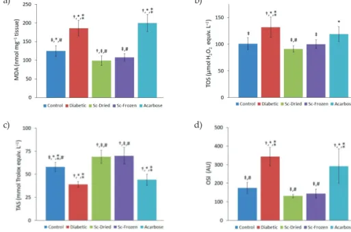
In the current study, liver MDA and TOS (Figs. 3a,b) were significantly elevated in the diabetic group in comparison with the control group (p < 0.05). MDA increased from the control group value of 125 ± 14 nmol mg–1 tissue to 186 ± 21 nmol mg–1 tissue in the
dia-betic group. However, Sc-Dried and Sc-Frozen treatment reduced MDA back to 99 ± 13 and 108 ± 10 nmol mg–1 tissue, resp. Besides, even acarbose administration caused an increase
in MDA (200 ± 23 nmol mg–1 tissue). TOS, another indicator of oxidative stress, increased
in the diabetic group compared to the control group, 132 ± 19 vs. 101 ± 11 µmol H2O2 equiv.
L–1. On the other hand, TOS value was found significantly lower in the Sc-Dried (91 ± 6
µmol H2O2 equiv. L–1) and Sc-Frozen (100 ± 8 µmol H2O2 equiv. L–1) administered groups
compared with the diabetic group. On the other hand, TOS was 119 ± 14 µmol H2O2 equiv.
L–1 in the acarbose group.
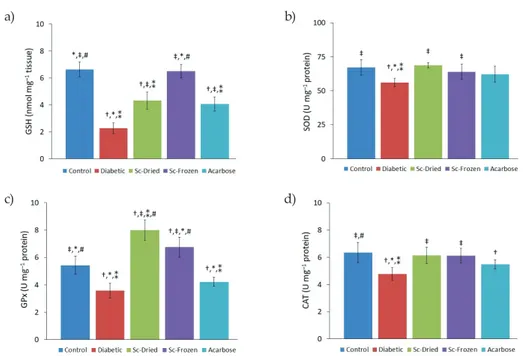
Oxidative status increases in diabetic conditions and leads to tissue damage (2). More-over, GSH concentration and SOD, GPx, and CAT activities of the liver were significantly decreased in diabetic rats compared to control group rats, indicating suppressed liver anti-oxidant defense against reactive oxygen species (ROS). GSH was significantly decreased (2.27 ± 0.4 vs. 6.63 ± 0.5 nmol mg–1 tissue) in the diabetic group compared to the control group.
Recently, diabetics were shown to have glutathione deficiency and reduced antioxidant
Fig. 3. a) MDA levels, b) TOS, c) OSI, d) TAS of control and diabetic rats (mean ± SD, n = 8).
Sc-Dried – dried Scorzonera cinerea administered group, Sc-Frozen – frozen Scorzonera cinerea admin-istered group, Ac – acarbose adminadmin-istered group. Significant difference at p < 0.05 vs.: † control group, ‡ diabetic group, * Sc-Dried group, ** Sc-Frozen group, # acarbose group.
a) b)
capacity (38), which is in accordance with this study. Due to increased blood glucose levels, competition for NADPH between aldose reductase in the polyol pathway and GSH-reduc-tase may occur (39). This results in GSH deficiency; consequently, this may cause other anti-oxidant enzymes, such as GPx, which uses GSH as a substrate, to decrease. However, Sc-Dried and Sc-Frozen administration increased the GSH level to 4.32 ± 0.6 and 6.50 ± 0.5 nmol mg–1 tissue, resp. Thus, Sc-Dried and Sc-Frozen co-treatment can significantly recuperate
liver antioxidant capacity. Overall, the administration of Sc-Dried and Sc-Frozen contributed to an increase in TAS and alleviation of TOS in liver tissue. SOD, GPx and CAT activities were lowest in the diabetic group 56 ± 3, 3.6 ± 0.5, and 4.8 ± 0.5 U mg–1 protein, resp.
How-ever, Sc-Dried administration increased these antioxidant activities to 69 ± 2, 8.0 ± 0.7, and 6.2 ± 0.6 U mg–1 protein, resp., whereas Sc-Frozen treatment showed similar values for SOD,
GPx and CAT of 64 ± 5, 6.8 ± 0.7, and 6.1 ± 0.5 U mg–1 protein, resp. Preventive effects of S.
sinensis extracts have been reported in the fatty diet by decreasing lipid peroxidation and
increasing SOD activity (40). In another study, although the SOD activity significantly de-creased in the CCl4-group, S. austriaca treatment increased the SOD activity (37). The
anti-oxidant capacity values are in good agreement with the data for genus Scorzonera. Polyphe-nolic compounds and minerals (such as Cu, Zn, and Mn) found in Scorzonera cinerea can provide protection against oxidative stress caused by hyperglycemia by strengthening the enzymatic and non-enzymatic antioxidant defense system.
Fig. 4. a) GSH concentration, b) SOD, c) GPx, d) CAT (D) activities of control and diabetic rats (mean ± SD, n = 8). Sc-Dried – dried Scorzonera cinerea administered group, Sc-Frozen – frozen Scorzonera cinerea administered group, Ac – acarbose administered group. Significant difference at p < 0.05 vs.: † control group, ‡ diabetic group, * Sc-Dried group, ** Sc-Frozen group, # acarbose group.
a) b)
CONCLUSIONS
The findings reveal that phytochemicals in S. cinerea may show liver protective effect by suppressing oxidative stress and boost antioxidant capacity. The study suggests that the phytochemical components in S. cinerea mediate its antidiabetic effect by increasing insulin secretion and inhibiting α-glucosidase activity. It is expected that S. cinerea may have anti-diabetic mechanisms of action by improving insulin sensibility, reducing glucose absorp-tion in the small intestine and increasing the uptake and bioavailability of glucose to the cells. Further studies may focus on these antidiabetic mechanisms of action for Scorzonera
cinerea.
Acknowledgements. – This research was supported by the Van Yüzüncü Yıl University Scientific Research Projects Foundation (grant number FYL-2017-6482).
REFERENCES
1. C. Steele, D. Steel and C. Waine, 6 – Pathophysiology of Diabetic Retinopathy, in Diabetes and the Eye (Eds. C. Steele, D. Steel and C. Waine), Butterworth-Heineman 2008, pp. 59–70; https://doi. org/10.1016/B978-0-08-045307-1.50011-3
2. O. R. Ayepola, N. L. Brooks and O. O. Oguntibeju, Antioxidant-antidiabetic Agents and Human Health, in Oxidative Stress and Diabetic Complications: The Role of Antioxidant Vitamins and Flavonoids, Intech Open, London 2014, pp. 25–58.
3. J. L. Ríos, F. Francini and G. T. Schinella, Natural products for the treatment of type 2 diabetes mellitus, Planta Med. 81 (2015) 975–994; https://doi.org/10.1055/s-0035-1546131
4. A. M. Donia, Phytochemical and pharmacological studies on Scorzonera alexandrina Boiss, J. Saud. Chem. Soc. 20 (2016) S433–S439; https://doi.org/10.1016/j.jscs.2013.01.001
5. L. Milella, A. Bader, N. De Tommasi, D. Russo and A. Braca, Antioxidant and free radical-scav-enging activity of constituents from two Scorzonera species, Food Chem. 160 (2014) 298–304; https:// doi.org/10.1016/j.foodchem.2014.03.097
6. Ö. B. Acıkara, G. S. Çitoğlu, S. Dall’Acqua, H. Özbek, J. Cvačka, M. Zemlička and K. Ŝmejkal, Bioassay-guided isolation of the antinociceptive compounds motiol and β-sitosterol from Scorzo-nera latifolia root extract, Pharmazie 69 (2014) 711–714; https://doi.org/10.1691/ph.2014.3920
7. E. K. Akkol, Ö. B. Acıkara, I. Süntar, B. Ergene and G. Saltan Çitoğlu, Ethnopharmacological evaluation of some Scorzonera species: In vivo anti-inflammatory and antinociceptive effects, J. Ethnopharmacol. 140 (2012) 261–270; https://doi.org/10.1016/j.jep.2012.01.015
8. E. K. Akkol, O. B. Acıkara, I. Süntar, G. Saltan Çitoğlu, H. Keleş and B. Ergene, Enhancement of wound healing by topical application of Scorzonera species: Determination of the constituents by HPLC with new validated reverse phase method, J. Ethnopharmacol. 137 (2011) 1018–1027; https:// doi.org/10.1016/j.jep.2011.07.029
9. Association of Official Analytical Chemists, Official Methods of Analysis of the AOAC International, (Ed. G. W. Latimer), 20th ed., AOAC International, Rockville 2016, pp. 570–655.
10. V. L. Singleton, R. Orthofer and R. M. Lamuela-Raventós, Analysis of total phenols and other oxidation substrates and antioxidants by means of folin-ciocalteu reagent, Methods Enzymol. 299 (1999) 152–178; https://doi.org/10.1016/S0076-6879(99)99017-1
11. J. Zhishen, T. Mengcheng and W. Jianming, The determination of flavonoid contents in mulberry and their scavenging effects on superoxide radicals, Food Chem. 64 (1999) 555–559; https://doi. org/10.1016/S0308-8146(98)00102-2
12. K. Mishra, H. Ojha and N. K. Chaudhury, Estimation of antiradical properties of antioxidants using DPPH• assay: A critical review and results, Food Chem. 130 (2012) 1036–1043; https://doi. org/10.1016/j.foodchem.2011.07.127
13. R. Re, N. Pellegrini, A. Proteggente, A. Pannala, M. Yang and C. Rice-Evans, Antioxidant activity applying an improved ABTS radical cation decolorization assay, Free Radic. Biol. Med. 26 (1999) 1231–1237; https://doi.org/10.1016/S0891-5849(98)00315-3
14. Y. M. Kim, Y. K. Jeong, M. H. Wang, W. Y. Lee and H. I. Rhee, Inhibitory effect of pine extract on α-glucosidase activity and postprandial hyperglycemia, Nutrition 21 (2005) 756–761; https://doi. org/10.1016/j.nut.2004.10.014
15. H. H. Draper and M. Hadley, Malondialdehyde determination as index of lipid peroxidation, Methods Enzymol. 186 (1990) 421–431; https://doi.org/10.1016/0076-6879(90)86135-I
16. E. Beutler, T. Gelbart and C. Pegelow, Erythrocyte glutathione synthetase deficiency leads not only to glutathione but also to glutathione-S-transferase deficiency, J. Clin. Invest. 77 (1986) 38–41; https://doi.org/10.1172/JCI112298
17. J. M. McCord, Analysis of superoxide dismutase activity, Curr. Protoc. Toxicol. 00 (1999) 7.3.1–7.3.9; https://doi.org/10.1002/0471140856.tx0703s00
18. W. A. Günzler, H. Kremers and L. Flohé, An improved coupled test procedure for glutathione peroxidase (EC 1-11-1-9-) in blood, Z. Klin. Chem. Klin. Biochem. 12 (1974) 444–448; https://doi. org/10.1515/cclm.1974.12.10.444.
19. H. Aebi, Catalase in vitro, Methods Enzymol. 105 (1984) 121–126; https://doi.org/10.1016/S0076-6879(84)05016-3
20. Ö. Erel, A novel automated direct measurement method for total antioxidant capacity using a new generation, more stable ABTS radical cation, Clin. Biochem. 37 (2004) 277–285; https://doi. org/10.1016/j.clinbiochem.2003.11.015
21. Ö. Erel, A new automated colorimetric method for measuring total oxidant status, Clin. Biochem.
38 (2005) 1103–1111; https://doi.org/10.1016/j.clinbiochem.2005.08.008
22. O. B. Acikara, J. Hošek, P. Babula, J. Cvačka, M. Budešínský, M. Dračinský, G. S. İşcan, D. Kadle-cová, L. Ballová and K. Šmejkal, Turkish Scorzonera species extracts attenuate cytokine secretion via inhibition of NF-κB activation, showing anti-inflammatory effect in vitro, Molecules 21 (2016) Article ID 43 (14 pages); https://doi.org/10.3390/molecules21010043
23. S. Dall’Acqua, G. Ak, S. Sut, I. Ferrarese, G. Zengin, E. Yıldıztugay, M. F. Mahomoodally, K. I. Sinan, and D. Lobine, Phenolics from Scorzonera tomentosa L.: Exploring the potential use in in-dustrial applications via an integrated approach, Ind. Crops Prod. 154 (2020) 112751–112760; https:// doi.org/10.1016/j.indcrop.2020.112751
24. Institute of Medicine, Dietary Reference Intakes: The Essential Guide to Nutrient Requirements, The National Academies Press, Washington DC 2006, pp. 286–402; https://doi.org/10.17226/11537 25. F. Taranto, A. Pasqualone, G. Mangini, P. Tripodi, M. M. Miazzi, S. Pavan and C. Montemurro,
Polyphenol oxidases in crops: Biochemical, physiological and genetic aspects, Int. J. Mol. Sci. 18 (2017) 377–393; https://doi.org/10.3390/ijms18020377
26. R. Khattaba, G. B. Celli, A. Ghanem and M. S. Brooks, Effect of frozen storage on polyphenol con-tent and antioxidant activity of haskap berries (Lonicera caerulea L.), J. Berry Res. 5 (2015) 231–242; https://doi.org/10.3233/JBR-150105
27. K. T. Kongstad, C. Ozdemir, A. Barzak, S. G. Wubshet and D. Staerk, Combined use of high-reso-lution α-glucosidase inhibition profiling and high-performance liquid chromatography-high-resolution mass spectrometry-solid-phase extraction-nuclear magnetic resonance spectroscopy for investigation of antidiabetic principles in crude plant extracts, J. Agric. Food Chem. 63 (2015) 2257–2263; https://doi.org/10.1021/jf506297k
28. D. Bagdas, B. C. Etoz, Z. Gul, S. Ziyanok, S. Inan, O. Turacozen, N. Y. Gul, A. Topal, N. Cinkilic, S. Tas, M. O. Ozyigit and M. S. Gurun, In vivo systemic chlorogenic acid therapy under diabetic conditions: Wound healing effects and cytotoxicity/genotoxicity profile, Food Chem. Toxicol. 81 (2015) 54–61; https://doi.org/10.1016/j.fct.2015.04.001
29. W. Blaschek, Natural products as lead compounds for sodium glucose cotransporter (SGLT) in-hibitors, Planta Med. 83 (2017) 985–993; https://doi.org/10.1055/s-0043-106050
30. C. Schulze, A. Bangert, G. Kottra, K. E. Geillinger, B. Schwanck, H. Vollert, W. Blaschek and H. Daniel, Inhibition of the intestinal sodium-coupled glucose transporter 1 (SGLT1) by extracts and polyphenols from apple reduces postprandial blood glucose levels in mice and humans, Mol. Nutr. Food Res. 58 (2014) 1795–1808; https://doi.org/10.1002/mnfr.201400016
31. S. Meng, J. Cao, Q. Feng, J. Peng and Y. Hu, Roles of chlorogenic acid on regulating glucose and lipids metabolism: A review, Evid-Based Complement. Alternat. Med. 2013 (2013) Article ID 801457 (11 pages); https://doi.org/10.1155/2013/801457
32. V. R. Punithavathi, P. S. M. Prince, R. Kumar and J. Selvakumari, Antihyperglycaemic, antilipid peroxidative and antioxidant effects of gallic acid on streptozotocin induced diabetic Wistar rats, Eur. J. Pharmacol. 650 (2011) 465–471; https://doi.org/10.1016/j.ejphar.2010.08.059
33. A. Bashta, N. Ivchuk and O. Bashta, Yacón and Scorzonera as functional enrichment of food, Ukrainian J. Food Sci. 3 (2015) 13–22.
34. N. Petkova, Characterization of inulin from black salsify (Scorzonera hispanica L.) for food and pharmaceutical purposes, Asian J. Pharm. Clin. Res. 11 (2018) 221–225; https://doi.org/10.22159/ajp-cr.2018.v11i12.28262
35. M. Rao, C. Gao, L. Xu, L. Jiang, J. Zhu, G. Chen, B. Y. K. Law and Y. Xu, Effect of inulin-type car-bohydrates on insulin resistance in patients with type 2 diabetes and obesity: A systematic review and meta-analysis, J. Diabetes Res. 2019 (2019) Article ID 5101423 (13 pages); https://doi. org/10.1155/2019/5101423
36. P. I. Ingaramo, M. T. Ronco, D. E. A. Francés, J. A. Monti, G. B. Pisani, M. P. Ceballos, M. Galleano, M. C. Carrillo and C. E. Carnovale, Tumor necrosis factor alpha pathways develops liver apopto-sis in type 1 diabetes mellitus, Mol. Immunol. 48 (2011) 1397–1407; https://doi.org/10.1016/j.mo-limm.2011.03.015
37. T. Zhang, Y. Xie, Z. Zhang and G. Wang, Study on hepatoprotective effects of total flavonoids in Scorzonera austriaca Wild in vivo and in vitro, Chin. J. Biochem. Pharm. 35 (2015) 6–9.
38. F. K. Lutchmansingh, J. W. Hsu, F. I. Bennett, A. V. Badaloo, N. McFarlane-Anderson, G. M. Gor-don-Strachan, R. A. Wright-Pascoe, F. Jahoor and M. S. Boyn, Glutathione metabolism in type 2 diabetes and its relationship with microvascular complications and glycemia, PLoS ONE 13 (2018) e0198626 (12 pages); https://doi.org/10.1371/journal.pone.0198626
39. L. J. Yan, Redox imbalance stress in diabetes mellitus: Role of the polyol pathway, Animal Model Exp. Med. 1 (2018) 7–13; https://doi.org/10.1002/ame2.12001
40. H. Yang and C. Zeng, Effects of water extract from Scorzonera sinensis Lipsch, Pteridium aquilinum and Sonchus oleraceus L. on plasma-lipids metabolism in mice fed high fats diet, Food Res. Dev. 36 (2015) 11–13; https://doi.org/10.3969/j.issn.1005-6521.2015.06.003