• Sonuç bulunamadı

A new marker in acute ischemic stroke patients: monocyte/hdl ratio

N/A
N/A
Protected

Academic year: 2021

Share "A new marker in acute ischemic stroke patients: monocyte/hdl ratio"

Copied!
6
0
0

Yükleniyor.... (view fulltext now)

Tam metin

(1)

ORIGINAL

ARTICLE

Aybala Neslihan Alagoz

1

Bilgehan Atılgan Acar

2

Belma Dogan Gungen

3

Yesim Guzey Aras

2

Turkan Acar

3

1The Department of Neurology, Kocaeli University Medical School, Kocaeli, Turkey. 2The Department of Neurology, Sakarya University Medical School, Sakarya, Turkey. 3The Department of Neurology, Istinye University Istanbul, Turkey.

Corresponding Author: Aybala Neslihan Alagöz

The Department of Neurology, Kocaeli University Medical School,

Kocaeli, Turkey E-mail:aybalaalagoz@hotmail.com Tel:+90 5336174804 Received: 13.09.2019 Acceptance: 13.02.2020 DOI: 10.18521/ktd.632555

Konuralp Medical Journal

e-ISSN1309–3878

konuralptipdergi@duzce.edu.tr konuralptipdergisi@gmail.com www.konuralptipdergi.duzce.edu.tr

A New Marker in Acute Ischemic Stroke Patients:

Monocyte / HDL Ratio

ABSTRACT

Objective: Stroke is a major cause of global morbidity and mortality. Ischemic stroke

is usually marked by cell death associated with inflammation and oxidative stress, and the role of inflammation in neurological diseases has become increasingly obvious. In our study, we studied the monocyte / HDL ratio (MHR) which can support the theory of inflammation in stroke and can be used in clinical practice. MHR can be used a prognostic marker in stroke patients base on this theory.

Methods: Our study registered 91 acute ischemic stroke patients (47 females and 44

males) and 50 healthy controls.

Results: MHR was elevated in the patient group and correlated with high-sensitivity

C-reactive protein (hsCRP). MHR was higher in patients with high a National Institutes of Health Stroke Scale (NIHSS) than those with a low NIHSS (p=0.050) as well as in patients with a larger infarct area and in than those with a smaller area (p=0.050). The MHR correlated with both the clinical condition and infarct area in acute ischemic stroke patients, which underscores the value of MHR as a new marker.

Conclusions: This cross-sectional study of 91 acute ischemic stroke patients indicates

that inflammation markers and MHR are correlated with clinical status and even radiological parameters.

Keywords: Acute Ischemic Stroke, Monocyte/HDL Ratio, İnflammation, New

Marker

Akut İskemik İnme Hastalarında Yeni Bir Markır:

Monosit / HDL Oranı

ÖZET

Amaç: İnme, dünyada major bir morbidite ve mortalite sebebidir. İskemik inmede

genellikle inflamasyon ve oksidatif strese bağlı hücre ölümü görülür. Nörolojik hastalıklarda inflamasyonun rolü giderek daha da belirginleşmektedir. Çalışmamızda inmede inflamasyon teorisini destekleyebilen ve klinik uygulamada kullanılabilecek monosit / HDL oranını (MHO) inceledik.

Gereç ve Yöntem: Çalışmamızda 91 akut iskemik inme hastası (47 kadın ve 44

erkek) ve 50 sağlıklı kontrol kaydedildi.

Bulgular: Hasta grubunda MHO yüksek idi ve yüksek duyarlılıkta C-reaktif protein

(hsCRP) ile korele idi. MHO, Ulusal Sağlık Enstitüleri İnme Ölçeği (NIHSS) skoru yüksek olan hastalarda, düşük NIHSS'li (p = 0.050) olanların yanı sıra, ve de daha büyük bir infarkt alanı olanlarda daha küçük infarkt alanına sahip olanlara (p = 0.050) göre daha yüksekti. MHO, yeni bir belirteç olarak, akut iskemik inme hastalarında, hem klinik durum hem de infarkt alanı ile koreledir.

Sonuç: 91 akut iskemik inme hastasının, bu cross-sectional çalışması, inflamasyon

belirteçlerinin ve MHO'nin; klinik durum ve hatta radyolojik parametrelerle körele olduğunu göstermektedir.

Anahtar Kelimeler: Akut İskemik İnme,Monosit/HDL Oranı,İnflamasyon,Yeni

(2)

INTRODUCTION

Stroke is defined by an abrupt onset of neurological deficits attributable to a focal vascular cause. Stroke is a major cause of morbidity and mortality worldwide. This may result from brain infarction or hemorrhage. The development of ischemic stroke is most commonly attributed to atherosclerosis and particularly carotid atherosclerosis (1).

Ischemic stroke usually initiates inflammation and oxidative stress leading to neuronal death (2). The role of inflammation in neurological disorders is increasingly recognized, and inflammatory processes are associated with the etiology and clinical progression of migraine,

psychiatric conditions,

epilepsy, cerebrovascular diseases, dementia, and neurodegeneration including that in Alzheimer's and Parkinson's diseases (3).

Atherosclerosis is an inflammatory disease within the arterial wall that is responsible for several important adverse vascular events including coronary artery disease, myocardial infraction, stroke, and peripheral artery disease. Both innate and adaptive immunity plays an important role in the development of atherosclerosis. In particular, monocytes and macrophages, which are the surrogate cells of innate immunity, have important pro-atherogenic effects (4).

Macrophages and monocytes are the most important cell types for the secretion of pro-inflammatory and pro-oxidant cytokines at the site of inflammation. In addition, high-density lipoprotein cholesterol (HDL-C) can defend endothelial cells against the unfavorable effects of low-density lipoprotein (LDL) and can inhibit oxidation of LDL molecules. Thus, HDL-C is thought to exhibit both anti-inflammatory and antioxidant actions. Therefore, monocytes exert pro-inflammatory and pro-oxidant effects, but HDL-C can reverse these processes (5,6).

In recent studies; monocyte count to HDL ratio (MHR) as a new marker may be a prognostic marker for cardiovascular disease and mortality is associated with MHR in patients with acute ischemic stroke (7). With this marker ‘’MHR’’; the criteria which were not studied in the literature before were included in our study. This study aimed to evaluate the usefulness of MHR as a marker in determining the clinical status and infarct area in acute ischemic stroke patients.

MATERIALS AND METHODS

The cross-sectional study was approved by the Institutional Ethics Committee at Sakarya University, Medical School; protocol number is: 16214662/050.01.04/46. It was conducted in accordance with the Declaration of Helsinki. Informed constent form was completed for all participitants.

The study registered 91 acute ischemic stroke patients: 47 women (51.6%) and 44 men (48.4%) as well as 50 healthy control individuals (31 women

(62%) and 19 men (38%)). Patients were not recommended in another vascular event in the last 1 year (cardiovascular, cerebrovascular or peripheral vascular, etc.). Exclusion criteria included recent infection, hematologic disease, blood dyscarias, malignancies, autoimmune or inflammatory diseases, renal and hepatic failure, severe valvular disease/heart failure, and the use of corticosteroid and non-steroidal anti-inflammatory drugs in the past 3 months.

Routine blood and urine analyses, echocardiography, carotis-vertebral Doppler ultrasonography, and cranial diffusion-weighted magnetic resonance imaging (DW-MRI) were performed in the first 24 hours of the acute cerebral event. Measurements of the infarct area in cerebral DW MRI were conducted by a neurologist blinded to the clinical conditions of the patients. In the DWI sequence, the hyperintense area with restricted DWI was measured manually. The lesion area was found by multiplying it by the slice thickness (+interslice gap).

Based on the etiological Trial of ORG 10172 in Acute Stroke Treatment (TOAST) classification, the patients were classified as follows: 1) large vessel atherosclerosis, 2) cardioembolic, 3) small vessel occlusion, 4) stroke associated with other causes, and 5) uncertain cause (9). The clinical status of the patients was determined according to the National Institutes of Health Stroke Scale (NIHSS) (10). Patient’s infarct volumes were calculated using the DW MRIs performed up to the 24th hour of the event. Grouping by infarct area was as follows: Group 1: 0-1.5 cm2, Group 2: 1.5-5 cm2; Group 3: 5-10 cm2; and Group 4: over 10 cm2.

Statistical Analysis: Descriptive values

were presented as the mean, standard deviation, percent, and count. The relations between categorical variables were analyzed using Fisher-Freeman-Halton test and those between numerical features with correlation analysis. Additionally, one-way variance analysis employed in-group comparisons. ROC analysis was used for diagnostic success of MHR between patients and controls. P values were accepted as statistically significant if less than 0.05.

RESULTS

Sociodemographic data of the patient and control groups are presented in Table 1. Gender distribution and alcohol consumption of patients and control individuals were similar; however, the presence of hypertension (HT), presence of diabetes mellitus (DM), smoking, and mean body mass index (BMI) were significantly higher in the patient group. The mean age was also similar in the patient and control groups, but BMI was significantly higher in the patient group (Table 2).

The MHR, white blood cell (WBC) count, and C-reactive protein (CRP) levels were higher and HDL level was lower in the patient group although the difference in the monocyte count was not statistically significant (Table 3).

(3)

Table 1. Sociodemographic data about the patient and control groups

Control Ischemic Stroke P

n % n % Sex M 19 38,0 44 48,4% 0.289 F 31 62,0 47 51,6% HT No 46 92,0 20 22,0 <0.0001 Yes 4 8,0 71 78,0 DM No 50 100,0 56 61,5 <0.0001 Yes 0 0,0 35 38,5 Smoking No 50 100,0 70 76,9 <0.0001 Yes 0 0,0 21 23,1 Alcohol consumption No 50 100,0 88 96,7 0.552 Yes 0 0,0 3 3,3

Table 2. Age and BMI values of the patient and control groups

Control Ischemic Stroke P

n Mean SD n Mean SD

Age 50 69,32 7,776 91 68,70 12,793 0.757

BMI 50 26,826 2,3215 91 28,092 4,1329 0.021

Table 3. Comparison between MHR, WBC count, CRP levels and HDL levels of patient and control groups

Group P

Control Ischemic Stroke

n mean SD n mean SD MHR 50 10,97 5,08 91 15,32 6,86 <0.0001 WBC 50 6,83 1,84 91 7,78 2,32 0.014 Monocyte 50 517,44 172,76 91 554,14 207,74 0.290 CRP 50 3,49 ,80 90 12,30 16,96 0.004 HDL 50 50,95 14,47 91 38,23 8,29 0.001

ROC analysis assessed the MHR in differentiating between patients and control individuals. The sensitivity and specificity were 71.4% and 64% with a cut off value of 11.3 (Graph

1). Thus, the MHR could differentiate between patients and controls at a statistically significant rate.

(4)

Acute ischemic stroke patients were allocated to four groups based on the infarct areas calculated from the cerebral DW-MRI images taken in the first 24 hours after the event: Group 1: 0-1.5 cm2, Group 2: 1.5-5 cm2; Group 3: 5-10 cm2; and Group 4: over 10 cm2. The clinical status of the patients was evaluated according to the NIHSS

values. Group 1 had a significantly lower mean NIHSS (n=33, 36.3%) than the other three groups (Table 4). This indicates that patients with a smaller infarct area have a better clinical status. Also, a significant positive correlation was established between NIHSS values and MHR (p=0.050) (Table 5).

Table 4. Comparison between the Infarct areas-Groups and NIHSS Infarct areas

Groups

NIHSS

n mean SD minimum maximum P

1 33 4,94 2,499 2 15

0.011

2 40 7,45 5,188 2 20

3 16 7,50 4,033 2 15

4 2 13,50 13,435 4 23

Table 5.Comparison between the MHR and NIHSS

NIHSS n=91

MHR r 0,201

p 0,050

Group 4 had significantly higher values for WBC count, monocyte count, and MHR in relation to infarct areas for all four groups (p values of 0.049, 0.007, and 0.050, respectively) (Table 6). This confirms that patients with larger infarct areas have higher monocyte counts and MHR’s.

Table 6. Comparison between the WBC count, monocyte count, MHR and Infarct areas-Groups

INFARCT AREAS-GROUPS

P

1 2 3 4

n mean SD n mean SD n mean SD n mean SD

WBC 33 7,98 2,32 40 7,18 1,51 16 8,47 3,47 2 10,81 1,27 0.049

Monocyte 33 578,8 180,07 40 534,9 170,31 16 494,7 223,20 2 1006,0 684,48 0.007

MHR 33 15,30 5,93 40 15,57 6,98 16 13,42 6,69 2 25,49 16,37 0.050

DISCUSSION

Atherosclerosis and carotis atherosclerosis in particular plays a major role in the development of ischemic stroke (1). Increasing evidence points to the inflammatory character of atherosclerosis, and several parameters of inflammation have been proposed as cerebrovascular risk markers (11). Numerous studies have evaluated the erythrocyte sedimentation rate, high-sensitivity CRP (hsCRP), leukocyte count, fibrinogen, etc. as serum inflammatory parameters. Although plasma hsCRP is elevated in response to inflammation caused by brain infarction, the association of CRP with

clinical outcomes after acute

ischemic stroke remains uncertain. In a study of 3653 acute ischemic stroke patients, Matsuo et al. found that high plasma hsCRP was independently associated with unfavorable clinical outcomes after acute ischemic stroke (12). MHR studies showed significant correlations between this ratio and levels of hsCRP as an inflammation marker (5, 13). In our study, CRP was found to be statistically significantly higher in stroke patients compared to the control group (p<0.004). Similarly, MHR levels in patients with ischemic stroke were as high as CRP levels to support inflammation (p<0.0001). Markers that can demonstrate the relationship between atherosclerosis and disease are important for monitoring clinical progression and treatment.

Inflammation and oxidative stress are

well-known mechanisms underlying the development and progression of atherosclerosis. Monocytes are the key player of this process. Activated monocytes interact with damaged or activated endothelium leading to overexpression of pro-inflammatory cytokines/adhesion molecules. Thereafter, monocytes differentiate into the macrophages that ingest oxidized LDL cholesterol and form dangerous foam cells (14). In contrast, HDL molecules counteract the migration of macrophages and promote efflux of oxidized cholesterol from these cells. Recent studies also indicate a role for HDL in controlling monocyte activation, adhesiveness, and inflammation (15,16) and in controlling the proliferation of progenitor cells that give rise to monocytes (5,17).

Monocytes exercise pro-inflammatory and pro-oxidant actions in the atherosclerotic process, and HDL-C reverses these actions (5). HDL-C has other effects on monocyte activation. HDL-C interrupts monocyte to macrophage differentiation, suppresses the inflammatory response, and withholds the inflammatory process. Ongoing studies focus on a new aspect of the anti-inflammatory properties of HDL particles that act within the hematopoietic system by suppressing hematopoietic stem cells and multi-potential progenitor cell proliferation, mobilization, and monocyte production. This decreases inflammation.

(5)

Elevated HDL levels or reconstituted HDL infusion can suppress the production of IL-23 and granulocyte-CSF. This interrupts monocyte production and differentiation in the extra-medullary hematopoiesis ultimately preventing exuberant monocytopoiesis (12,18,19,20).

Qiao et al. investigated atherosclerosis in macrophage colony-stimulating factor deficient mice and reported a gene dosage-related reduction in atherosclerosis to be correlated with a decrease in blood monocyte counts. Previous animal studies also show a significant relation between blood monocyte counts and the extent of atherosclerosis (21).

Monocytes play an important role in the pathogenesis of atherosclerosis inside the arterial wall and in circulation. Circulating monocytes are a source of numerous factors and receptors that interact primarily with platelets and endothelial cells leading to inflammation, thrombosis, and atherosclerosis (22,23). Palmerini et al. examined monocyte-derived tissue factor in vitro using stent thrombosis samples. They reported that there was a significant increase in monocyte-tissue factor expression with stent implantation. Extracting monocytes from the milieu led to diminished fibrin deposition by 45% and decreased the tissue factor level of the thrombus by 83% versus the control group (24). Cetin and colleagues evaluated MHR in another study of patients who developed stent thrombosis. MHR was found to be a novel marker of inflammation and seemed to be an independent predictor of definite stent thrombosis after primary percutaneous coronary intervention for ST-segment elevation myocardial infarction (13).

In the study conducted by Kanbay et al. with 340 patients with chronic renal failure followed up in terms of cardiovascular terms; MHR was increased with decreasing eGFR in predialytic patients. MHR was associated with poor cardiovascular profile and arose as independent predictors of major cardiovascular events during follow up (7). In another study conducted by Cicek et al. with of 760 patients, MHR is associated independently and significantly with short term and long-term mortality in ST-elevation myocardial infarction patients who undergo primary percutaneous coronary intervention (25).

In a retrospective study conducted by Bolayır et al. with 466 acute ischemic stroke patients and 408 controls, monocyte count was higher, HDL levels were lower and MHR was higher in the patient group compared to the control group. MHR value above 17.52 seem to be an independent risk factor or predicting the 30-day mortality of patient with acute ischemic stroke (8). In another study conducted with 75 stroke patients; Glasgow Coma Score, NIHSS score, infarct volume (cm3), neutrophil count, platelet count, WBC count,

albumin, MHR and Ca+ were significant for the prediction of one-month mortality. MHR were significantly associated with short term mortality (26). In our study, levels of MHR, WBC, CRP and HDL were statistically and significantly supported inflammation in ischemic stroke patients compared to the control group (p<0.0001; 0.014; 0.004; 0.001).

There are publications indicating stroke volume in ischemic stroke patients is in correlation with NIHSS score (27). In another study, ischemic neural tissue volume showed significant results at 90% sensitivity and 60% specifity for one-month mortality (26). In our study, stroke volume was calculated as area (m2). As the infarct area increased, the NIHSS scores of the patients increased in a statistically significant way (p=0.011). NIHSS was significantly higher in Group 4 patients with an infarct area greater than 10 cm2, but it could not be fully evaluated due to the small number of patients in this group. In addition, WBC, monocyte count and MHR were significantly higher as inflammation indicator with groups created in accordance with the size of infarct area (p=0.049; 0.007; 0.050).

To summarize; we found that blood monocyte count and HDL-C levels were independently and significantly correlated with the clinical status and infarct area of acute ischemic stroke patients. In addition, MHR was found to significantly correlate with hsCRP and was elevated in acute ischemic stroke patients with a high NIHSS and larger infarct area. This suggests inflammation and poor prognosis.

CONCLUSION

This study is limited in that it is a single center study. The control group was selected from individuals with high BMI; this is another limitation of the study because it is a factor that increases the risk of stroke. More patients with longer follow-up could increase the validation of MHR as a new marker. Still, this cross-sectional study of 91 acute ischemic stroke patients indicates that inflammation markers and MHR are correlated with clinical status and even radiological parameters.

Acknowledgements: Author contributions:

Concept; A.N.A., design; A.N.A., B.A. supervision; A.N.A., resource; A.N.A and B.D.G, materials; A.N.A. and Y.G.A., data collection &/or processing; B.D.G., Y.G.A. and T.A., analysis &/or interpretation; A.N.A. and B.A., literature search; A.N.A., writing; A.N.A., critical review; A.N.A., B.A. The authors have no financial or personal conflict of interests related to this work.

Conflict of Interests: The authors declared,

they do not have anything to disclose regarding conflict of interest with respect to this manuscript.

(6)

REFERENCES

1. Sengupta D, Bardhan J, Baran A, et al. Correlation between lipid profile and carotid intima media thickness in cerebral ischemia. Indian J PhysiolPharmacol. 2014 Oct-Dec;58(4):354-64.

2. Chehaibi K, Trabelsi I, Mahdouani K, et al. Correlation of oxidative stress parameters and inflammatory markers in ischemic stroke patients. J Stroke Cerebrovasc Dis. 2016 Nov;25(11):2585-2593.

3. Lénárt N, Brough D, Dénes Á. Inflammasomes link vascular disease with neuroinflammation and brain disorders. J Cereb Blood Flow Metab. 2016 Oct;36(10):1668-1685.

4. Taleb S. Inflammation in atherosclerosis. Arch Cardiovasc Dis. 2016 Aug 29. pii: S1875-2136(16)30112-7. 5. Canpolat U, Çetin EH, Cetin S, et al. Association of monocyte-to-HDL cholesterol ratio with slow coronary

flow is linked to systemic inflammation. ClinApplThrombHemost. 2016 Jul;22(5):476-82.

6. Ancuta P, Wang J, Gabuzda D. CD16+ monocytes produce IL-6, CCL2, and matrix metalloproteinase-9 upon interaction with CX3CL1-expressing endothelial cells. J Leukoc Biol. 2006; 80(5):1156-1164.

7. Kanbay M, Solak Y, Unal HU, et al. Monocyte count/HDL-C ratio and cardiovascular events in patients with chronic kidney disease. IntUrolNephrol. 2014;46(8): 1619-1625.

8. Bolayır A,Gokce SF, Cigdem B, et al. Monocyte/high density lipoprotein ratio predicts the mortality in ischemic stroke patients. Neurol Neurochir Pol. 2018 Mar;52(2):150-155. doi: 10.1016/j.pjnns.2017.08.011. Epub 2017 Aug 24.

9. Adams HP, Bendixen BH, Kappelle LJ, et al. Classification of subtype of acute ischemic stroke. Definitions for use in a multicenter clinical trial. TOAST. Trial of ORG 10172 in Acute Stroke Treatment. Stroke. 1993; 24:35–41.

10. Brott T, Adams HP, Jr., Olinger CP et al. Measurements of acute cerebral infarction: a clinical examination scale. Stroke 1989;20(7):864-70.

11. Minić GA. Leucocyte count indicates carotid plaque instability in stroke patients. Vojnosanit Pregl. 2016 Jun;73(6):515-25.

12. Matsuo R, Ago T, Hata J, et al. Plasma C-reactive protein and clinical outcomes after acute ischemic stroke: A prospective observational study. PLoS One. 2016 Jun 3;11(6):e0156790.

13. Leuschner F, Rauch PJ, Ueno T et al. Rapid monocyte kinetics in acute myocardial infarction are sustained by extramedullary monocytopoiesis. J Exp Med. 2012 Jan 16;209(1):123-37.

14. Ghattas A, Griffiths HR, Devitt A, et al. Monocytes in coronary artery disease and atherosclerosis: Where are we now? J Am CollCardiol. 2013;62(17):1541-1551.

15. Murphy AJ, Chin-Dusting JP, Sviridov D, et al. The anti-inflammatory effects of high density lipoproteins. Curr Med Chem. 2009;16(6):667-675.

16. Murphy AJ, Woollard KJ. High-density lipoprotein: A potent inhibitor of inflammation. ClinExpPharmacol Physiol. 2010;37(7): 710-718.

17. Yvan-Charvet L, Pagler T, Gautier EL, et al. ATP-binding cassette transporters and HDL suppress hematopoietic stem cell proliferation. Science. 2010;328(5986):1689-1693.

18. Murphy AJ, Woollard KJ, Hoang A et al. High-density lipoprotein reduces the human monocyte inflammatory response. Arterioscler Thromb Vasc Biol. 2008 Nov; 28(11): 2071-7.

19. Wang N, Lan D, Chen W, et al. ATP-binding cassette transporters G1 and G4 mediate cellular cholesterol efflux to high-density lipoproteins. Proc Natl Acad Sci USA. 2004 Jun 29;101(26):9774-9.

20. Westerterp M, Gourion-Arsiquaud S, Murphy AJ, et al. Regulation of hematopoietic stem and progenitor cell mobilization by cholesterol efflux pathways. Cell Stem Cell. 2012 Aug 3;11(2):195-206.

21. Qiao JH, Tripathi J, Mishra NK, et al. Role of macrophage colony-stimulating factor in atherosclerosis: studies of osteopetrotic mice. Am J Pathol. 1997 May;150(5):1687-99.

22. Mestas J, Ley K. Monocyte-endothelial cell interactions in the development of atherosclerosis.Trends Cardiovasc Med. 2008 Aug;18(6):228-32.

23. Woollard KJ, Geissmann F. Monocytes in atherosclerosis: Subsets and functions. Nat Rev Cardiol. 2010 Feb;7(2):77-86. Rev.

24. Palmerini T, Coller BS, Cervi V, et al. Monocyte-derived tissue factor contributes to stent thrombosis in an in vitro system. J Am Coll Cardiol. 2004 Oct 19;44(8):1570-7.

25. Cicek G, Kundi H, Bozbay M, et al. The relationship between admission monocyte HDL-C ratio with short-term and long-short-term mortality among STEMI patients treated with successful primary PCI. Coron Artery Dis. 2016 May;27(3):176-84.

26. Algin A, Inan I. The role of radiologic, clinical and biochemical parameters in prediction of stroke mortality. Neurosciences (Riyadh). 2019 Apr;24(2):110-114.

27. Yaghi S, Herber C, Boehme AK, et al. The Association between Diffusion MRI-Defined Infarct Volume and NIHSS Score in Patients with Minor Acute Stroke. J Neuroimaging 2017; 27: 388-391.

Şekil

Table 3. Comparison between MHR, WBC count, CRP levels and HDL levels of patient and control groups
Table 4. Comparison between the Infarct areas-Groups and NIHSS  Infarct areas

Referanslar

Benzer Belgeler

Older patients presenting with elevated levels of preoperative white blood cell count and lymphocyte/monocyte ratio, excessive tobacco consumption, prolonged

/lymphocyte (MLR), and the platelet/lymphocyte ratios (PLR) measured in blood samples taken at admission and clinical outcomes (CO) on the 90th day in patients, who were diagnosed

Accordingly, when patients with cellulitis were divided into two groups as ≥65 years and &lt;65 years, a statistically sig- nificant difference was noted among the WBC, NLR, and

In this study, it has been shown that; monocyte/HDL cholesterol ratio were statistically higher in patients having hemodialysis than in the ones followed up with

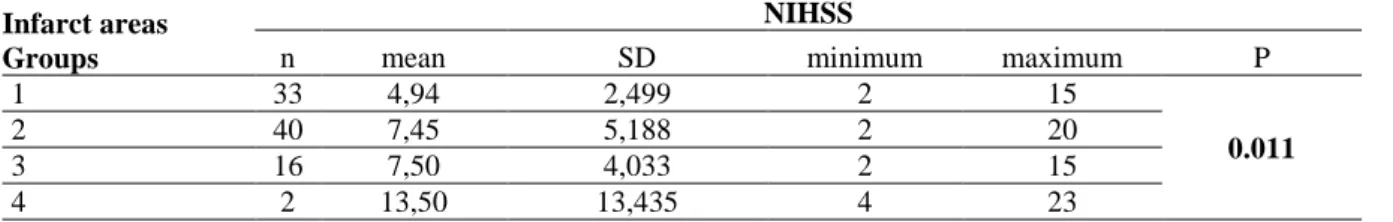
When patients are analyzed in regards to Bamford classification, NLR and CRP were significantly higher in total anterior circulation compared to other groups

One study reported that increased PD is significantly associated with elevated CRP concentrations, and that the surface area or volume of periodontal lesion in AgP patients

The patient’s clinical characteristics including age, gender, preoperative monocyte count, HDL-C, creatinine, and blood urea nitrogen (BUN) levels, left ventricular ejection

Monocyte to HDL Cholesterol Ratio and its association with cardio-metabolic risk factors in Primary Hyperparathyroidism.. Primer Hiperparatiroidili hastalarda monosit sayısının