107 Case Report / Olgu Sunumu
Turkish Journal of Thoracic and Cardiovascular Surgery 2019;27(1):107-110 http://dx.doi.org/doi: 10.5606/tgkdc.dergisi.2019.16885
A new surgical approach to a patient with anomalous origin of left
coronary artery from pulmonary artery: A case report
Pulmoner arterden çıkan sol koroner arter anomalisi olan bir hastaya yeni cerrahi yaklaşım: Olgu sunumu
Emin Can Ata, Korhan Erkanlı, Erkan Yıldız, Halil Türkoğlu
ÖZ
Ana pulmoner arterden çıkan sol ana koroner arter anormalisi, çocuklarda çok nadir görülen bir doğuştan kalp hastalığıdır. Ölümcül iskemik ve aritmik komplikasyonlar genellikle pulmoner vasküler direncin düştüğü ve sol koroner arter akımının azaldığı erken bebeklik döneminde ortaya çıkar. Yaygın kollateral koroner arter dolaşım gelişirse, hastaların yaklaşık %10’u çocukluk ve yetişkinlik çağına ulaşır. Bu yazıda, 10 yaşındaki bir hastada sol internal mamaryan arterin sol ana koroner artere uygulanan uç uca anastomoz tekniğimiz sunuldu.
Anah tar söz cük ler: ALCAPA; pulmoner arterden çıkan sol koroner
arter anomalisi; doğuştan kalp hastalığı; koroner arter.
ABSTRACT
Anomalous origin of the left main coronary artery from the main pulmonary artery is a very rarely seen congenital heart disease in children. Fatal ischemic and arrhythmic complications usually occur during early infancy, when pulmonary vascular resistance falls and left coronary artery flow decreases. Nearly 10% of patients reach childhood and adulthood, if extensive collateral coronary artery circulation develops. In this article, we report our end-to-end anastomosis technique of the left internal mammary artery to the left main coronary artery in a-10-year-old patient.
Keywords: ALCAPA; Anomalous origin of left coronary artery from the
pulmonary artery; congenital heart disease; coronary artery.
Received: July 09, 2018 Accepted: October 18, 2018
Department of Cardiovascular Surgery, Medipol Mega University Hospital, İstanbul, Turkey
Correspondence: Emin Can Ata, MD. Medipol Mega Üniversite Hastanesi Kalp ve Damar Cerrahisi Kliniği, 34214 Bağcılar, İstanbul, Turkey.
Tel: +90 505 - 389 34 76 e-mail: [email protected]
©2019 All right reserved by the Turkish Society of Cardiovascular Surgery.
Ata EC, Erkanlı K, Yıldız E, Türkoğlu H. A new surgical approach to a patient with anomalous origin of left coronary artery from pulmonary artery: A case report. Turk Gogus Kalp Dama 2019;27(1):107-110
Cite this article as: Since it was first described in 1933 by Bland, White and Garland, anomalous origin of left coronary artery from the pulmonary artery (ALCAPA) is also known as Bland-White-Garland syndrome. ALCAPA composes 0.5% of all congenital heart anomalies with an incidence of 1/300.000 among live-born infants. If untreated, ALCAPA has a fairly high mortality, and approximately 90% of cases die within the first year due to congestive heart failure and dilated cardiomyopathy, despite medical treatment. A few cases with extensive collateral arteries can reach the adult age, although they are still at risk of heart failure and sudden death.
Due to its fatality, immediate surgical correction is strongly advised in patients with a confirmed diagnosis. Most common surgical techniques in pediatric cases
are Takeuchi operation (intrapulmonary baffle) and direct reimplantation of the left main coronary artery (LMCA) to the aortic root.[1,2] Simple ligation of the LMCA has been also applied for some patients, although long-term results have not been published. In adult patients, the left internal mammary artery (LIMA) to the left anterior descending (LAD) artery and saphenous vein to the circumflex (Cx) artery bypass is mostly favored.
In this article, we present a female case of ALCAPA treated by end-to-end anastomosis of the LIMA to the LMCA under cardiopulmonary bypass (CPB). To the best of our knowledge, there is no study in the literature describing our technique in pediatric patients.
108
Turk Gogus Kalp Dama 2019;27(1):107-110
CASE REPORT
A 10-year-old female ALCAPA patient with increasing complaints of shortness of breath and fatigue was referred our clinic for surgical correction by the pediatric cardiologist. On admission, her blood pressure was 105/70 mmHg, heart rate was 110 bpm, and blood oxygen saturation was 98%. Her body weight was 27 kg and height was 130 cm. There was 2/6 degree of systolic murmur at the left parasternal third intercostal space. Transthoracic echocardiography showed moderate mitral regurgitation, reverse flow toward main pulmonary
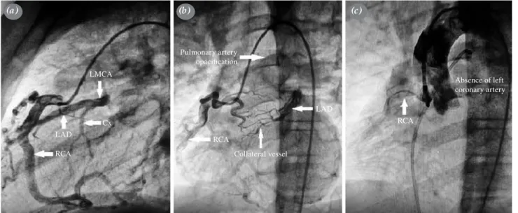
artery (MPA), normal left ventricular function, and a pulmonary artery pressure of 30 mmHg. Cardiac computed tomography (CT) angiography showed dominant right coronary artery (RCA) originating from normal aortic position and LMCA originating posterior of the MPA (Figure 1a, b). Coronary angiography revealed RCA originating from the right Valsalva sinus and running normal anatomic route and left-sided coronary arteries filling from dominant RCA by prominent collateral arteries between the RCA and LAD. On aortography, LMCA was absent (Figure 2a-c).
Figure 1. Computed tomography angiography. (a) RCA originating from ascending aorta. (b) LMCA originating from posterior MPA. RCA: Right coronary artery; MPA: Main pulmonary artery; LMCA: Left main coronary artery.
RCA MPA MPA LMCA Ascending aorta Ascending aorta (a) (b)
Figure 2. Coronary angiography. (a) Main coronary arteries. (b) Pulmonary artery opacification and prominent collateral vessels. (c) Absent left-sided coronary artery in aortography.
LMCA: Left main coronary artery; LAD: Left anterior descending artery; Cx: Circumflex artery; RCA: Right coronary artery.
Cx LMCA LAD RCA RCA LAD RCA Collateral vessel Pulmonary artery opacification Absence of left coronary artery (a) (b) (c)
109 Ata et al.
A new surgical approach to a patient with anomalous origin of left coronary artery from pulmonary artery
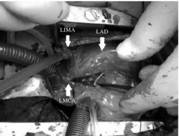
The patient was prepared for surgical correction. A written informed consent was obtained from each parent of the patient. Median sternotomy was performed under general anesthesia. The LIMA was inspected, the pulsation was excellent, and the diameter was found to be close to the LMCA. The LIMA was, then, harvested in a pedicular fashion. Heparin (200 IU/kg) was administered and CPB was initiated, after aortic and right atrial double-stage cannula were inserted (Figure 3). The LMCA was transected from where the pulmonary artery was connected. After confirmation of the LIMA and LMCA diameters were about 1.75 mm and ideal for anastomosis, the fish mouth-shaped end-to-end anastomosis technique was performed using 7/0 polypropylene suture as heart beating. Total CPB time was 28 min and the total operation time was 69 min. No postoperative complications were observed during follow-up and the patient was discharged uneventfully on the fifth postoperative day. Improved physical exercise capacity and mild-to-moderate mitral regurgitation were observed at six months after operation.
DISCUSSION
Anomalous origin of left coronary artery from the pulmonary artery is a rare congenital cardiac malformation. In the fetal phase, there are no findings, as the pressure and oxygen saturations in the aorta and pulmonary artery are equal. The decrease of pulmonary artery pressure and oxygen saturation after birth leads to left ventricular ischemia and dysfunction due to inability to meet oxygen requirement of the
left ventricle. Majority of patients present with signs and symptoms of heart failure caused by myocardial ischemia in early infancy. In our case, the symptoms were seen, until the end of the first school year, as the right coronary artery and collateral vessels were well developed and the pulmonary artery pressure was within normal range.
Various surgical techniques are used in the treatment. The main goal of the original operations is to establish the dual coronary system. The initial technique for this purpose is the Takeuchi procedure which involves creation of an aortopulmonary window and an intrapulmonary tunnel that baffles the aorta to the ostium of the anomalous left coronary artery. Although the long-term survival is acceptable, complication rates such as baffle leak, pulmonary artery stenosis and need for reoperation following the procedure are common.[1] The literature shows the direct reimplantation (coronary button technique) have similar survival and freedom from reoperation rates, compared to the Takeuchi procedure.[3,4] However, this technique requires CPB, cross-clamping and cold cardioplegic cardiac arrest, while pulmonary artery transection may be also required in most of cases.
In our case, the LIMA configuration was close to the adult and compatible with the LMCA diameter, which was why we preferred this method. The LIMA was also preferred, due to its known best patency rates and excellent growth potentials in pediatric patients.[5] Owing to RCA dominance and well-established collateral vessels, no myocardial ischemia was expected during the LIMA-LMCA anastomosis. We believe that the proximal blood flow from the LIMA provides more physiological coronary blood distribution both for the LAD artery and Cx artery than LMCA ligation and simple LIMA-LAD anastomosis. The use of a saphenous vein as a conduit may lead to catastrophic outcomes due to late intimal hyperplasia in pediatric patients.
Moderate mitral insufficiency in our case was mainly based on ischemia and might have contributed to the appearance of symptoms. Evidence shows that establishment of a two-coronary circulation without mitral valve repair leads to improved LV function, irrespective of the age at the time of repair, and mitral regurgitation decreases over time in surviving patients.[5,6] In our case, we did not intervene moderate mitral regurgitation; therefore, echocardiographic evaluation showed minimal relief of regurgitation at six months after the operation.
Figure 3. Operation view. LIMA and LMCA prepared for
anastomosis. No cross-clamping was applied.
LIMA: Left internal mammary artery; LAD: Left anterior descending; LMCA: Left main coronary artery.
LIMA LAD
110
Turk Gogus Kalp Dama 2019;27(1):107-110
In conclusion, this technique is easy-to-use without requiring cross-clamping and cardioplegic cardiac arrest. Using cardiopulmonary bypass may reduce excessive blood loss and prevents hemodynamic compromise. End-to-end left internal mammary artery to left main coronary artery anastomosis also makes it needless to perform two separate graft anastomosis to the left anterior descending and circumflex arteries. We consider that this technique may be alternative to reimplantation for both children and adult patients. However, life-long follow-up and long-term outcomes are still lacking.
Declaration of conflicting interests
The authors declared no conflicts of interest with respect to the authorship and/or publication of this article.
Funding
The authors received no financial support for the research and/or authorship of this article.
REFERENCES
1. Hoashi T, Kagisaki K, Okuda N, Shiraishi I, Yagihara T, Ichikawa H. Indication of Takeuchi technique for patients with anomalous origin of the left coronary artery from the pulmonary artery. Circ J 2013;77:1202-7.
2. Ceylan Ö, Örün UA, Koç M, Özgür S, Doğan V,
Karademir S, et al. Anomalous coronary artery originating from the pulmonary artery: a report of four cases. Turk Gogus Kalp Dama 2013;21:122-6.
3. Naimo PS, Fricke TA, d’Udekem Y, Cochrane AD, Bullock A, Robertson T, et al. Surgical intervention for anomalous origin of left coronary artery from the pulmonary artery in children: A long-term follow-up. Ann Thorac Surg 2016;101:1842-8.
4. Sarıoğlu T, Yalçınbaş YK, Erek E, Arnaz A, Türkekul Y, Avşar MK. Anomalous left coronary artery originating from pulmonary artery: recovery of left ventricular function after dual coronary system restoration and clinical results. Turk Gogus Kalp Dama 2013;21:1-6.
5. Kitamura S, Seki T, Kawachi K, Morita R, Kawata T, Mizuguchi K, et al. Excellent patency and growth potential of internal mammary artery grafts in pediatric coronary artery bypass surgery. New evidence for a “live” conduit. Circulation 1988;78:I129-39.
6. Georgiev SG, Lazarov SD, Mitev ID, Latcheva AZ, Christov GA, Velkovski IG, et al. Left ventricular and mitral valve function long after repair of left anomalous coronary artery from the pulmonary artery: Recovery years after severe ischemia. World J Pediatr Congenit Heart Surg 2012;3:321-7.
7. Lange R, Cleuziou J, Krane M, Ewert P, Pabst von Ohain J, Beran E, et al. Long-term outcome after anomalous left coronary artery from the pulmonary artery repair: a 40-year single-centre experience. Eur J Cardiothorac Surg 2018;53:732-9.