https://doi.org/10.1177/2515841420971936 https://doi.org/10.1177/2515841420971936 Ther Adv Ophthalmol
2020, Vol. 12: 1–7 DOI: 10.1177/ 2515841420971936 © The Author(s), 2020. Article reuse guidelines: sagepub.com/journals-permissions
Therapeutic Advances in Ophthalmology
journals.sagepub.com/home/oed 1
Introduction
Diabetic macular edema (DME) is the main cause of vision loss in diabetic patients.1 Ten years after
diabetes mellitus was first diagnosed, 20% of the patients were found to be affected by DME.2 In
recent years, the development of different tech-nologies, such as optical coherence tomography (OCT), can demonstrate the presence of DME. Several patterns of DME, such as serous macular detachment (SRD), cystoid macular edema (CME), and diffuse retinal thickening (DRT), have been identified on OCT images.3–6 DME can
also be a mixture of these three patterns. SRD is different from others due to its pathogenesis, which is caused by breakdown of the outer
blood-retinal barriers. DRT and CME are mainly caused by dysfunction of the inner blood-retinal barriers.7–10 Due to differences in pathogenic
mechanisms, DME can be divided in two groups: (1) SRD and (2) non-SRD (DRT CME).
Several studies have shown that the DME pat-terns can affect treatment outcomes. Liu and colleagues compared intravitreal triamcinolone acetonide with intravitreal bevacizumab in DME patients with SRD. In that study, the triamci-nolone acetonide group has better functional and anatomical response than bevacizumab group.11
They said this outcome was associated with anti-inflammatory effects of triamcinolone acetonide.
Effects of dexamethasone treatment on
serous retinal detachment in
ranibizumab-resistant diabetic macular edema
Alper Halil Bayat and Mustafa Nuri Elçioğlu Abstract
Purpose: To evaluate outcome of intravitreal dexamethasone implant (IDI) treatment on serous
retinal detachment (SRD) in patients with ranibizumab-resistant diabetic macular edema (DME).
Materials and methods: Forty-eight eyes of 48 patients with DME resistant to ranibizumab
were enrolled in this retrospective and comparative study. Patients were divided into two groups according to presence of serous retinal detachment: (1) SRD or (2) non-SRD groups. All patients had at least three monthly ranibizumab injections, after which they were treated with IDI. The best-corrected visual acuity (BCVA), central retinal thickness (CRT), use of antiglaucomatous drugs, and presence of cataract progression were noted at 1, 3, and 6 months post-IDI treatment.
Results: There was not any statistically significant difference in terms of baseline
characteristics of the patients. The mean CRT was declined in both groups at 1, 3, and
6 months (p < 0.001). After IDI treatment, the mean BCVA was improved in both groups at 1, 3, and 6 months (p < 0.001). When groups were compared, the change in CRT was higher in the SRD group (p = 0.018), while there was no statistically significant difference between groups in terms of BCVA changes (p = 0.448).
Conclusion: The presence of SRD resulted in higher anatomical gain. SRD had no effects on
visual changes after dexamethasone treatment in patients with ranibizumab-resistant DME. Keywords: dexamethasone implantation, diabetic macular edema, optical coherence tomography, ranibizumab, serous retinal detachment
Received: 15 May 2020; revised manuscript accepted: 16 October 2020.
Correspondence to:
Alper Halil Bayat Department of Ophthalmology, Esenler Hospital, Medipol University, Birlik mah. Bahçeler cad. No:5, Esenler, 34083 İstanbul, Turkey
Mustafa Nuri Elçioğlu
Department of Ophthalmology, Okmeydanı Training and Research Hospital, University of Health Sciences, İstanbul, Turkey
Shimura and colleagues studied patients with DME who were treated with ranibizumab. In their study, SRD patients had worse functional and anatomical outcomes with ranibizumab treat-ment than non-SRD patients.12 Demircan and
colleagues13 demonstrated that dexamethasone
implantation was more useful than ranibizumab in DME with SRD. However, all of the these studies had patients who had treatment-naive DME. In the current study, we aimed to explain the influence of SRD on the outcome of intravit-real dexamethasone implant (IDI) in patients with ranibizumab-resistant DME.
Methods
This retrospective and comparative study was per-formed accordance with Declaration of Helsinki. Written informed consents were obtained from all patients. All necessary authorizations were obtained from the Institutional Review Board of Okmeydanı Research and Training Hospital, İstanbul, Turkey with number 751.
Forty-eight eyes of 48 patients with DME resist-ance to ranibizumab were enrolled in this study. Patients were divided in two groups according to presence of SRD observed on OCT scans: (1) SRD and (2) non-SRD groups. All patients had at least three monthly ranibizumab injections. Reduction of less than 20% central retinal thick-ness (CRT) on SD-OCT 1 month after third ranibizumab injections was defined as resistant to ranibizumab therapy and treated with intravitreal dexamethasone treatment. Best-corrected visual acuity (BCVA), CRT use of antiglaucomatous drugs, and presence of cataract progression were noted at 1, 3, and 6 months post-IDI treatment. Inclusion criteria were consisted of several param-eters: (1) OCT > 250 µm, (2) age > 18 years old, (3) at least three monthly ranibizumab injections, and (4) ranibizumab resistance. Patients with sev-eral conditions were excluded as follows: (1) a history of glaucoma, (2) steroid induced ocular hypertension, (3) vitrectomy, (4) other vitroreti-nal diseases and retinopathies, (5) corneal opac-ity, and (6) laser photocoagulation within 6 months prior to study enrollment.
All of the patients had standard ophthalmic exami-nations pre- and post-treatment (1-, 3-, and 6-month follow-ups and final visit). The examina-tions included slit-lamb microscopy, BCVA, tonometry, spectral domain OCT (SD-OCT), and indirect ophthalmoscopy. BCVA was measured
with the Snellen chart, and the decimal visual acu-ity was converted to the logarithm of the minimal angle of resolution (logMAR) units for statistical analyses. The OCT was performed on an SD-OCT (Spectralis, Heidelberg Engineering, Heidelberg, Germany). The CRT was computed 1 mm central diameter area by using OCT mapping software. All injections were performed in the operating room under aseptic condition with topical anes-thesia (0.4% benoxinate). A dexamethasone implant (0.7 mg) (Ozurdex, Allergan Inc. Bayer, Berlin, Germany) was injected with a 22-gauge applicator through the pars plana at 3.5–4 mm posterior of limbus. After injections topical moxi-flaxacin were given as therapy for one week. After IDI, the patients did not receive any additional treatment for 6 months. IDI was performed 1 month after last ranibizumab injection.
Statistical analyses were performed using the SPSS software version 21. Descriptive analyses were presented using means and standard devia-tions for normally distributed variables. The change in CMT and BCVA by the time of investi-gation was performed using repeated measures analysis of variance (ANOVA). A Greenhouse– Geisser correction was used when the sphericity assumption was violated. The Kruskal–Wallis test was conducted to compare non-parametric parameters among the groups. A one-way ANOVA was used to compare parametric parameters among the groups. A p-value < 0.05 was consid-ered to show a statistically significant result. Results
Twenty-two eyes in the SRD group and 26 eyes in the non-SRD group were examined. The mean age of the patients was 61.4 ± 9.1 years in the SRD group and 64.8 ± 8.9 in the non-SRD group (p = 0.229). The mean number of previous ranibizumab injections was 5 ± 1.8 and 5.0 ± 2.8 in the SRD and non-SRD groups, respectively (p = 0.883). There was no statistically significant difference between groups in terms of initial BCVA, CMT, glycosylated hemoglobin (HbA1c) levels, and gender (p = 0.185, 0.148, 0.209, and 0.662, respectively). The baseline characteristics of the patients are shown in Table 1.
The mean initial CRT was 503 ± 114 µm in SRD group and 446 ± 89 µm in the non-SRD group (p = 0.883). The mean CRT declined to 270 ± 56, 257 ± 54, and 309 ± 107 µm in the SRD group
and 328 ± 63, 296 ± 69, and 353 ± 101 µm in the non-SRD group at 1, 3, and 6 months, respec-tively (p < 0.001). When the groups were com-pared, the change in CRT was higher in the SRD group (p = 0.018). The changes in CRT are dis-played in Figure 1 and summarized in Table 2. The mean BCVAs in the SRD and non-SRD groups were 0.72 ± 0.47 and 0.97 ± 0.64 logMAR, respectively (p = 0.185). After IDI treatment, the mean BCVA was 0.62 ± 0.45, 0.56 ± 0.42, and 0.59 ± 0.53 logMAR at 1, 3, and 6 months, respec-tively, in the SRD group, while it was 0.67 ± 0.52, 0.61 ± 0.51, and 0.65 ± 0.58 logMAR in the non-SRD group at the same time points (p < 0.001). There was no statistically significant difference between groups in terms of BCVA changes (p = 0.448). The changes in BCVA are displayed in Figure 2 and summarized in Table 2.
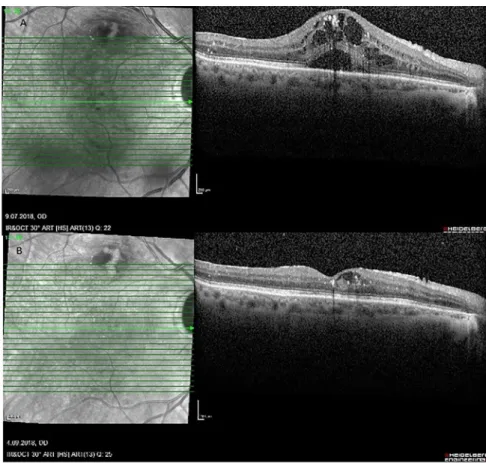
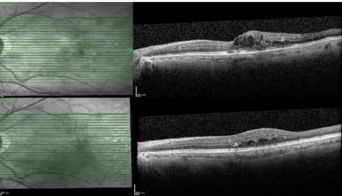
OCT samples of the patients in SRD and non-SRD groups were displayed in Figures 3 and 4, respectively.
Two eyes in the SRD group and three eyes in the non-SRD group had to use anti-glaucomatous drugs because of an intraocular pressure elevation.
One eye in each group had cataract progression at the end of the 6-month follow-up.
Discussion
In the current study, we found that IDI was effec-tive in both SRD and non-SRD patterns of DME resistance to ranibizumab treatment with respect to both anatomical and functional gain. SRD patients were found to have faster responses to IDI than non-SRD patients, and the SRD patients presented a better anatomical gain. In terms of visual acuity changes, there was no statistically sig-nificant difference between SRD and non-SRD patients.
Previous studies have shown different types of DME patterns, such as SRD, DRT, and CME on OCT scans.3–6 Each pattern of DME may have a
different pathogenesis. For example, SRD is defined as fluid accumulation in the subretinal space as a result of retinal pigment epithelium dys-function and damage to the external limiting membrane.9,10 DRT results from
ischemia-induced intracytoplasmic swelling of Müller cells, and if Müller cells have necrosis with cavity for-mation, CME results.7,8
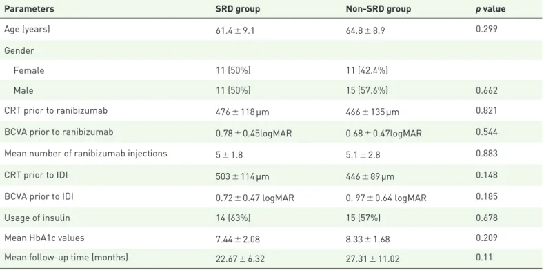
Table 1. The baseline characteristics of the patients.
Parameters SRD group Non-SRD group p value
Age (years) 61.4 ± 9.1 64.8 ± 8.9 0.299
Gender
Female 11 (50%) 11 (42.4%)
Male 11 (50%) 15 (57.6%) 0.662
CRT prior to ranibizumab 476 ± 118 µm 466 ± 135 µm 0.821
BCVA prior to ranibizumab 0.78 ± 0.45logMAR 0.68 ± 0.47logMAR 0.544
Mean number of ranibizumab injections 5 ± 1.8 5.1 ± 2.8 0.883
CRT prior to IDI 503 ± 114 µm 446 ± 89 µm 0.148
BCVA prior to IDI 0.72 ± 0.47 logMAR 0. 97 ± 0.64 logMAR 0.185
Usage of insulin 14 (63%) 15 (57%) 0.678
Mean HbA1c values 7.44 ± 2.08 8.33 ± 1.68 0.209
Mean follow-up time (months) 22.67 ± 6.32 27.31 ± 11.02 0.11
Several studies in the current literature aim to explain the effects of treatment modalities on SRD and non-SRD patients. Kim and colleagues studied patients with different DME types who underwent bevacizumab treatment. They reported that bevacizumab injections were more effective in DRT patients than those with SRD or CME.14 In agreement with their results, Shimura
and colleagues12 also reported that bevacizumab
has the lowest effects in the SRD type of DME.
Seo and colleagues5 reported that DRT patients
had good responses to ranibizumab injections, while the SRD group had the worst visual acuity. They demonstrated that SRD patients had poorer visual gain because of damage to ellipsoid zone integrity. Ozdemir and colleagues15 reported that
SRD was a good predictive factor for DME dur-ing intravitreal triamcinolone acetonide treat-ment. In a recent study, Demircan and colleagues compared ranibizumab and dexamethasone in Figure 1. The changes in CRT with time after intravitreal dexamethasone implantation.
CRT, central retinal thickness; SRD, serous retinal detachment.
Table 2. Comparison of monthly changes in CRT and BCVA between groups.
Parameter SRD group Non-SRD group p value
Initial CRT 503 ± 114 µm 446 ± 89 µm 0.148
First month CRT 270 ± 56 µm 328 ± 63 µm 0.009*
Third month CRT 257 ± 54 µm 296 ± 69 µm 0.096
Sixth month CRT 309 ± 107 µm 353 ± 101 µm 0.244
Initial BCVA 0.72 ± 0.47 logMAR 0.97 ± 0.64 logMAR 0.185
First month BCVA 0.62 ± 0.45 logMAR 0.67 ± 0.52 logMAR 0.770
Third month BCVA 0.56 ± 0.42 logMAR 0.61 ± 0.51 logMAR 0.742
Sixth month BCVA 0.59 ± 0.53 logMAR 0.65 ± 0.58 logMAR 0.775
BCVA, best-corrected visual acuity; CRT, central retinal thickness; SRD, serous retinal detachment. *Statistically significant.
Figure 2. The changes in BCVA with time after intravitreal dexamethasone implantation.
BCVA, best-corrected visual acuity; SRD: serous retinal detachment.
Figure 3. Optical coherence tomography images of a patient with serous retinal detachment before and after intravitreal dexamethasone implantation.
DME with SRD at the 1 month follow-up period. They reported that IDI was found to be more effective in reduction of CRT and SRD height.13
However, their study was done only at the 1-month follow-up period. Our study has a 6-month follow-up time, and IDI was found to be effective in CRT reduction at the end of the 6 months. Previous studies have shown that IDI has maximum effects at two months while its effects decrease slowly from months 4 to 6.16
Thus, in order to explain the effects of IDI, the 6-month follow-up point is necessary. Our study has the advantage over longer times compared to Demircan and colleagues. In another recent study, Ozdemir and colleagues studied 24 eyes of patients with SRD related to DME. They reported that IDI caused an increase in BCVA and a reduc-tion in SRD and CRT.17 However, their study
lacked a control group. In our study, we had a control group (non-SRD patients) and found that although IDI was effective in visual acuity changes, there were no statistically significant dif-ferences between SRD and non-SRD patients. All of these studies had treatment-naive DME patients. In the current literature, there is only one study that aims to explain effects of dexa-methasone implantation with ranibizumab- resistant DME based on OCT patterns. Kaldırım and colleagues studied 35 eyes of 31 patients with ranibizumab-resistant DME. They reported that the SRD group had better BCVA than DRT and CME groups, but the difference was not statisti-cally significant at 4 months. At the end of the 6 months, DRT groups had better BCVA than
other groups because they administered anti-vas-cular endothelial growth factor (VEGF) agents at 4 months.18 As mentioned above, in order to
bet-ter understand the effects of IDI, a 6-month fol-low-up point is necessary.16 In the current study,
we observed IDI effects on SRD and non-SRD patients over the 6-month follow-up time.
Kim and colleagues19 reported that SRD or CME
patients had higher concentrations of inflamma-tory cytokines, such as interleukins (ILs)-6 and -8 and platelet-derived growth factor, in the aqueous humor of DME rather than DRT patients. These differences can explain why steroids were found to be more effective in SRD patients, while anti-VEGF agents were found to be more effective in non-SRD patients.12–14
There are some limitations to our study, such as retrospective dosing and small sample size. In order to better explain effects of SRD on dexa-methasone treatment in patients with DME, a larger sample size and prospective dosing studies are needed.
In conclusion, the presence of SRD resulted in higher anatomical gain, but it had no effects on visual acuity changes with respect to dexametha-sone treatment in patients with ranibizumab-resistant DME.
Conflict of interest statement
The authors declared no potential conflicts of interest with respect to the research, authorship, and/or publication of this article.
Figure 4. Optical coherence tomography images of a patient without serous retinal detachment before and after intravitreal dexamethasone implantation.
Funding
The authors received no financial support for the research, authorship, and/or publication of this article.
ORCID iD
Alper Halil Bayat https://orcid.org/0000-0003- 1827-968X
References
1. Romero-Aroca P. Managing diabetic macular edema: the leading cause of diabetes blindness.
World J Diabetes 2011; 2: 98–104.
2. Early Treatment Diabetic Retinopathy Study Research Group: photocoagulation for diabetic macular edema: ETDRS report no.4. Int
Ophtahlmol Clin 1987; 27: 265–272.
3. Otani T, Kishi S and Maruyama Y. Patterns of diabetic macular edema with optical coherence tomography. Am J Ophthalmol 1999; 127: 688–693.
4. Kim BY, Smith SD and Kaiser PK. Optical coherence tomographic patterns of diabetic macular edema. Am J Ophthalmol 2006; 142: 405–412.
5. Seo KH, Yu SY, Kim M, et al. Visual and morphologic outcomes of intravitreal ranibizumab for diabetic macular edema based on optical coherence tomography patterns. Retina 2016; 36: 588–595.
6. Giocanti-Auregan A, Hrarat L, Qu LM, et al. Functional and anatomical outcomes in patients with serous retinal detachment in diabetic macular edema treated with ranibizumab. Invest
Ophthalmol Vis Sci 2017; 58: 797–800.
7. Yanoff M, Fine BS, Brucker AJ, et al. Pathology of human cystoid macular edema. Surv
Ophthalmol 1984; 28: 505–511.
8. Daruich A, Matet A, Moulin A, et al. Mechanisms of macular edema: beyond the surface. Prog Retin Eye Res 2018; 63: 20–68. 9. Vujosevic S, Torresin T, Berton M, et al.
Diabetic macular edema with and without subfoveal neuroretinal detachment: two different morphologic and functional entities. Am J
Ophthalmol 2017; 181: 149–155.
10. Kaya M, Kaya D, Idiman E, et al. A novel biomarker in diabetic macular edema with serous retinal detachment: serum chitinase-3-like protein 1. Ophthalmologica 2019; 241: 90–97. 11. Liu Q, Hu Y, Yu H, et al. Comparison of
intravitreal triamcinolone acetonide versus intravitreal bevacizumab as the primary treatment of clinically significant macular edema. Retina 2015; 35: 272–279.
12. Shimura M, Yasuda K, Yasuda M, et al. Visual outcome after intravitreal bevacizumab depends on the optical coherence tomographic patterns of patients with diffuse diabetic macular edema.
Retina 2013; 33: 740–747.
13. Demircan A, Ozkaya A, Alkin Z, et al. Comparison of the effect of ranibizumab and dexamethasone implant on serous retinal detachment in diabetic macular edema. J Fr
Ophtalmol 2018; 41: 733–738.
14. Kim M, Lee P, Kim Y, et al. Effect of intravitreal beva-cizumab based on optical coherence tomography patterns of diabetic macular edema.
Ophthalmologica 2011; 226: 138–144.
15. Ozdemir H, Karacorlu M and Karacorlu SA. Regressionof serous macular detachment after intravitreal triamcinoloneacetonide in patients with diabetic macular edema. Am J Ophthalmol 2005; 140: 251–255.
16. Chang-Lin JE, Attar M, Acheampong AA, et al. Pharmacokinetics and pharmacodynamics of a sustained release dexamethasone intravitreal implant. Invest Ophthalmol Vis Sci 2011; 52: 80–86.
17. Ozdemir MH, Elbay A, Kirik F, et al. Regression of serous macular detachment after intravitreal dexamethasone implant in patients with diabetic macular edema. J Ocul Pharmacol Ther 2019; 35: 558–564.
18. Kaldırım H, Yazgan S, Atalay K, et al. Intravitreal dexamethasone implantation in patients with different morphological diabetic macular edema having insufficient response to ranibizumab.
Retina 2018; 38: 986–992.
19. Kim M, Kim Y and Lee SJ. Comparison of aqueous concentrations of angiogenic and inflammatory cytokines based on optical coherence tomography patterns of diabetic macular edema. Indian J Ophthalmol 2015; 63: 312–317.
Visit SAGE journals online journals.sagepub.com/ home/oed