Background: Several studies have demonstrated an association between obesity, periodontitis, and exercise. Aims: This study aimed to investigate the effects of regular exercise on obese women with periodontal disease, using serum, saliva, and gingival crevicular fluid (GCF) samples. A before–after study design was adopted to evaluate the effects of 12 weeks of regular exercise on obese women grouped according to periodontal status, without a control group (no exercise). The study sample comprised of 15 patients without periodontitis (NP group) and 10 patients with chronic periodontitis (CP group), from whom periodontal parameters were measured and serum, saliva, and GCF samples were collected. Body mass index (BMI), anthropometric measurements, somatotype-motoric tests, and maximal oxygen consumption (VO2max) were recorded at baseline and after exercise. Subjects and Methods: Med Calc was used for statistical analysis. Results: After exercise, a significant decrease in BMI and a significant increase in VO2max were observed in both groups. A significant decrease in probing depth and clinical attachment loss, serum leptin, GCF tumor necrosis factor-α(TNF-α) and leptin, and a significant increase in GCF resistin were observed in the CP group. A significant decrease in serum TNF-α and leptin levels and a significant increase in serum resistin and GCF TNF-α, leptin, resistin, and adiponectin levels were observed in the NP group. Significant correlations between bleeding on probing and levels of interleukin-1β and leptin in GCF were observed in the CP group. Conclusions: This study showed that regular exercise exerts different impacts with respect to clinical and biochemical aspects of periodontal and systemic conditions in obese women.
Keywords: Adipokines, chronic periodontitis, exercise, gingival crevicular fluid, obesity, saliva
Effects of Exercise on Periodontal Parameters in Obese Women
B Alkan, E Guzeldemir‑Akcakanat1, B Odabas‑Ozgur2, T Ozgur3, A Demirdizen‑Taskiran4, HM Kir5, N Alpay6,
E Cayci‑Akkan7
Address for correspondence: Prof. E Guzeldemir‑Akcakanat, Department of Periodontology, Faculty of Dentistry, Kocaeli University, 41190, Basiskele Kocaeli, Turkey. E‑mail: [email protected]
Department ofPeriodontology, Faculty of Dentistry, Istanbul Medipol University, Istanbul, Turkey (Previously Department of Periodontology, Faculty of Dentistry, Kocaeli University, Kocaeli, Turkey), 1Department of
Periodontology, Faculty of Dentistry, Kocaeli University, Kocaeli, Turkey, 2Sport Sciences PhD, New
Jersey, United States (Previously Department of Physical Education and Sports, Faculty of Sports Sciences, Kocaeli University, Kocaeli, Turkey), 3Biobehavioral
Sciences, Teacher College, Columbia University, New York, United States (Previously Department of Coaching, Faculty of Sports Sciences, Kocaeli University, Kocaeli, Turkey), 4Department
of Physical Education and Sports Teaching, Faculty of Sports Sciences, Istanbul Gedik University, Istanbul, Turkey (Previously Department of Coaching, Faculty of Sports Sciences, Kocaeli University, Kocaeli, Turkey), 5Department of
Medical Biochemistry, Faculty of Medicine, Kocaeli University, Kocaeli, Turkey, 6Life Sciences
Application and Research Center, Gazi University, Ankara, Turkey,
7Specialist in Periodontology,
Private Practice, Kocaeli, Turkey (Previously Department of Periodontology, Faculty of Dentistry, Kocaeli University, Kocaeli, Turkey)
Abstract
This is an open access journal, and articles are distributed under the terms of the Creative Commons Attribution‑NonCommercial‑ShareAlike 4.0 License, which allows others to remix, tweak, and build upon the work non‑commercially, as long as appropriate credit is given and the new creations are licensed under the identical terms.
For reprints contact: [email protected]
How to cite this article: Alkan B, Guzeldemir-Akcakanat E, Odabas-Ozgur B, Ozgur T, Demirdizen-Taskiran A, Kir HM, et al. Effects of exercise on periodontal parameters in obese women. Niger J Clin Pract 2020;23:1345-55.
Access this article online
Quick Response Code:
Website: www.njcponline.com DOI: 10.4103/njcp.njcp_627_19 PMID: ******* Received: 15-Nov-2019; Revision: 04-Jan-2020; Accepted: 30-Mar-2020; Published: 12-Oct-2020
Introduction
O
besity, which is characterized by excess fat accumulation in the body, is among the most serious public health problems.[1] The prevalence of obesity has increased since the 1980s, and current estimates indicate that >10% of adults worldwide are obese.In obesity, excessive production of inflammatory cytokines and hormones, called adipokines, leads to chronic low-grade inflammation.[2] A high prevalence of periodontal disease in obese participants has been reported in earlier literature.[3]
Periodontitis is a chronic inflammatory disease of the periodontal tissues.[4] It involves the local production of inflammatory mediators that exacerbate current inflammation in other organ systems.[5] Abnormal production of adipokines appears to be instrumental in the relationship between obesity, chronic disease, and periodontitis.[3]
Maintenance of physical activity is an effective
strategy for preventing obesity.[1] Even in the
absence of significant weight loss, regular exercise has been associated with improvements in systemic
health.[6] A correlation between reduced physical
activity and periodontitis prevalence has also been reported.[7]
The present study hypothesized that regular aerobic exercise (RE) may have positive effects on the systemic, oral, and periodontal health of obese women. Hence, the study’s aim was to assess the possible relationship between periodontal status, obesity, and exercise in women.
Subjects and Methods
The study protocol was approved according to the Declaration of Helsinki of 1975, as revised in 2000 (Ethical identification number: 36290600). The approval from the ethics committee had been declared and the date of the approval has been added. (Ethical identification number / Date: 36290600 / 11 December 2013).
Study population
A total of 527 participants registered for the 12 weeks aerobic exercise program organized between February and May 2015. After the study protocol was explained to them, eligible participants willing to participate in the study were evaluated with respect to body mass index (BMI), medical history, and periodontal status, and their written informed consent was taken.
Participants were excluded if they were not obese (BMI˂30 kg/m2) according to the classification of the World Health Organization[8]; if they required
antibiotic prophylaxis; if they had received antibiotic therapy within the preceding 3 months or during the exercise program, if they had any contagious diseases, if they were lactating or possibly pregnant, if they had uncontrolled systemic disease; if they had undergone periodontal treatment within the preceding 6 months; if they had fewer than 14 teeth; and if they were smokers who smoked >10 cigarettes per day.
Sociodemographic and dental habit data
All participants answered a sociodemographic and dental habits questionnaire that assessed age, educational and marital status, interdental cleaning methods, bruxism, frequency of dentist visits (e.g. in need, once a year), smoking (no/yes (number)), and self-perceived periodontal health (bad, average, good).
Periodontal evaluation
The whole-mouth clinical periodontal parameters were recorded as follows; plaque index (PI) and gingival index (GI) were recorded from four sites (disto-buccal, mid-buccal, mesio-buccal, and mid-palatal), and probing depth (PD), bleeding on probing (BOP), gingival recession (GR), and clinical attachment loss (CAL) were recorded from 6 sites (disto-buccal, mid-buccal, mesio-buccal, disto-palatal, mid-palatal, and mesio-palatal). All assessments were performed using Williams’ periodontal probe (Hu-Friedy, Chicago, IL, USA) by the same researcher (BA). The extent and severity of alveolar bone loss were determined via clinical and radiographical examination for all individuals. All patients were diagnosed according to the criteria of the 1999 International Workshop for a Classification of Periodontal Diseases and Conditions.[9]
Maximal oxygen consumption, somatotype‑motoric tests, anthropometric measurements, and exercise program
Maximal oxygen consumption (VO2max), as an
indicator of physical fitness, was calculated using
the Rockport walking analysis.[10] Smedley-type
dynamometers (Tokyo, Japan) were used to measure hand-grip and back-leg strength, which was expressed in kilograms. All anthropometric measurements, including BMI, circumferences (girth of abdomen, neck, shoulder, calf, arm, mid-thigh, waist, hip, and bust), skinfolds (skinfolds of biceps, triceps, subscapular, iliac crest, abdominal, front thigh, and medial calf), and diameters of body parts (length of arm, forearm, and hand and breadth of bi-epicondylar humerus, bi-epicondylar femur, bi-acromial, and bi-iliocristal) were recorded by a professional instructor. Qualified fitness instructors conducted the exercise program for one hour per day, 3 days a week, over 12 weeks.
Individuals who did not attend the program for more than 7 days were removed from the study.
Sample collections
All samples were obtained in the morning following an overnight fast. GCF sampling was performed 1 day after the clinical measurements were taken. All measurements and samplings were performed both before and after the exercise program. No attempt was made to change participants’ oral hygiene habits.
Unstimulated saliva was collected in a disposable Falcon tube (Kartell, Milan, Italy), mixed for 5 min, left to stand at room temperature for 5 min, and centrifuged at 4000 rpm for 10 min at 4°C. The supernatant was isolated.
Venous blood samples were collected in serum tubes (BD Vacutainer Co., NJ, USA). The tubes were gently rinsed several times, left to stand at room temperature for 5 min, and centrifuged at 2000 × g for 5 min. The serum was isolated.
In patients with chronic periodontitis (CP group), GCF samples were collected from teeth with alveolar bone loss and CAL ≥4 mm, PD ≥4 mm, and BOP (+) sites. In patients without periodontitis (NP group), GCF samples were taken from one side of a tooth, exhibiting PD ≤3 mm without BOP and CAL. In order to avoid saliva contamination, the test sites were gently air-dried and kept dry using cotton rolls. The supragingival plaque was gently removed using a curette without touching the gingiva. The surface was gently dried for 2 s, and a standardized periopaper strip (OraFlow, NY, USA) was carefully inserted into the pocket/sulcus and left there for 30 s. Blood- or saliva-contaminated pieces were discarded. The fluid volume was measured using a precalibrated electronic gingival fluid–measuring device (OraFlow, NY, USA). Readings from the device were converted into microliters according to the standard curve using a software program (Periotron MLCONVRT. EXE software, NY, USA).
All samples were collected in safe-lock tubes (Eppendorf Co., Hamburg, Germany), sealed with parafilm to avoid evaporation, and stored in a refrigerator at −80°C until analysis.
Biochemical analysis
The samples were transported to the laboratory on dry ice and analyzed immediately. The strips were pooled into 100-µL phosphate-buffered solution (pH 7.4), mixed, and centrifuged at 1000 rpm for 1 min. Serum, saliva, and GCF samples were analyzed for the quantitative assessment of human IL-1β (Invitrogen, CA, USA), TNF-α (Invitrogen, CA, USA), leptin (EMD
Millipore, MO, USA), resistin (EMD Millipore, MO, USA), and adiponectin (EMD Millipore, MO, USA) using commercially available enzyme-linked immunosorbent (ELISA) assay kits.
Statistical analyses
Continuous variables were presented using descriptive statistics, for example, mean, median, standard deviation, and range (min, max). The distributions of the variables were analyzed for normality using the Shapiro–Wilk test. Normally distributed variables were analyzed using the Student’s t-test. Independent and nonparametric variables were analyzed using the Mann–Whitney U test. The relationship between discrete variables was analyzed using the Chi-square test or Fisher’s exact test. Pearson’s correlation coefficients were calculated to study the relation between pairs of normally distributed continuous variables. If the variables were nonparametric, their correlation was analyzed using Spearman’s rho test. P < 0.05 was considered statistically significant. A software program was used to perform statistical analyses (MedCalc Statistical Software version 12.7.7, Ostend, Belgium).
Results
Study population
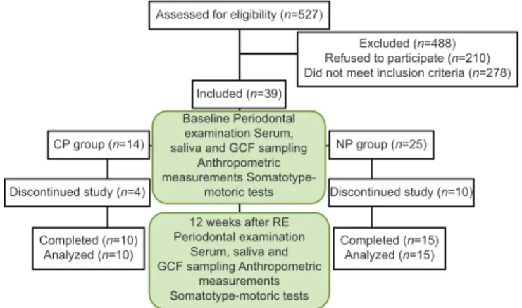
A total of 527 participants applied to participate in the exercise program. During the interview, 210 stated that they would be unable to manage the continuity of the exercise program, while 278 failed to meet the inclusion criteria [Figure 1]. A total of 39 women (age range 32–61 years), including the NP group (n = 25) and the CP group (n = 14), participated in the study.
In total 25 obese participants (10 in the CP group and 15 in the NP group; mean ages, 49.10 and 45.93 years; range, 36–60 and 32–61 years) completed the program. A total of 14 subjects (4 from the CP group and 10 from the NP group) were excluded from the
Assessed for eligibility (n=527)
Excluded (n=488) Refused to participate (n=210) Did not meet inclusion criteria (n=278) Included (n=39)
CP group (n=14)
Baseline Periodontal examination Serum, saliva and GCF sampling
Anthropometric measurements
Somatotype-motoric tests
NP group (n=25)
Discontinued study (n=4) Discontinued study (n=10)
Completed (n=10)
Analyzed (n=10) Completed (n=15)Analyzed (n=15) 12 weeks after RE
Periodontal examination Serum, saliva and GCF sampling Anthropometric
measurements Somatotype-motoric tests Figure 1: Flowchart of the study
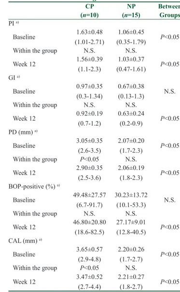
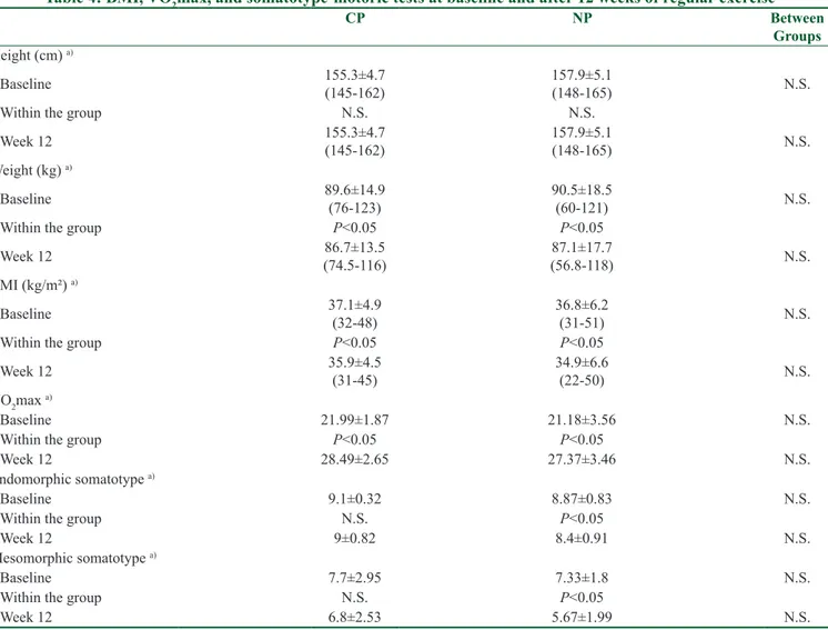
study, as they were unable to guarantee their regular attendance at the exercise program. Initially, descriptive and sociodemographic determinants [Table 1], GI and BOP parameters [Table 2], saliva and serum variables [Table 3], BMI, VO2max, anthropometric measurements, and somatotype-motoric tests [Table 4] showed no significant (P > 0.05) difference between the groups.
Otherwise, the CP group had higher PI, PD, CAL [Table 2], GCF IL-1β, resistin, and adiponectin [Table 3] levels compared with the NP group at baseline (P < 0.05). Significant improvements in periodontal parameters were observed only in the CP group (P < 0.05) [Table 2].
BMI, VO
2max,
somatotype‑motoric
tests, and anthropometric results
After RE, a significant increase in VO2max and a decrease in BMI (P < 0.05) [Table 4], mid-thigh girth, breadth of the bi-epicondylar femur, and forearm and hand lengths were seen in both groups (data not shown). Moreover, both endomorphic and mesomorphic somatotypes, girths of neck, arm, bust, waist, and abdomen, breadth of bi-epicondylar humerus, and skinfolds of triceps and subscapular in the NP group and breadth of bi-iliocristal and skinfolds of biceps and abdomen in the CP group exhibited statistically significant decreases (data not shown).
Table 1: Comparison of sociodemographic and descriptive determinants of the groups at baseline
CP NP P (n=10) (n=15) Age (years) a) 49.1±7.9 45.9±8.3 N.S. (36-60) (32 - 61) Education status n (%) N.S. Non-literate 2 (20) 1 (6.67) Primary school 5 (50) 8 (53.33) Secondary school 1 (10) 2 (13.33) High school 2 (20) 3 (20) University 0 (0) 1 (6.67) Marital Status n (%) N.S. Single 0 (0) 1 (6.7) Married 10 (100.0) 14 (93.3) Brushing n (%) N.S. 1 per day 3 (30) 8 (53.33) 2 per day 2 (20) 3 (20) 3 per day 2 (20) 1 (6.67) Sometimes 3 (30) 3 (20) Interdental cleaning n (%) N.S. No 6 (60) 9 (60) Toothpick 3 (30) 4 (26.67) Interdental brush 1 (10) 2 (13.33) Bruxism n (%) N.S. No 4 (40) 6 (40) Sometimes 6 (60) 9 (60) Dentist frequency n (%) N.S. In need 10 (100) 14 (93.33) Once a year 0 (0) 1 (6.67) Smoking n (%) N.S. No 9 (90) 15 (100) Yes 1 (10) 0 (0)
Describe your gingiva n (%)
N.S. Bad 3 (30) 3 (20) Average 4 (40) 10 (66.67) Good 3 (30) 2 (13.33) P: statistically significant, N.S.: not significant, n: number, TL: Turkish Lira, %: percentage. a)mean±standard deviation values (min - max)
Table 2: Periodontal parameters at baseline and after 12 weeks of regular exercise
CP NP Between (n=10) (n=15) Groups PI a) Baseline 1.63±0.48 (1.01-2.71) 1.06±0.45 (0.35-1.79) P˂0.05 Within the group N.S. N.S. Week 12 1.56±0.39 (1.1-2.3) 1.03±0.37 (0.47-1.61) P˂0.05 GI a) Baseline 0.97±0.35 (0.3-1.34) 0.67±0.38 (0.13-1.3) N.S. Within the group N.S. N.S. Week 12 0.92±0.19 (0.7-1.2) 0.63±0.24 (0.2-0.9) P˂0.05 PD (mm) a) Baseline 3.05±0.35 (2.6-3.5) 2.07±0.20 (1.7-2.3) P˂0.05 Within the group P˂0.05 N.S. Week 12 2.90±0.35 (2.5-3.6) 2.06±0.19 (1.8-2.3) P˂0.05 BOP-positive (%) a) Baseline 49.48±27.57 (6.7-91.7) 30.23±13.72 (10.1-53.3) N.S. Within the group N.S. N.S. Week 12 46.80±20.80 (18.6-82.5) 27.17±9.01 (12.8-40.5) P˂0.05 CAL (mm) a) Baseline 3.65±0.57 (2.9-4.8) 2.20±0.26 (1.7-2.7) P˂0.05 Within the group P˂0.05 N.S. Week 12 3.47±0.52 (2.7-4.4) 2.21±0.27 (1.8-2.7) P˂0.05
P˂0.05 statistically significant, N.S.: not significant, n: number,
%: percentage, PI: plaque index, GI: gingival index, PD: probing depth, BOP: bleeding on probing, CAL: clinical attachment level. a)mean±standard deviation values (min-max)
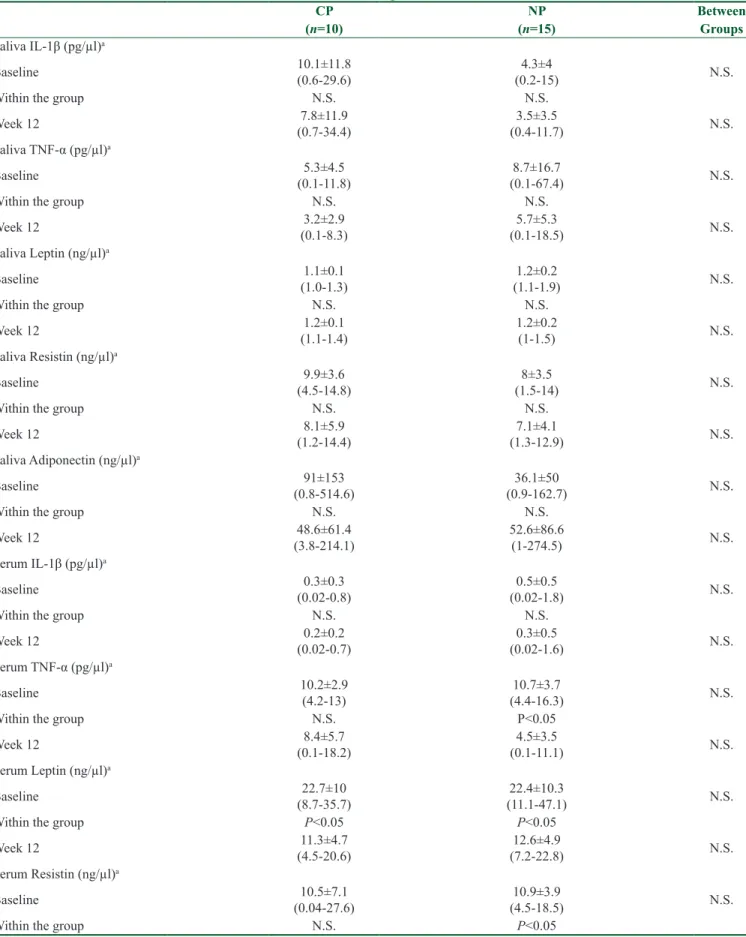
Table 3: Saliva, serum, total amount (pg)/(ng) and concentration (pg/µl)/(ng/µl) of GCF parameters at baseline and after 12 weeks of regular exercise
CP NP Between (n=10) (n=15) Groups Saliva IL-1β (pg/µl)a Baseline 10.1±11.8 (0.6-29.6) (0.2-15)4.3±4 N.S. Within the group N.S. N.S. Week 12 (0.7-34.4)7.8±11.9 (0.4-11.7)3.5±3.5 N.S. Saliva TNF-α (pg/µl)a Baseline (0.1-11.8)5.3±4.5 (0.1-67.4)8.7±16.7 N.S. Within the group N.S. N.S. Week 12 (0.1-8.3)3.2±2.9 (0.1-18.5)5.7±5.3 N.S. Saliva Leptin (ng/µl)a Baseline (1.0-1.3)1.1±0.1 (1.1-1.9)1.2±0.2 N.S. Within the group N.S. N.S. Week 12 (1.1-1.4)1.2±0.1 1.2±0.2 (1-1.5) N.S. Saliva Resistin (ng/µl)a Baseline (4.5-14.8)9.9±3.6 (1.5-14)8±3.5 N.S. Within the group N.S. N.S. Week 12 (1.2-14.4)8.1±5.9 (1.3-12.9)7.1±4.1 N.S. Saliva Adiponectin (ng/µl)a Baseline (0.8-514.6)91±153 (0.9-162.7)36.1±50 N.S. Within the group N.S. N.S. Week 12 (3.8-214.1)48.6±61.4 52.6±86.6 (1-274.5) N.S. Serum IL-1β (pg/µl)a Baseline (0.02-0.8)0.3±0.3 (0.02-1.8)0.5±0.5 N.S. Within the group N.S. N.S. Week 12 (0.02-0.7)0.2±0.2 (0.02-1.6)0.3±0.5 N.S. Serum TNF-α (pg/µl)a Baseline 10.2±2.9 (4.2-13) (4.4-16.3)10.7±3.7 N.S. Within the group N.S. P˂0.05 Week 12 (0.1-18.2)8.4±5.7 (0.1-11.1)4.5±3.5 N.S. Serum Leptin (ng/µl)a Baseline (8.7-35.7)22.7±10 (11.1-47.1)22.4±10.3 N.S. Within the group P˂0.05 P˂0.05 Week 12 (4.5-20.6)11.3±4.7 (7.2-22.8)12.6±4.9 N.S. Serum Resistin (ng/µl)a Baseline (0.04-27.6)10.5±7.1 (4.5-18.5)10.9±3.9 N.S. Within the group N.S. P˂0.05 Contd...
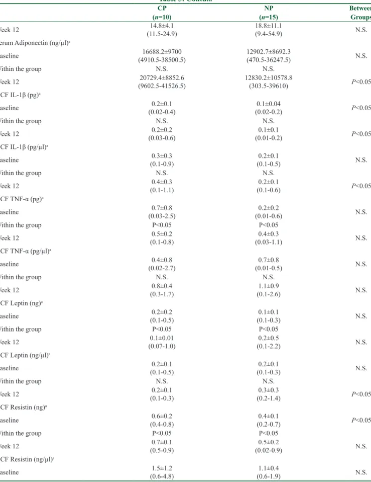
Table 3: Contd... CP NP Between (n=10) (n=15) Groups Week 12 (11.5-24.9)14.8±4.1 18.8±11.1 (9.4-54.9) N.S. Serum Adiponectin (ng/µl)a Baseline (4910.5-38500.5)16688.2±9700 12902.7±8692.3 (470.5-36247.5) N.S. Within the group N.S. N.S. Week 12 (9602.5-41526.5)20729.4±8852.6 12830.2±10578.8 (303.5-39610) P˂0.05 GCF IL-1β (pg)a Baseline (0.02-0.4)0.2±0.1 (0.02-0.2)0.1±0.04 P˂0.05 Within the group N.S. N.S. Week 12 (0.03-0.6)0.2±0.2 (0.01-0.2)0.1±0.1 P˂0.05 GCF IL-1β (pg/µl)a Baseline (0.1-0.9)0.3±0.3 (0.1-0.5)0.2±0.1 N.S. Within the group N.S. N.S. Week 12 (0.1-1.1)0.4±0.3 (0.1-0.6)0.2±0.1 P˂0.05 GCF TNF-α (pg)a Baseline (0.03-2.5)0.7±0.8 (0.01-0.6)0.2±0.2 N.S. Within the group P˂0.05 P˂0.05 Week 12 (0.1-0.8)0.5±0.2 (0.03-1.1)0.4±0.3 N.S. GCF TNF-α (pg/µl)a Baseline (0.02-2.7)0.4±0.8 (0.01-0.5)0.7±0.8 N.S. Within the group N.S. N.S. Week 12 (0.3-1.7)0.8±0.4 (0.1-2.6)1.1±0.9 N.S. GCF Leptin (ng)a Baseline (0.1-0.5)0.2±0.2 (0.1-0.3)0.1±0.1 N.S. Within the group P˂0.05 P˂0.05 Week 12 (0.07-1.0)0.1±0.01 (0.1-2.2)0.2±0.5 N.S. GCF Leptin (ng/µl)a Baseline (0.1-0.5)0.2±0.1 (0.1-0.3)0.2±0.1 N.S. Within the group N.S. N.S. Week 12 (0.1-0.3)0.2±0.1 (0.2-1.4)0.3±0.3 P˂0.05 GCF Resistin (ng)a Baseline (0.4-0.8)0.6±0.2 (0.2-0.7)0.4±0.1 P˂0.05 Within the group P˂0.05 P˂0.05 Week 12 (0.5-0.9)0.7±0.1 (0.02-0.9)0.5±0.2 N.S. GCF Resistin (ng/µl)a Baseline (0.6-4.8)1.5±1.2 (0.6-1.9)1.1±0.4 N.S. Contd...
Table 3: Contd... CP NP Between (n=10) (n=15) Groups Within the group N.S. N.S. Week 12 (1.1-2.1)1.4±0.4 (0.1-2.1)1.3±0.6 N.S. GCF Adiponectin (ng)a Baseline (0.2-5.2)1.8±2 (0.1-2.9)0.4±0.7 P˂0.05 Within the group N.S. P˂0.05 Week 12 1.8±1.2 (0.2-4) (0.1-9.1)1.7±2.6 N.S. GCF Adiponectin (ng/µl)a Baseline (0.4-8.3)2.8±2.5 (0.3-7.7)1.1±1.9 P˂0.05 Within the group N.S. P˂0.05 Week 12 (0.7-6.6)3.4±1.9 (0.5-17.3)4.5±5.8 N.S. P˂0.05: statistically significant, N.S.: not significant, n: number, GCF: gingival crevicular fluid, IL-1β: interleukin 1 beta, TNF-α: tumor necrosis factor-alpha. a)mean±standard deviation values (min-max)
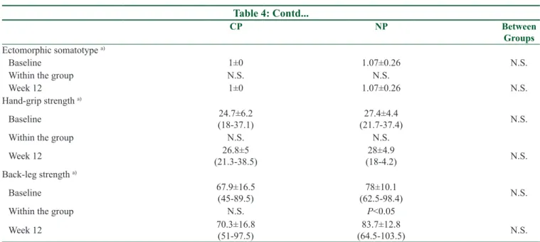
Table 4: BMI, VO2max, and somatotype-motoric tests at baseline and after 12 weeks of regular exercise
CP NP Between Groups Height (cm) a) Baseline 155.3±4.7 (145-162) 157.9±5.1 (148-165) N.S. Within the group N.S. N.S. Week 12 155.3±4.7 (145-162) 157.9±5.1 (148-165) N.S. Weight (kg) a) Baseline 89.6±14.9 (76-123) 90.5±18.5 (60-121) N.S. Within the group P˂0.05 P˂0.05 Week 12 (74.5-116)86.7±13.5 (56.8-118)87.1±17.7 N.S. BMI (kg/m²) a) Baseline 37.1±4.9 (32-48) 36.8±6.2 (31-51) N.S. Within the group P˂0.05 P˂0.05 Week 12 35.9±4.5 (31-45) 34.9±6.6 (22-50) N.S. VO2max a) Baseline 21.99±1.87 21.18±3.56 N.S. Within the group P˂0.05 P˂0.05 Week 12 28.49±2.65 27.37±3.46 N.S. Endomorphic somatotype a) Baseline 9.1±0.32 8.87±0.83 N.S. Within the group N.S. P˂0.05 Week 12 9±0.82 8.4±0.91 N.S. Mesomorphic somatotype a) Baseline 7.7±2.95 7.33±1.8 N.S. Within the group N.S. P˂0.05 Week 12 6.8±2.53 5.67±1.99 N.S. Contd...
Biochemical analysis
No significant changes were observed in saliva adipokines in either group (P > 0.05) [Table 3].
Table 3 presents the differences in adipokine serum levels for both groups. A significant (P < 0.05) decrease in TNF-α and an increase in resistin levels were observed in the NP group, while leptin levels decreased significantly (P < 0.05) in both groups.
The alterations in adipokines in GCF showed a significant (P < 0.05) decrease in TNF-α and leptin levels in the NP group; a significant (P < 0.05) increase in TNF-α, leptin, and adiponectin levels in the CP group; and an increase in resistin levels in both groups after RE [Table 3].
Correlations
The correlation coefficients between periodontal parameters and BMI, VO2 max, and anthropometric and adipokine parameters were demonstrated with variations according to the groups (data not shown). Hand length and neck and bust girths were negatively correlated with PI in the CP group. Similarly, the girth of the hip and GI, BMI, and CAL were shown to be negatively correlated with one another. No correlation was found
between periodontal parameters and VO2 max, but
TNF-α levels in GCF were demonstrated to have a significant negative correlation with VO2 max and resistin levels in saliva following completion of the RE. The levels of Il-1β in GCF and PI and BOP; leptin levels in GCF and BOP and PD; TNF-α levels in serum and PD and CAL; resistin levels in GCF and IL-1β levels in serum; leptin levels in serum; and IL-1β levels
in GCF showed significant positive correlations in the CP group. Furthermore, negative correlations were observed between BMI and CAL, leptin and adiponectin levels in GCF, and leptin levels in serum and resistin levels in GCF. Back-leg strength and BOP and skinfolds of the front thigh and medial calf and PI showed significant negative correlations, whereas girth of neck and PD, breadth of bi-iliocristal, and GI showed positive correlations in the NP group. The periodontal parameters and BMI or VO2max showed no correlation with one another. However, a negative correlation was observed
between BMI and TNF-α levels in GCF. VO2max
presented negative correlations with resistin levels in GCF and IL-1β and leptin levels in serum. Positive correlations were also found between GI and leptin levels in both saliva and GCF, TNF-α levels in GCF and PD and CAL, adipokine levels in serum and GI, and PD and BOP among the groups. Among the adipokine parameters, positive correlations were observed between GCF and saliva TNF-α levels, resistin levels in GCF and leptin levels in serum, adiponectin levels in GCF and leptin levels in saliva, and leptin levels in GCF and adiponectin levels in serum, whereas TNF-α and leptin levels in saliva were negatively correlated with one another.
Discussion
The mechanism underlying the relationship between obesity and periodontal disease hasyet to be revealed. However, the most widely held belief is that excessive fat adipokine secretion negatively affects periodontal health via systemic circulation.[3] It has been shown Table 4: Contd... CP NP Between Groups Ectomorphic somatotype a) Baseline 1±0 1.07±0.26 N.S. Within the group N.S. N.S. Week 12 1±0 1.07±0.26 N.S. Hand-grip strength a) Baseline (18-37.1)24.7±6.2 (21.7-37.4)27.4±4.4 N.S. Within the group N.S. N.S. Week 12 (21.3-38.5)26.8±5 (18-4.2)28±4.9 N.S. Back-leg strength a) Baseline 67.9±16.5 (45-89.5) (62.5-98.4)78±10.1 N.S. Within the group N.S. P˂0.05 Week 12 70.3±16.8 (51-97.5) (64.5-103.5)83.7±12.8 N.S.
P˂0.05 statistically significant, N.S.: not significant, n: number, BMI: body mass index, VO2max : maximal oxygen consumption. a) mean±standard deviation values (min-max)
that TNF-α, which locally increased in fat tissue of obese rats, also increased in systemic circulation.[11] This suggests that fatty tissue is not only an energy source but is also an endocrine organ.Increased macrophage infiltration in the fat mass is associated with low-grade inflammation, which isa characteristic feature of obesity.[12] The possible pathophysiological link among low-grade inflammation, obesity, periodontitis, and other chronic diseases is related to inflammatory adipokines.[2,3] Earlier studies of periodontal diseases and exercise have generally focused on the correlation between physical form and periodontitis[13] or physical activity and periodontitis.[7] A limited number of clinical studies have focused on the association between regular exercise and periodontal status and suggested that biochemical changes may contribute to improvements in periodontal health.[14,15] This novel study has clinically and biochemically evaluated the relationship between obesity in women, regular exercise, and periodontal disease. One of the essential aspects of the present study is the evaluation of two study groups with five different adipokine types in three bodily fluids (serum, saliva, and GCF). According to the available literature, this is the first study to evaluate the effects of regular exercise on saliva, serum and GCF TNF-α, leptin, resistin, and adiponectin.
The results of this study demonstrated that regular exercise significantly decreased BMI and increased VO2 max in obese women. These results were compatible with those of several other studies, confirming that exercise programs of 12 weeks’ duration or longer can improve BMI and VO2 max in different study groups.[16-19]
An earlier study demonstrated a significant decrease in PD and CAL in the CP group with no significant reduction in GCF IL-1β levels in either group. Mendoza-Nunez et al.[15] reported a significant decrease in the periodontal disease index but no differences in the amount of oral debris and calculus in a group of elderly participants who practiced Tai Chi exercises for six months. Park et al.[14] demonstrated that 4 weeks of adherence to a specific diet and a regular exercise program was correlated with a significant reduction in PI and GCF IL-1β levels in young, obese, and overweight participants. This reduction in PI may be related to the participants’ calorie-restricted diet, younger age, and BMI differences.
In this study, a significant decrease in serum TNF-α and an increase in serum resistin levels were observed after regular exercise in the NP group. Furthermore, serum leptin levels were significantly decreased in both groups.
Limprasertkul et al.[20] reported that serum IL-1β level was not affected by 12 weeks of aerobic exercise training in older participants. The alteration of serum TNF-α levels after regular exercise in overweight and obese female participants differed across several studies.[17,18,21] Kondo et al.[17] reported a significant reduction in serum TNF-α level after regular exercise, whereas other studies have indicated no significant difference in the serum level of this adipocytokine.[18,21] Similar to the present study, some earlier studies observed that regular exercise training decreased serum leptin levels.[16-18,22] In the literature, the relationship between regular exercise and the serum/plasma resistin level is unclear; whether the resistin levels have decreased,[21,23] increased,[24,25] or showed no significant changes after exercise,[22,26,27] and it needs further exploration. These different results may suggest that regular exercise does not have a precise role in plasma/serum resistin concentration. Moreover, regular exercise alone, without dietary control, does not appear to affect circulating adiponectin levels in obese participants.[16,21,22,26] However, the combination of a calorie-restricted diet and exercise training program has resulted in a significant increase in the level of circulating adiponectin.[17,19,24] It may be necessary to enhance weight reduction through diet and exercise to observe the alterations of circulating adiponectin levels. An earlier study among females diagnosed with anorexia nervosa reported that an initial increase in serum adiponectin levels was observed during the first month of weight recovery and that it is subsequently declined gradually after 5 months of refeeding.[28] While the adiponectin hormone was not directly associated with exercise-induced weight loss, it may have a protective role in balancing energy during recovery from malnutrition.
In the present study, no significant decreases in salivary adipokine levels were observed in either group. Mendoza-Nunez et al.[15] reported a significant decrease in IL-1β levels in the saliva of elderly participants practicing Tai Chi for 6 months. Aguilar-Cordero et al.[29] reported that leptin levels in saliva normalized after one year’s participation in a physical activity program for overweight and obese boys. Roupas et al.[30] compared elite athletes with a sedentary control group and they found that, while salivary resistin levels showed no significant difference between the groups, adiponectin levels were higher in the athletes than in the control group. Insufficient standardization of analytical methods for the determination of salivary adipokine levels, diversity of subject classifications, inclusion/exclusion criteriain research’ protocols, duration, frequency, type, and intensity of workouts may affect results differently.
The present study demonstrated that RE could have positive effects on the systemic, oral, and periodontal health of obese women. However, our study had some limitations. As patients who used antibiotics during the research for any reason or who had to quit the exercise program due to injuries were removed from the study, the number of participants was lower than expected. The subjects were informed about healthy nutrition at the beginning, but it was not possible to control their daily diet during the exercise program. Overall, the homogeneity of age, BMI, and gender within the groups was this study’s key strength.
Conclusion
In conclusion, this study demonstrated a correlation between periodontal parameters and adipokine levels in serum, saliva, and GCF in obese women with varying periodontal conditions and that this was affected differentially by 12 weeks of RE. In addition, it revealed that the effects of regular exercise on adipokine levels may differ locally and systemically in obese women. Further studies with higher numbers of obese participants are required to understand the relationship between exercise, obesity, and periodontal disease.
Acknowledgements
This research was supported kindly by a grand from TUBITAK (The Scientific and Technological Research Council of Turkey), (114S144, [EG-A]), and the Kocaeli University Scientific Research Projects Unit (KOU 2014/061 [EG-A]). The authors thank TUBITAK and Kocaeli University for their generous support. The authors thank Meral Gunhan, DDS, Ph.D., Professor, Ankara, Turkey, for kindly providing Periotron for this study.
Declaration of patient consent
The authors certify that they have obtained all appropriate patient consent forms. In the form the patient(s) has/have given his/her/their consent for his/ her/their images and other clinical information to be reported in the journal. The patients understand that their names and initials will not be published and due efforts will be made to conceal their identity, but anonymity cannot be guaranteed.
Financial support and sponsorship
This research was supported by a grand from TUBITAK (The Scientific and Technological Research Council of Turkey), (114S144, [EG-A]), and the Kocaeli University Scientific Research Projects Unit (KOU 2014/061 [EG-A]). Also, the authors thank Meral Gunhan, DDS, Ph.D., Professor, Ankara, Turkey, for kindly providing Periotron for our study.
Conflicts of interest
There are no conflicts of interest.
References
1. Giannopoulou C, Kamma JJ, Mombelli A. Effect of inflammation, smoking and stress on gingival crevicular fluid cytokine level. J Clin Periodontol 2003;30:145-53.
2. Trayhurn P, Wood IS. Adipokines: Inflammation and the pleiotropic role of white adipose tissue. Br J Nutr 2004;92:347-55.
3. Genco RJ, Grossi SG, Ho A, Nishimura F, Murayama Y. A proposed model linking inflammation to obesity, diabetes, and periodontal infections. J Periodontol 2005;76:2075-84.
4. Offenbacher S. Periodontal diseases: Pathogenesis. Ann Periodontol 1996;1:821-78.
5. Haynes WG, Stanford C. Periodontal disease and atherosclerosis: From dental to arterial plaque. Arterioscler Thromb Vasc Biol 2003;23:1309-11.
6. Donnelly JE, Blair SN, Jakicic JM, Manore MM, Rankin JW, Smith BK. American College of Sports Medicine Position Stand. Appropriate physical activity intervention strategies for weight loss and prevention of weight regain for adults. Med Sci Sports Exerc 2009;41:459-71.
7. Al-Zahrani MS, Borawski EA, Bissada NF. Increased physical activity reduces prevalence of periodontitis. J Dent 2005;33:703-10.
8. World Health Organization: Obesity and Overweight. Fact Sheet No 311, [updated 16 February 2018]. Available
from: https://www.who.int/news-room/fact-sheets/detail/
obesity-and-overweight.
9. Armitage GC. Development of a classification system for periodontal diseases and conditions. Ann Periodontol 1999;4:1-6. 10. Kline GM, Porcari JP, Hintermeister R, Freedson PS, Ward A,
McCarron RF, et al. Estimation of VO2 max from a one-mile track walk, gender, age, and body weight. Med Sci Sports Exerc 1987;19:253-9.
11. Hotamisligil GS, Shargill NS, Spiegelman BM. Adipose expression of tumor necrosis factor-alpha: Direct role in obesity-linked insulin resistance. Science 1993;259:87-91. 12. Cancello R, Henegar C, Viguerie N, Taleb S, Poitou C,
Rouault C, et al. Reduction of macrophage infiltration and chemoattractant gene expression changes in white adipose tissue of morbidly obese subjects after surgery-induced weight loss. Diabetes 2005;54:2277-86.
13. Eberhard J, Stiesch M, Kerling A, Bara C, Eulert C, Hilfiker-Kleiner D, et al. Moderate and severe periodontitis are independent risk factors associated with low cardiorespiratory fitness in sedentary non-smoking men aged between 45 and 65 years. J Clin Periodontol 2014;41:31-7.
14. Park HS, Nam HS, Seo HS, Hwang SJ. Change of periodontal inflammatory indicators through a 4-week weight control intervention including caloric restriction and exercise training in young Koreans: Apilot study. BMC Oral Health 2015;15:109. 15. Mendoza-Nunez VM, Hernandez-Monjaraz B, Santiago-Osorio E,
Betancourt-Rule JM, Ruiz-Ramos M. Tai Chi exercise increases SOD activity and total antioxidant status in saliva and is linked to an improvement of periodontal disease in the elderly. Oxid Med Cell Longev 2014;2014:1-6.
16. Polak J, Klimcakova E, Moro C, Viguerie N, Berlan M, Hejnova J, et al. Effect of aerobic training on plasma levels and subcutaneous abdominal adipose tissue gene expression of adiponectin, leptin, interleukin 6, and tumor necrosis factor alpha
in obese women. Metabolism 2006;55:1375-81.
17. Kondo T, Kobayashi I, Murakami M. Effect of exercise on circulating adipokine levels in obese young women. Endocr J 2006;53:189-95.
18. Akbarpour M. The effect of aerobic training on serum adiponectin and leptin levels and inflammatory markers of coronary heart disease in obese men. Biol Sport 2013;30:21-7. 19. Bruun JM, Helge JW, Richelsen B, Stallknecht B. Diet and
exercise reduce low-grade inflammation and macrophage infiltration in adipose tissue but not in skeletal muscle in severely obese subjects. Am J Physiol Endocrinol Metab 2006;290:961-7. 20. Limprasertkul A, You T, Fisher N, Awad A, Pendergast DR.
Exercise-induced plasma cytokines between older and younger adults. Int Sport Med J. 2013;14:16-28.
21. Kadoglou NP, Perrea D, Iliadis F, Angelopoulou N, Liapis C, Alevizos M. Exercise reduces resistin and inflammatory cytokines in patients with type 2 diabetes. Diabetes Care 2007;30:719-21.
22. Giannopoulou I, Fernhall B, Carhart R, Weinstock RS, Baynard T, Figueroa A, et al. Effects of diet and/or exercise on the adipocytokine and inflammatory cytokine levels of postmenopausal women with type 2 diabetes. Metabolism 2005;54:866-75.
23. Rashidlamir A, Gholamian S, Atri AE, Seyyedalhoseyni M, Kooshki MH. Effect of regular aerobic exercise on plasma levels of resistin and adiponectin in active young females. J Mazand Univ Med Sci 2013;23:73-83.
24. Elloumi M, Ben Ounis O, Makni E, Van Praagh E, Tabka Z,
Lac G. Effect of individualized weight-loss programmes on adiponectin, leptin and resistin levels in obese adolescent boys. Acta Paediatr 2009;98:1487-93.
25. Tofighei A, Samadian Z, Mehdizadeh A, Zolfagharei M. The response of serum resistin to aerobic exercise and its possible association with metabolic indices in women with type 2 diabetes. Med J Tabriz Univ Med Sci Health Serv 2014;36:18-25. 26. Kelly AS, Steinberger J, Olson TP, Dengel DR. In the absence
of weight loss, exercise training does not improve adipokines or oxidative stress in overweight children. Metabolism 2007;56:1005-9.
27. Shadid S, Stehouwer CD, Jensen MD. Diet/Exercise versus pioglitazone: Effects of insulin sensitization with decreasing or increasing fat mass on adipokines and inflammatory markers. J Clin Endocrinol Metab 2006;91:3418-25.
28. Modan-Moses D, Stein D, Pariente C, Yaroslavsky A, Ram A, Faigin M, et al. Modulation of adiponectin and leptin during refeeding of female anorexia nervosa patients. J Clin Endocrinol Metab 2007;92:1843-7.
29. Aguilar-Cordero MJ, Sánchez-López AM, Padilla-López CA, González-Mendoza JL, Mur-Villaf N, Peronay JS, et al. Influence of a program of physical activity in children and adolescents obese evaluation of physiological stress by compounds in saliva; study protocol. Nutr Hosp 2013;28:705-8.
30. Roupas ND, Mamali I, Armeni AK, Markantes GK, Theodoropoulou A, Alexandrides TK, et al. The influence of intensive physical training on salivary adipokine levels in elite rhythmic gymnasts. Horm Metab Res 2012;44:980-6.