• Sonuç bulunamadı

Comparison of the Anti-inflammatory Effects of Proanthocyanidin, Quercetin, and Damnacanthal on Benzo(a)pyrene Exposed A549 Alveolar Cell Line

N/A
N/A
Protected

Academic year: 2021

Share "Comparison of the Anti-inflammatory Effects of Proanthocyanidin, Quercetin, and Damnacanthal on Benzo(a)pyrene Exposed A549 Alveolar Cell Line"

Copied!
8
0
0

Yükleniyor.... (view fulltext now)

Tam metin

(1)

ORIGINAL ARTICLE

Comparison of the Anti-inflammatory Effects

of Proanthocyanidin, Quercetin, and Damnacanthal

on Benzo(a)pyrene Exposed A549 Alveolar Cell Line

Ersin Günay,

1,6,7

Sefa Celik,

2

Sevinc Sarinc-Ulasli,

1

Arzu Özyürek,

3

Ömer Hazman,

3

Sibel Günay,

4

Mehmet Özdemir,

5

and Mehmet Ünlü

1

Abstract—Phytochemical compounds are emerging as a new group of anti-inflammatory, antioxidant, and anti-cancer agents that help minimize toxicity in patients with pulmonary diseases. The goal of this study was to investigate the potential curative effects of Quercetin (QC), Damnacanthal (DAM), and Proanthocyanidine (PA) on inflammatory mediators and oxidative stress parameters and to examine the viability of the A549 cell line treated with benzo(a)pyrene (BaP) in vitro. The A549 cell line was treated with BaP, a BaP/QC combination, a BaP/DAM combination, and BaP/PA combination. Inflammatory markers, oxidative stress parameters, mRNA expression levels of apoptotic and antiapoptotic proteins, and cell viability were assessed, and the results were compared. There were higher levels of lactate dehydrogenase after BaP treatment of A549 cell lines. Interferon-γ level significantly decreased in the QC, DAM, and PA-treated group (P<0.001). IL-1β and TNF-α levels significantly decreased after PA and QC treatments (P<0.001). Some of the oxidative stress markers (NO, MDA, TOS) and OSI decreased, while antioxidant (GSH) levels increased after treatment with QC, DAM, and PA. The QC and DAM treatments profoundly upregulated apoptotic gene expression and downregulated antiapoptotic gene expression. Viability of QC, DAM, and PA-treated cells was found to be significantly higher in comparison to the control and BaP-treated groups (p<0.001). Our results revealed that A549 cell lines treated with BaP-stimulated necrosis produced higher level of inflammatory cytokines and oxidative stress parameters. Treatments with PA, QC, and DAM reduced inflammatory response induced by BaP exposure.

KEY WORDS: inflammation; cell culture; oxidative stress; antioxidants; COPD.

INTRODUCTION

Polycyclic aromatic hydrocarbons (PAHs) are ubiq-uitous environmental cytotoxic and genotoxic compounds found particularly in tobacco. Benzo(a)pyrene (BaP) (C20H12) is the best known and most studied PAH [1,2].

In many studies, it has been reported that BaP exposure occurs in both those living in urban areas with air pollution and tobacco users. As a result of this exposure, it has been shown to lead many chronic lung diseases, especially lung cancer and chronic obstructive lung disease (COPD) [1–4]. BaP like other PAHs is converted to highly toxic reactive products with various metabolic activation processes inside the cells [5,6].

1Department of Chest Diseases, Faculty of Medicine, Afyon Kocatepe

University, Afyonkarahisar, Turkey

2Department of Medical Biochemistry, Faculty of Medicine, Afyon

Kocatepe University, Afyonkarahisar, Turkey

3

Department of Chemistry, Faculty of Science and Arts, Afyon Kocatepe University, Afyonkarahisar, Turkey

4

Chest Diseases Clinic, Afyon State Hospital, Afyonkarahisar, Turkey

5

Department of Nutrition and Dietetics, Fethiye School of Health, Mugla Sitki Kocman University, Mugla, Turkey

6

Afyon Kocatepe Universitesi Tip Fakultesi Arastirma ve Uygulama Hastanesi C Blok Gogus Hastaliklari A.D., 03200 Afyonkarahisar, Turkey

7

To whom correspondence should be addressed at Afyon Kocatepe Universitesi Tip Fakultesi Arastirma ve Uygulama Hastanesi C Blok Gogus Hastaliklari A.D., 03200 Afyonkarahisar, Turkey. E-mail: ersingunay@gmail.com

0360-3997/16/0200-0744/0#2016 Springer Science+Business Media New York

(2)

Epithelial cells of the airways—the first barrier for protection—contact inevitably with a variety of inhaler agents (particles and BaP). In toxicological reactions caused by these malicious agents, respiratory epithelial cells have an important role. Due to the exposure of BaP, increase in the level of inflammatory mediators and oxida-tive stress were reported in human alveolar epithelial cells (A549) in the literature [2].

In recent years, in vivo and in vitro studies were conducted to demonstrate favorable effects of many plant-derived extracts and components on treatment of lung cancer and inflammatory processes. These studies were conducted particularly to determine treatment effica-cy of plant extracts and their active ingredients that previ-ously determined antioxidants and anticarcinogens [7–9].

In this study, we aimed to investigate and compare the anti-inflammatory, antioxidant, and apoptotic effect of Q u e r c e t i n ( Q C ) , P r o a n t h o c y a n i d i n ( PA ) , a n d Damnacanthal (DAM) on BaP exposed A549 cell line.

METHODS

Major Reactives

We obtained human lung cell lines A549 from Dr. J. Mazella (CNRS, Valbonne, France). Dulbecco’s modified Eagle’s medium (DMEM), fetal bovine serum, and BaP were obtained from Sigma-Aldrich (USA). QC was pur-chased from Cayman Chemical (USA). PA was purpur-chased from Santa Cruz Biotechnology (Germany). DAM was purchased from Merck Calbiochem (Germany).

Cell Culture and Experimental Procedures

A549 cell line, distal respiratory epithelium (type II pneumocytes), was maintained in DMEM supplemented with 10 % of fetal calf serum (FCS), 2 mML-glutamine,

100 units/ml penicillin, and 100μg/ml streptomycin. Pre-pared cell line was incubated under 5 %CO2in atmosphere

at 37 °C.

A549 cells were seeded in 75-cm2culture flasks and allowed to grow for 1–2 days before experiments [2]. When 70–80 % confluence was achieved, the culture medium was replaced to a new fresh medium containing 20μM BaP (C20H12>96 % pure) for 48 h [10]. BaP was

first dissolved in dimethyl sulfoxide (DMSO) and then added to the culture medium with the final concentration of DMSO less than 0.1 %. The medium was changed daily to maintain the stable concentration of BaP [2,10].

Treatment with Quercetin, Damnacanthal, and Proanthocyanidin

After 24 h incubation of A549 cell line with BaP, culture medium containing BaP was removed, and fresh medium containing 10μM QC (BaP+QC) [11], 50μM DAM (BaP+DAM) [12], and 50μg/ml PA (BaP+PA) [8] alone were added into the flasks according to the groups. As control group, A549 cells were incubated with medium containing vehicle (DMSO) at the same concentration. After 48 h incubation with agents, medium was removed and the cells in all four groups were used to perform the experiments described as follows.

Utmost care for sterilization was taken at every stages of the experiment. Bacterial contamination was prevented with Penicillin and Streptomycin combination treatment in cell culture media. All measurements were performed in triplicate and each experiment was repeated three times.

Protein Analysis

Cells from all groups were washed twice with ice-cold phosphate-buffered saline (PBS) and then removed by scraping. The removed cells were lysed in lysis buffer (100 mM NaH2PO4, 1 % Triton X-100, 1 M HEPES, and

1 % protease inhibitor cocktail). The homogenate was centrifuged at 13,000g at 4 °C for 40 min. The supernatants were collected, and protein concentrations were deter-mined using Bradford method [13]. In this study, cells which were obtained in the same passage were used in all groups.

Cell Viability Measurements Test and MTT Assay The number of viable A549 cells after treatments was evaluated by the MTT (3-[4,5-methylthiazol-2-yl]-2,5-diphenyl-tetrazolium bromide) assay. In summary, A549 cells (2×10⁴cells/well) were seeded in a 24-well plate and kept overnight for attachment. The next day, the medium was replaced with fresh medium with BaP and cells were let to grow for 48 h. After completion of incuba-tion, therapeutic agents were added in each well, and after completion of second incubation, 100 μl of MTT (10 mg/ml, Sigma, USA) was added in each well, followed by 4 h incubation at 37 °C. Later, medium was removed and 150 μl DMSO was added to each well. The plate was then shaken for 10 min in the dark at room temperature. The absorbance value at 490 nm was quantified using a UV/VIS spectrophotometer (T70, PG Instruments). The results were expressed as the percentage of treated cells relative to controls.

(3)

Analysis of Lactate Dehydrogenase Activity and Cytokine Production

Lactate dehydrogenase (LDH) activity was mea-sured in the supernatant prior to the BaP application and after 2 h incubation of cells with BaP and com-bined treatments applied [(BaP+QC), (BAP+DAM), and (BaP+PA)] in accordance with the proposal of manufacturer to examine whether necrotic cell death occurred or not. The effect of QC, DAM, and PA on release of cytokine levels (IFN-γ, IL-1β, TNF-α) in cell lysate was analyzed by ELISA technique. The results were expressed as pg/mg protein.

Measurement of Nitric Oxide Production

Nitric oxide (NO) level in cell culture medium and cell lysate were measured by Giress reaction [14]. Speci-mens were deproteinized with 75 mmol zinc-sulphate. Total nitrite was measured by a spectrophotometer at 546 nm after alteration of nitrate to nitrite by using vanadium-III-chloride. The values were expressed in μmol/L in culture medium and μmol/g protein in cell lysate.

Evaluation of Glutathione Level

Glutathione (GSH) level in cell lysate was evaluated by the method defined by Buetler et al. [15]. The results were expressed asμmol/g protein.

Measurement of Malondialdehyde Level in Cell Lysate Malondialdehyde (MDA) level was measured by the method described by Yoshioka et al. [16]. In this method, the formation of a pink color under the acidic condition upon the reaction of MDA and thiobarbituric acid is essen-tial. The absorbance values were read on a spectrophotom-eter at 535 nm. After computation of the values, the results were implied as nmol/g protein.

Analysis of Total Oxidant Status, Total Antioxidant Status, and Oxidative Stress Index

Total oxidant status (TOS) of cell lysate was mea-sured by using automated colorimetric method described by Erel et al. [17]. The results were expressed as μmol H2O2Eq/g protein. Total antioxidant status (TAS) of cell

lysate was determined by another automated direct color-imetric measurement method described Erel et al. [18]. Results were expressed as mmol Trolox equivalent/g pro-tein. Oxidative stress index (OSI) which indicates the

degree of oxidative stress was calculated via a formula described by Esen et al. [19] as follows:

OSI arbitrary unitð Þ ¼ TOS μmolH2O2Eq

. g protein

 .

TAS mmol Trolox Eq .

g protein


 100

Molecular Analysis (Total RNA Isolation and mRNA Expression Levels of Genes by Real-Time Reverse Transcription-Polymerase Chain Reaction (RT-PCR))

Molecular analyses were performed as described by Ulasli et al. [10]. A549 cells were seeded in 25 cm2culture flasks and were allowed to grow for 1–2 days before the analyses were performed. Cells were collected and washed with phosphate-buffered saline (PBS) after completion of incubations. Total RNA was isolated by RNAeasy kit (QIAGEN), and cDNA was generated with a First Strand cDNA Synthesis kit (Thermo Scientific) at a total volume of 20 μl in accordance with the manufacturer’s instruc-tions. Real-time quantitative PCR was performed in a Strategene Mx3005P QPCR system (USA). Expression levels of target gene were normalized to the housekeeping gene β-actin (ΔCt). Gene expression values were then calculated based on theΔΔCt method using the equation: RQ = 2−ΔΔCt. PCR amplification was performed with Maxima SYBR Green/ROXqPCR Master Mix (Thermo Scientific). The primer sequences are described in Table1. Each assay was performed in triplicate and repeat-ed three times.

Statistical Analysis

The statistical analyses of experimental data were performed by using SPSS (Statistical Package for Social Science for Windows, Version 18.0; Chicago, IL, USA). The normality of distributions of all variables was investi-gated by using Shapiro-Wilk test. The results were expressed as mean±standard deviation (SD) or median value (min-max range) according to distribution. One-way ANOVA or Kruskall-Wallis test was used to compare continuous parameters according to distribution among study groups. When an overall significance was present, pairwise post hoc tests were performed. The P value less than 0.05 was accepted as significance level.

(4)

RESULTS

Inflammatory Mediators

In groups which were treated with QC, DAM, and PA, IFN-γ levels were significantly decreased when compared with BaP exposed A549 cell line (P < 0.001). QC and PA-treated cells had significantly lower IL-1β and TNF-α level when compared with BaP-exposed cells (P < 0.001). IL-1β level was simi-lar in DAM-treated cells and only BaP-exposed cells (Table 2).

Oxidative Stress Parameters

In cells treated with QC, DAM, and PA, oxidative stress parameters were lower than only BaP-exposed cells. Oxidative stress index (OSI) was significantly lower in PA-treated cells than all other cells. Glutathione level was significantly higher in PA treated group (p<0.001). Oxida-tive stress results were outlined in Table3.

mRNA Expression Levels of Genes in A549 Cells QC and DAM upregulated TRAIL receptor 1 (TRAIL-R1) and TRAIL receptor 2 (TRAIL-R2) expressions. PA

Table 1. Oligonucleotide Primer Sequences and PCR Programs

Transcripts Primer sequences PCR programs Cycles

Bax F-5′cgcctcactcaccatctggaa3′ R-5′cctcaagaccactcttcccca3′ Initial: 95 °C-10 min; 94 °C-1 m/58 °C-1 m/72 °C-1 m 35 Bcl-2 F-5′gaggggctacgagtgggatgc3′ R-5′ggaggagaagatgcccggtgc3′ Initial: 95 °C-10 min; 94 °C-1 m/62 °C-1 m/72 °C-1 m 35 Bcl-xL F-5′cacatcaccccagggacagca3′ R-5′aaaggccacaatgcgacccca3′ Initial: 95 °C-10 min; 94 °C-1 m/61 °C-1 m/72 °C-1 m 40 IKK1 F-5′gctacagaagagcccctatgga3′ R-5′agatcaatggcacgctgttcc3′ Initial: 95 °C-10 min; 94 °C-1 m/57 °C-1 m/72 °C-1 m 35 TRAIL-R1 F-5′gagaagtccctgcaccacgac3′

R-5′ccggaaagttcctggtttgcac3′ Initial: 95 °C-10 min;94 °C-1 m/59 °C-1 m/72 °C-1 m

35 TRAIL-R2 F-5′tccttacctgaaaggcatctgc3′ R-5′gtcgttgtgagcttctgtcca3′ Initial: 95 °C-10 min; 94 °C-1 m/57 °C-1 m/72 °C-1 m 35 CYCLIN-D1 F-5′atgctggaggtctgcgaggaa3′ R-5′cgacaggaagcggtccaggta3′ Initial: 95 °C-10 min; 94 °C-1 m/60 °C-1 m/72 °C-1 m 35 P21 F-5′ccgtgagcgatggaacttcgac3′ R-5′tgggaaggtagagcttgggca3′ Initial: 95 °C-10 min; 94 °C-1 m/60 °C-1 m/72 °C-1 m 35 NFKB F-5′ggtgcggctcatgtttacagc3′ R-5′gcgtctgataccacgggttcc3′ Initial: 95 °C-10 min; 94 °C-1 m/59 °C-1 m/72 °C-1 m 35 β-Actin F-5′caccccagccatgtacgttgc3′ R-5′ccggagtccatcacgatgcca3′ Initial: 95 °C-10 min; 94 °C-1 m/61 °C-1 m/72 °C-1 m 35

Table 2. The Levels of Proinflammatory Cytokines (in cell lysates) and LDH Activity (in medium)

Groups IFN-γ (pg/mg protein) IL-1β (pg/mg protein) TNF-α (pg/mg protein) LDH (U/L) BaP 0.52±0.02 136.71±24.68 7.57±1.33 43.00±2.00 QC 0.31±0.02‡ 80.52±8.53† 4.19±2.08† 37.00±1.73† DAM 0.37±0.02‡ 135.47±26.24 6.39±1.79 32.67±0.58† PA 0.14±0.02‡ 65.18±17.00‡ 3.19±0.60† 31.33±3.05†

All experiments were performed in triplicate, and data was expressed as mean±SD for all parameters. One-way analysis of variance (ANOVA) with Bonferroni corrections

IFN-γ interferon gamma, IL-1β Interleukin-1 beta, LDH lactate dehydrogenase, TNF-α tumor necrosis factor alpha, BaP benzo(a)pyrene, QC Quercetin, DAM Damnacanthal, PA Proanthocyanidin

Significantly different compared to benzo(a)pyrene treated group at P<0.05Significantly different compared to benzo(a)pyrene treated group at P<0.001

(5)

upregulated TRAIL-R1 expression. PA and QC upregulated Bax gene expression. DAM downregulated Bax gene ex-pression. QC and DAM upregulated p21 gene exex-pression. All agents (QC, DAM, and PA) decreased the expression of cyclin D1 gene expression. mRNA expression levels of genes in A549 cells are presented in Table4.

Cell Viability

Cell viability was investigated and compared among groups (Fig.1). Viability of BaP-exposed cells was signif-icantly decreased when compared with control (P<0.05). Significantly increased cell viability was seen after QC, DAM, and PA treatment when compared with BaP exposure.

DISCUSSION

In this study, we observed significantly higher activity of LDH after BaP exposure in A549 cells when compared

with controls. Additionally, we observed significant decrease in cell viability in BaP-exposed cells. When these findings and increased inflammatory and oxida-tive stress levels were taken into account together, we supposed that in vitro BaP exposure in A549 cells results in a trend to cell death causing necrosis with a higher inflammatory process. After application of protective agents, increased cell viability, marked antiinflammatory, and antioxidant activity were ob-served, especially in PA and QC-treated cell lines.

Benzo(a)pyrene (BaP) is a prototype of PAH family. They are formed as a result of incomplete combustion of organic substances (such as biomass fuel or waste mate-rials). Additionally, BaP is also found in tobacco smoke [20,21]. BaP is an environmental pollutant, and it is known that it is associated with airway inflammation and damage in smokers [22, 23]. BaP exposure results in decreased cellular antioxidant level (glutathione, GSH) and increased oxidative stress [24]. In our study, increased inflammatory mediators and oxidative stress parameters were observed in BaP-exposed cell line when compared to control.

Table 3. Oxidative Stress Parameters of All Study Groups

Groups GSHa (μmol/g protein) NO- mediuma (μmol/L) NO- lysatea (μmol/g protein) MDAa (nmol/g protein) TASa (mmol Trolox equivalent/g protein) TOSa (μmol H2O2 Eq/g protein) OSIb BaP 24.38±6.86 7.83±0.85 1.47±0.41 7.53±0.46 0.52±0.16 7.04±0.87 12.97 (9.91–23.00) QC 24.10±1.33 9.24±1.85 1.19±0.37 5.30±0.25‡ 0.19±0.11† 3.98±0.28‡ 22.12 (11.15–90.20) DAM 24.07±3.04 5.63±1.02 1.14±0.21† 6.38±0.12‡ 0.47±0.18 6.37±1.07 13.10 (8.56–29.04) PA 60.22±2.70‡ 7.07±1.28 0.82±0.17† 2.45±0.27‡ 0.34±0.05 1.91±0.53‡ 4.81 (3.83–8.79)† All experiments were performed in triplicate, and the values reported as mean±SD or median (min.-max) according to the distribution status

GSH glutathione, MDA malondialdehyde, NO nitric oxide, OSI oxidative stress index, TAS total antioxidant status, TOS total oxidant status, BaP benzo(a)pyrene, QC Quercetin, DAM Damnacanthal, PA Proanthocyanidin

a

One-way analysis of variance (ANOVA) with Bonferroni corrections

b

Kruskall-Wallis Test and pairwise post hoc test with Mann-Whitney U test

Significantly different compared to benzo(a)pyrene treated group at P<0.05Significantly different compared to benzo(a)pyrene treated group at P<0.001

Table 4. mRNA Expression Levels of Genes in A549 Cells

Groups mRNA expression levels of genes (fold increase +/ decrease−)

BAX BCL-2 BCL-XL IKK1 TRAIL-R2 NF-κB P21 CYCLIN-D1 TRAIL-R1

BaPa (−) 1.1 (−) 2.0 (−) 2.3 (−) 1.74 (−) 1.41 (−) 1.11 (+) 1.33 (−) 2.5 (−) 1.15

QCb (+) 1.2 (−) 1.41 (+) 2.0 (−) 1.2 (+) 1.24 (−) 1.14 (+) 1.31 (−) 1.2 (+) 1.07

DAMb (−) 1.02 (−) 1.3 (+) 1.77 (+) 1.01 (+) 1.28 (+) 1.05 (+) 1.74 (−) 1.07 (+) 1.1

PAb (+) 2.3 (+) 53.44 (+) 2.82 (+) 12.46 (−) 4.6 (+) 3.31 (−) 2.82 (−) 1.54 (+) 5.3 QC Quercetin, DAM Damnacanthal, PA Proanthocyanidin

a

According to the control group

(6)

B i o f l a v o n o i d Qu e r c e t i n ( Q C ) ( 3 , 3 ′,4′,5,7-pentahydroxyflavone) is a polyphenolic compound found in some fruits and vegetable. It has been shown that QC has chemopreventive activity during carcinogenesis and also has cytotoxic activity against various in vitro and in vivo tumor cells [7,25]. Furthermore, in the previous studies, it has been reported that QC has protective activity on pul-monary toxicity and oxidative stress [26,27]. Additionally, QC has beneficial effect on lipid peroxidation and reducing pulmonary damage occurred after CCL4− and paraquate

[28–30]. In a recent study, Verma et al. [31] reported that QC ameliorates the development of bleomycin-induced pulmonary fibrosis in rats. In the present study, we ob-served a significant decrease in inflammatory mediators (IFN-γ, IL-1β, and TNF-α) and oxidative stress parame-ters (especially TAS and MDA level) when compared with BaP-exposed cell line.

Proanthocyanidin (PA) is a biologically active poly-phenolic bioflavonoid that was derived from many plants. Generally, PA is used as an enriched grape seed extract in traditional herbal medicine. In the literature, protective effects of PA on oxidative stress were demonstrated [32– 34]. PA has a cleaning effect on reactive oxygen substances (ROS), inhibitor effect on ischemia-reperfusion injury, and anti-inflammatory and inhibitory effects on oxidative stress produced by pesticides. In addition to these crucial func-tions, antioxidant, vasodilator, antithrombotic, cardioprotective, and anticancer effects of PA also reported

in the literature [32–34]. Agackiran et al. [34] reported that PA has a protective and anti-inflammatory effect in bleomycin-induced lung fibrosis model in rats. In the pres-ent study, we showed that PA has a statistically significant effect on reducing the level of inflammatory mediators (IFN-γ, IL-1β, and TNF-α) and oxidative stress indicators (GSH, NO, MDA, TOS, and oxidative stress index (OSI)) when compared with only BaP-exposed A549 cell line. These findings suggest that PA has a significant positive effect on oxidative stress and inflammation in A549 alve-olar epithelial cells exposed to BaP.

Damnacanthal (DAM), an anthraquinone compound, is extracted from the roots of Morinda citrifolia L. (noni), which has been used for traditional therapy in several chronic diseases. DAM is an activity inhibitor of tyrosine kinase. Kinases control cell division and mediate transduc-tion and intracellular and extracellular processing of sig-nals. These kinases are involved in oncogenesis. Impaired function of tyrosine kinase leads to the development of non-small cell lung cancer [35, 36]. Additionally, anti-inflammatory effect of DAM has been reported in the literature [37]. In this study, IFN-γ level was significantly

decreased after DAM treatment. Additionally, in our study, statistically significant reduction in NO and MDA levels were found in cells treated with DAM. Besides, in the present study, DAM showed its anti-inflammatory effect by increasing NF-κB gene expression which was previ-ously reported by Nualsanit et al. [37].

Fig. 1. Cell viability of A549 cells after treatment with benzo(a)pyrene (BaP), BaP + Quercetin (QC), BaP + Damnacanthal (DAM), BaP + Proanthocyanidin (PA). Cell viability was quantified by a MTT assay 72 h after exposure to BaP, following a 24-h treatment with other drugs. Significantly decreased in cell viability was seen after treatment with BaP alone when compared to control. QC, DAM, and PA showed significant protection against BaP-induced cell death/necrosis with significant increase in cell viability. *P<0.05 compared to the control group, and#p<0.001 compared to the BaP group. The graph is

(7)

When we consider the effects of therapeutic agents on apoptotic pathways in cell culture model, it has been shown that QC upregulated the apoptotic gene expression (Bax, p21, TRAIL-R1, and TRAIL-R2) and downregulated the antiapoptotic gene expression (bcl-2, NF-κB, IKK1, cyclin-D1). Meanwhile, DAM upregulated the apoptotic gene expression especially TRAIL-R1, TRAIL-R2, and p21 and decreased the expression of bcl-2 and cyclin-D1. Nevertheless, it has been shown that PA showed its apo-ptotic activity with upregulation of bax and TRAIL-R1 gene expression. Therefore, we suggest that QC and DAM show their protective effect on the cancer cells triggered by BaP by influencing apoptotic pathway. Nev-ertheless, further studies are warranted to understand their anticancer activity.

In conclusion, in this cell culture model with A549 alveolar epithelial cells exposed to BaP, we demonstrated that both PA and QC had higher anti-inflammatory and antioxidant activity than DAM. We concluded that al-though this is an in vitro study, these results will shed light on the further in vivo studies. Therefore, we suggest that PA and QC may be used in prevention and treatment of in-flammatory lung diseases caused by smoking in the near future.

ACKNOWLEDGMENTS

We thank Emma Hickey for English Editing of our manuscript.

COMPLIANCE WITH ETHICAL STANDARDS Disclosures. This project was supported by a grant from Kocatepe University Scientific Research Council, Afyonkarahisar, Turkey: Project number: 12.TIP.01.

REFERENCES

1. Jiang, Y., K. Rao, G. Yang, X. Chen, Q. Wang, A. Liu, et al. 2012. Benzo(a)pyrene induces p73 mRNA expression and necrosis in human lung adenocarcinoma H1299 cells. Environmental Toxicology 27: 202– 10.

2. Min, L., S. He, Q. Chen, F. Peng, H. Peng, and M. Xie. 2011. Com-parative proteomic analysis of cellular response of human airway epithelial cells (A549) to benzo(a)pyrene. Toxicology Mechanisms and Methods 21: 374–82.

3. Anandakumar, P., S. Kamaraj, S. Jagan, G. Ramakrishnan, S. Asokkumar, C. Naveenkumar, et al. 2012. Capsaicin inhibits

benzo(a)pyrene-induced lung carcinogenesis in an in vivo mouse mod-el. Inflammation Research 61: 1169–75.

4. Borm, P.J., A.M. Knaapen, R.P. Schins, R.W. Godschalk, and F.J. Schooten. 1997. Neutrophils amplify the formation of DNA adducts by benzo[a]pyrene in lung target cells. Environmental Health Perspec-tives 105: 1089–93.

5. Rubin, H. 2001. Synergistic mechanisms in carcinogenesis by polycy-clic aromatic hydrocarbons and by tobacco smoke: a bio-historical perspective with updates. Carcinogenesis 22: 1903–30.

6. Wang, Z., Y. Qi, Q. Chen, D. Yang, S. Tang, X. Jin, et al. 2009. Cyclin A is essential for the p53-modulated inhibition from benzo(a)pyrene toxicity in A549 cells. Toxicology 256: 1–6.

7. Kamaraj, S., R. Vinodhkumar, P. Anandakumar, S. Jagan, G. Ramakrishnan, and T. Devaki. 2007. The effects of quercetin on antioxidant status and tumor markers in the lung and serum of mice treated with benzo(a)pyrene. Biological and Pharmaceutical Bulletin 30: 2268–73.

8. Kim, H., J.Y. Kim, H.S. Song, K.U. Park, K.C. Mun, and E. Ha. 2011. Grape seed proanthocyanidin extract inhibits interleukin-17-induced interleukin-6 production via MAPK pathway in human pulmonary epithelial cells. Naunyn-Schmiedeberg’s Archives of Pharmacology 383: 555–62.

9. Lin, F.L., J.L. Hsu, C.H. Chou, W.J. Wu, C.I. Chang, and H.J. Liu. 2011. Activation of p38 MAPK by damnacanthal mediates apoptosis in SKHep 1 cells through the DR5/TRAIL and TNFR1/TNF-α and p53 pathways. European Journal of Pharmacology 650: 120–9. 10. Ulasli, S.S., S. Celik, E. Gunay, M. Ozdemir, O. Hazman, A. Ozyurek,

et al. 2013. Anticancer effects of thymoquinone, caffeic acid phenethyl ester and resveratrol on A549 non-small cell lung cancer cells exposed to benzo(a)pyrene. Asian Pacific Journal of Cancer Prevention 14: 6159–64.

11. Woo, H.D., B.M. Kim, Y.J. Kim, Y.J. Lee, S.J. Kang, Y.H. Cho, et al. 2008. Quercetin prevents necrotic cell death induced by co-exposure to benzo(a)pyrene and UVA radiation. Toxicology In Vitro 22: 1840–5. 12. Nualsanit, T., P. Rojanapanthu, W. Gritsanapan, S.H. Lee, D. Lawson, and S.J. Baek. 2012. Damnacanthal, a noni component, exhibits antitumorigenic activity in human colorectal cancer cells. Journal of Nutrition and Biochemistry 23: 915–23.

13. Bradford, M.M. 1976. A rapid and sensitive for the quantitation of microgram quantities of protein utilizing the principle of protein-dye binding. Analytical Biochemistry 72: 248–54.

14. Miranda, K.M., M.G. Espey, and D.A. Wink. 2001. A rapid, simple spectrophotometric method for simultaneous detection of nitrate and nitrite. Nitric Oxide 5: 62–71.

15. Buetler, E., O. Dubon, and B.M. Kelly. 1963. Improved method for the determination of blood glutathione. Journal of Laboratory and Clinical Medicine 61: 882–8.

16. Yoshioka, T., K. Kawada, T. Shimada, and M. Mori. 1979. Lipid peroxidation in maternal and cord blood and protective mechanisms against activated-oxygen toxicity in the blood. American Journal of Obstetrics and Gynecology 135: 372–5.

17. Erel, O. 2005. A new automated colorimetric method for measuring total oxidant status. Clinical Biochemistry 38: 1103–11.

18. Erel, O. 2004. A novel automated direct measurement method for total antioxidant capacity using a new generation, more stable ABTS radical cation. Clinical Biochemistry 37: 277–85.

19. Esen, C., B.A. Alkan, M. Kırnap, O. Akgül, S. Işıkoğlu, and O. Erel. 2012. The effects of chronic periodontitis and rheumatoid arthritis on serum and gingival crevicular fluid total antioxidant/oxidant status and oxidative stress index. Journal of Periodontology 83: 773–9. 20. Piccardo, M.T., A. Stella, and F. Valerio. 2010. Is the smokers

expo-sure to environmental tobacco smoke negligible? Environmental Health 9: 5.

(8)

21. Qamar, W., R. Khan, A.Q. Khan, M.U. Rehman, A. Lateef, M. Tahir, et al. 2012. Alleviation of lung injury by glycyrrhizic acid in benzo(a)pyrene exposed rats: probable role of soluble epoxide hydro-lase and thioredoxin reductase. Toxicology 291: 25–31.

22. Podechard, N., V. Lecureur, E. Le Ferrec, I. Guenon, L. Sparfel, D. Gilot, et al. 2008. Interleukin-8 induction by the environmental con-taminant benzo(a)pyrene is aryl hydrocarbon receptor-dependent and leads to lung inflammation. Toxicology Letters 177: 130–7. 23. Xie, J.G., Y.J. Xu, Z.X. Zhang, W. Ni, and S.X. Chen. 2004. Smoking,

the level of DNA adducts and chronic obstructive pulmonary diseases. Zhonghua Jie He He Hu Xi Za Zhi 27: 469–73.

24. Lin, T., and M.S. Yang. 2007. Benzo[a]pyrene-induced elevation of GSH level protects against oxidative stress and enhances xenobiotic detoxification in human HepG2 cells. Toxicology 235: 1–10. 25. Hung, H. 2007. Dietary quercetin inhibits proliferation of lung

carci-noma cells. Forum of Nutrition 60: 146–57.

26. Formica, J.V., and W. Regelson. 1995. Review of the biology of quercetin and related bioflavonoids. Food and Chemical Toxicology 33: 1061–80.

27. Hayashi, Y., M. Matsushima, T. Nakamura, M. Shibasaki, N. Hashi-moto, K. Imaizumi, et al. 2012. Quercetin protects against pulmonary oxidant stress via heme oxygenase-1 induction in lung epithelial cells. Biochemical and Biophysical Research Communications 417: 169– 74.

28. Park, H.K., S.J. Kim, Y. Kwon do, J.H. Park, and Y.C. Kim. 2010. Protective effect of quercetin against paraquat-induced lung injury in rats. Life Sciences 87: 181–6.

29. Taslidere, E., M. Esrefoglu, H. Elbe, A. Cetin, and B. Ates. 2014. Protective effects of melatonin and quercetin on experimental lung injury induced by carbon tetrachloride in rats. Experimental Lung Research 40: 59–65.

30. Terao, J., and M.K. Piskula. 1999. Flavonoids and membrane lipid peroxidation inhibition. Nutrition 15: 790–1.

31. Verma, R., L. Kushwah, D. Gohel, M. Patel, T. Marvania, and S. Balakrishnan. 2013. Evaluating the ameliorative potential of quercetin against the bleomycin-induced pulmonary fibrosis in Wistar rats. Pulmonary Medicine 2013: 921724.

32. Yamagishi, M., M. Natsume, N. Osakabe, K. Okazaki, F. Furukawa, T. Imazawa, et al. 2003. Chemoprevention of lung carcinogenesis by cacao liquor proanthocyanidins in a male rat multi-organ carcinogen-esis model. Cancer Letters 191: 49–57.

33. Song, X., N. Siriwardhana, K. Rathore, D. Lin, and H.C. Wang. 2010. Grape seed proanthocyanidin suppression of breast cell carcinogenesis induced by chronic exposure to combined 4-(methylnitrosamino)-1-(3-pyridyl)-1-butanone and benzo[a]pyrene. Molecular Carcinogene-sis 49: 450–63.

34. Agackiran, Y., H. Gul, E. Gunay, N. Akyurek, L. Memis, S. Gunay, et al. 2012. The efficiency of proanthocyanidin in an experimental pulmonary fibrosis model: comparison with taurine. Inflammation 35: 1402–10.

35. Anekpankul, T., M. Goto, M. Sasaki, P. Pavasanta, and A. Shotipruk. 2007. Extraction of anti-cancer damnacanthal from roots of Morinda citrifolia by subcritical water. Separation and Purification Technology 55: 343–9.

36. Taşkin, E.I., K. Akgün-Dar, A. Kapucu, E. Osanç, H. Doğruman, H. Eraltan, et al. 2009. Apoptosis-inducing effects of Morinda citrifolia L. and doxorubicin on the Ehrlich ascites tumor in Balb-c mice. Cell Biochemistry and Function 27: 542–6.

37. Nualsanit, T., P. Rojanapanthu, W. Gritsanapan, T. Kwankitpraniti, K.W. Min, and S.J. Baek. 2011. Damnacanthal-induced anti-inflammation is associated with inhibition of NF-κB activity. Inflam-mation & Allergy Drug Targets 10: 455–63.

Şekil

Table 1. Oligonucleotide Primer Sequences and PCR Programs
Table 3. Oxidative Stress Parameters of All Study Groups
Fig. 1. Cell viability of A549 cells after treatment with benzo(a)pyrene (BaP), BaP + Quercetin (QC), BaP + Damnacanthal (DAM), BaP + Proanthocyanidin (PA)

Referanslar

Benzer Belgeler

In the current study, treatment with pravastatin inhibited markedly the L-NAME- induced high total and LDL cholesterol levels although statins are demonstrated to have no lipid

In conclusion, pneumoperitoneum performed after thoracotomy does not increase the inflammatory reaction and oxidative stress caused by thoracotomy and is an effective

No significant difference was found between lesio- nal tissues and control tissues in terms of TNF- α and IFN-γ but Fas R was statistically significantly higher in the patient

*p&lt;0.05 significant compared to control and # : p&lt;0.05 significant compared to standard, MELR: Methanolic extract of Limnophila repens, SEM: Standard error of the mean..

Cytotoxic activity of plant extract on different cancer cell lines The cytotoxicity of the crude ethanolic extract from the flowering parts of C.. solstitialis at

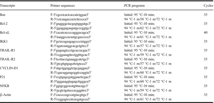
In this study, we measured the expression levels of P53, BAX, BCL-2, CAS3, CAS9, BIRC, and PPIA (housekeeping) genes related to apoptosis on A549 human lung carcinoma

Similarly, in our study, TNF-α and IL-6 levels were found to be higher in patients with influenza than in the control group and were even higher in fatal patients1. IL-1β levels

Results: It was determined that HP extract treatment increased the expression of apoptotic proteins (AIF, GADD153, GRP78, caspase-3, Bax) and cell cycle G2 / M checkpoint