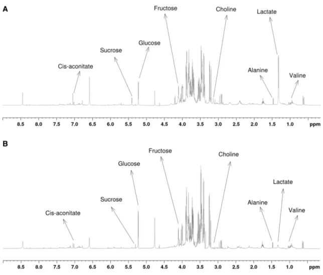
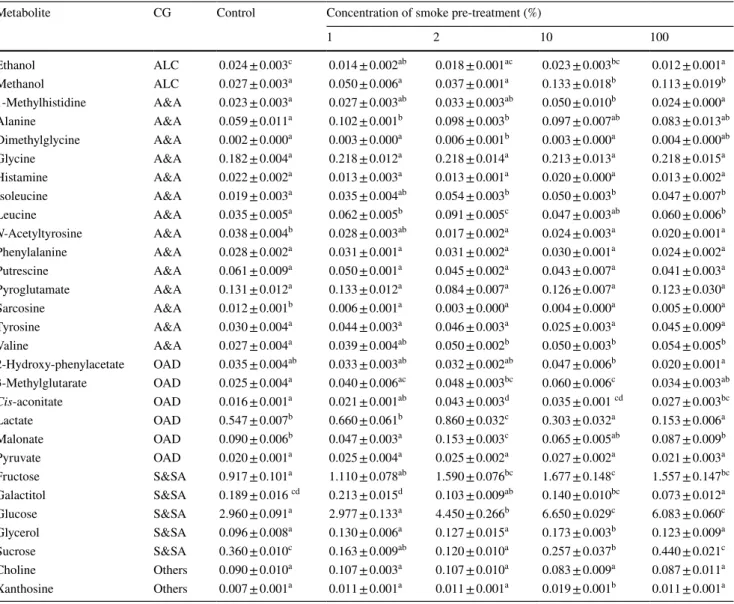
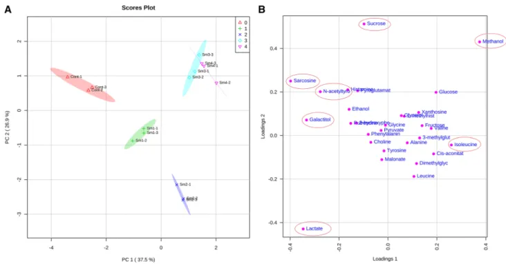
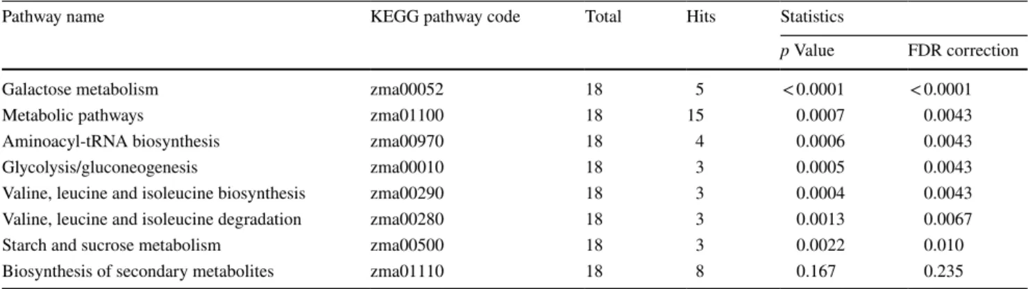
NMR-based metabolomics reveals that plant-derived smoke stimulates root growth via affecting carbohydrate and energy metabolism in maize
Tam metin
Şekil

Benzer Belgeler
2 Niğde Üniversitesi, Tarım Bilimleri ve Teknolojileri Fakültesi, Hayvansal Üretim ve Teknolojileri Bölümü, TR-51240 Niğde - TÜRKİYE 3 Gaziosmanpasa Üniversitesi,
McCaslin’in (1990), “Sınıfta Yaratıcı Drama” (Creative Drama in The Classroom) başlıklı çalışmasında, Meszaros’un (1999), “Eğitimde Yaratıcı Dramanın
Cümlelerde geçen sıfatların ( ön ad ) altını çizelim. "……..…….……düşük enerji üreten bir elektrik kay- a) Akıllı çocuk soruların tamamını bildi. "
İki bölümden oluşan bir tavuk çiftliğinde birinci bölümde 159, ikinci bölümde ise 158 tavuk bulunmaktadır. Hafta sonu 94 tavuk satıldığına göre çiftlikte kaç
18 Üçüncü Yol, devletin varlığını ancak ve ancak devletin serbest piyasaların örgüt yapısına kendisini uyarlaya- bilme ve hem devletin kendi dönüşümü hem de
Kent tarihi, kent- kültür ilişkisi, kentlilik olgusu, kent hakkı, kent kültürünün dönüşümü ve kent kültü- rünün Türkiye’deki durumu başlıklar hâlinde
Following that, the sub-dimen- sions of the ethical environment, which are thought to control the impact between machiavellian tendencies and whistleblowing intention, were included
Araştırmamızda saptadığımız, hasta grubunun ortalama serum lipid düzeylerinin tüm mevsimlerde düşük olması, yaş ortalamalarının da düşük olmasına bağlı
