www.biodicon.com Biological Diversity and Conservation
ISSN 1308-8084 Online; ISSN 1308-5301 Print 12/3 (2019) 111-118
Research article/Araştırma makalesi DOI: 10.5505/biodicon.2019.07379 Morphological, anatomical and palynological investigations on endemic Silene tunicoides Boiss.
(Caryophyllaceae)
Büşra DARICI 1, Hüseyin DURAL 1, Burcu YILMAZ ÇITAK *1 ORCID: 0000-0003-3336-6466; 0000-0002-5579-5037; 0000-0003-3703-7731
1 University of Selçuk, Faculty of Science, Department of Biology, Konya, Turkey
Abstract
The current study deals with anatomical, palynological and micromorphological aspects of endemic Silene tunicoides for the first time. With morphological analysis, the descriptions of species were enlarged as different from previous studies in point of calyx and corolla dimensions, androecium and gynoecium dimensions. With anatomical analysis, the shape of leaves of cross sections, presence of starch grains in pith cells are found different. Pollen and seed morphology can be useful to distinguishing species. Pollen dimensions, pore shape and structure, microechinae number, ornamentation are the most important were determined as pollen traits. Seed shape, suture structure, surface, teeth shape and dimensions are the most valuable seed characteristics.
Key words: anatomy, Caryophyllaceae, pollen, seed, Silene, SEM --- ---
Endemik Silene tunicoides Boiss. (Caryophyllaceae) üzerine morfolojik, anatomik ve palinolojik araştırmalar Özet
Mevcut çalışma, endemik Silene tunicoides’i anatomik, palinolojik ve mikromorfolojik yönlerden ilk defa incelemektedir. Morfolojik analizler ile türün deskripsiyonu, kaliks ve korolla boyutları, erkek ve dişi organ boyutları açısından önceki çalışmalardan farklı olarak genişletilmiştir. Anatomik analizler ile yaprak enine kesitlerinin şekli ve öz hücrelerinde nişasta tanelerinin varlığı farklı bulunmuştur. Polen ve tohum morfolojisi türleri ayırmada faydalıdır. Polen boyutları, por şekli ve yapısı, mikroekina sayısı ve ornamentasyonu polen özelliği bakımından en önemli olarak belirlenmiştir. Tohum şekli, sutur, yapısı, diş şekli ve boyutları en önemli tohum karakterleridir.
Anahtar kelimeler: anatomi, Caryophyllaceae, polen, SEM, tohum 1. Introduction
Silene L., the largest genus in the family Caryophyllaceae with 700 species [1], is represented in Southwest Asia, with 119 species (34 endemic) in Turkey [2], about 139 species (48 endemic) in Iran, and 37 species in Iraq [3,4]. After Coode & Cullen’s (1967) study, and with addition of new species the total number of Silene genus have been increased to 166 in Turkey [2,5,6,7,8].
Silene genus has been subjected many times in palynological and micromorphological studies but little times in anatomical ones [9,10,11,12,13,14,15,16,17]. The main consensus of these researches that the shape of leaf, starch in pith, the pore dimensions and numbers, microechinae structure, ornamentation, seed shape, testa cell types, ridge features can be useful to distinguish species in taxonomic levels.
Silene tunicoides is an endemic, distinct, semi-shrub plant only grows in Kemer, Antalya province which is its type locality from section Tunicoideae (Boiss.) Chowdhuri. This species can be distinguished from the other two relatives (S. ozyurtii and S. brevicalyx) by the shape of the capsule, calyx teeth and petal limbs, length of calyx and petal. It has been no study conducting the anatomy, palynology and micromorphology of this Turkish species. The aim of this paper
*Corresponding author / Haberleşmeden sorumlu yazar: Tel.: +903322231886; Fax.: +903322231886; E-mail: [email protected] © Copyright 2019 by Biological Diversity and Conservation - Available online at www.biodicon.com/Tüm hakları saklıdır BioDiCon. 797-1218
to investigate the mentioned features of Silene tunicoides and to discuss these properties whether important for species taxonomy.
2. Materials and methods 2.1. Plant material
The materials used in this study were collected from type locality of species (Kemer, Antalya) during the first author master thesis field excursion. These collected materials were stored as herbarium materials and some of them were put in 70% ethyl-alcohol as anatomical materials. The species were photographed in nature (Figure 1).
2.2. Morphological methods
2.2.1. Macro-morphological investigations
In macromorphological investigations, the collected plant materials were prepared according to standard herbarium techniques and were identified using Davis (1988) literature [18]. The herbarium materials were stored in KNYA herbarium. When the descriptions of the species were made, such as stem height, leaves and flowers dimensions were measured at least 20 times for each characters.
2.2.2. Micro-morphological investigations
In micromorphological investigations, the mature fruit and seed were selected under a stereomicroscope and were stored in paper bag. At least 20 samples of fruits and seeds were measured by several characteristics such as fruit length, and seed length. Then these fruits and seeds were transferred to the aluminium stab and, were covered with gold and were micro photographed aid of a Zeiss Jeol Evo LS 10 SEM.
2.3. Anatomical analysis
For anatomical investigations, the paraffin method were applied to root, stem and leaf of species. The cross sections were taken using a microtome [19]. The woody and herbaceous stems cross sections were taken by hand using a razor blade. The paraffin sections were stained with safranin-fast green and they were mounted with entellan. Slides were observed Leica DM1000 light microscope and were photographed with a Canon EOS 450D camera. Measurements were made with Cameram 21 programme.
2.4. Palynological analysis
The pollen slides were made according to Wodehouse (1935) technique [20]. The pollen grains were directly taken the glass-slides and were washed with ethyl-alcohol. A few drops glycerine-jelly with safranin were added the heated slides and they were left to dry [20]. The pollen slides were examined under a light microscope (Leica DM 1000) and were photographed with Canon EOS 450 D camera. For SEM investigations, non-acetolysis pollen grains were put aluminium stabs and were micro photographed at several magnifications using a Zeiss SEM. Punt et al. (2007) was followed for pollen terminology [21].
All of the measurements were given in minimum, maximum and mean values.
3. Results
3.1. Macro-morphological Results
Perennial, with many erect, slender stems arising from a very woody branched at base. Stems up to 40 cm, diameter 0.63-0.85 mm, with short, retrorse indumentum below, glabrous and viscid above. Cauline leaves linear, setaceous, callous, 7-13 mm, margins setose. Inflorescence paniculate, the branches wide-spreading and cymose. Calyx 5×1.5 mm, teeth 1.5×1 mm, glabrous, ovoid, bracts 0.4-0.8 mm, Petals cream-transparent with an entire limb. Anthophore 1.5 mm. Anther 1 mm, filaments 3.5 mm. Ovary 1 mm, style 0.2 mm, stigma capitate. Capsule 3-4 mm, included in the calyx. Fl. 6. Cliffs, pine forests, c. 100 m.
Figure 1. A. The general view of Silene tunicoides in nature conditions. B. Flowers and capsule
3.2. Micro-morphological Results 3.2.1. Fruit micromorphology
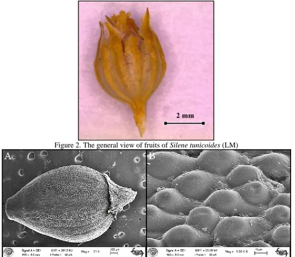
The fruits are capsule, 4.16-4.4× 2.6-2.99 mm, globose-ovoid, tuberculate in ornamentation. Ovoid at base, carpels are not separated, narrowed and truncate in apex (Figs. 2-3).
Figure 2. The general view of fruits of Silene tunicoides (LM)
3.2.1. Seed micromorphology
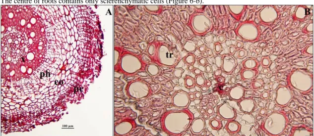
The seeds of S. tunicoides are reniform, dark-brown and shiny, 0.81‒1.2 × 1.04‒1.33 mm, the ratio length-width being 1.18, seed outline is rectangular-orbicular, chalazal pole is obtuse, micropylar pole is emarginate, seed surface type convex-sometimes flat, shape of tubercle rounded, seed surface granulation fine, suture is sinuous, ridge is absent or indisctinct, flat, the number of teeth is 10, teeth shape is rectangular, 8.21-10.85 × 9.45-11.05 µm, testa cells 117.08-146.22 × 109.72-138.91 µm, the ratio length-width of testa cells 1.05 (Figs. 4-5).
Figure 4. The general view of seeds of Silene tunicoides (LM).
Figure 5. A. The general view of seeds of Silene tunicoides (SEM) B. Ornamentation
3.3. Anatomical Results Root anatomy
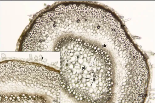
Peridermis which is characterized many layered, partly broken with phellem cells is an outermost layer of roots in S. tunicoides (Figure 6-a). Cortex cells are parenchymatic and their shape is defined as rectangular-oval. The vascular tissue composed of phloem and xylem elements is well-developed. Trachea cells were measured as 47.16 µm in mean values. The centre of roots contains only sclerenchymatic cells (Figure 6-b).
Figure 6. The root cross sections of Silene tunicoides A. General view of roots pe: peridermis, co: cortex, ph: phloem, x: xylem. B. the centre of roots tr: trachea, c: center of root.
Woody stem anatomy
The cross sections taken from woody stems of S. tunicoides were showed that secondary structure developed. The outermost surface of roots is limited by peridermis (Figure 7-a). Cortex is characterized with sclerenchymatic cells.
Endodermis is one layered. Phloem and xylem follow endodermis towards the centre. The centre of stem is covered with parenchymatic pith cells containing abundant starch grains (Figure 7-b).
Figure 7. The cross sections of woody stem of Silene tunicoides pe: peridermis, sc: sclerenchyma, en: endodermis, ph: phloem, x: xylem, p: parenchyma cell, pi: pith region, s: starch.
Herbaceous stem anatomy
The outermost surface of stem is limited with a single layered epidermis with a few cells gland-trichomes (Figure 8-a,b). The cortex which is characterized by parenchymatous cells is oval shaped with chloroplasts. Sclerenchyma is covered large area in stem. Phloem and xylem are well-developed. The pith is composed of parenchymatic cells with starch grains (Figure 8-a).
Figure 8. The cross sections of herbaceous stem of Silene tunicoides ep: epidermis, co: cortex parenchyma, sc: sclerenchyma, ph: phloem, x: xylem, p: parenchyma cell, pi: pith region, s: starch, t: trichome.
Leaf anatomy
The cross sections of leaves clearly show the three main parts: epidermis, mesophyll, and vascular tissue (Figure 989). The epidermis in a single layer is covered the leaf. Stomata occurs both upper and lower epidermis. Also, the hairs are positioned on epidermis of both sides. Mesophyll tissue is composed of two types of cells as palisade and sponge parenchyma. Druse crystals are present into the leaf mesophyll. Central vascular bundle is larger and collateral in type (Figure 9).
Figure 9. The cross sections of leaves of Silene tunicoides ue: upper epidermis, le: lower epidermis, pp: palisade parenchyma, sp: spongy parenchyma, vb: vascular bundle, sc: sclerenchyma, dr: druse crystal, t: trichome.
3.3. Pollen morphology
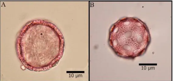
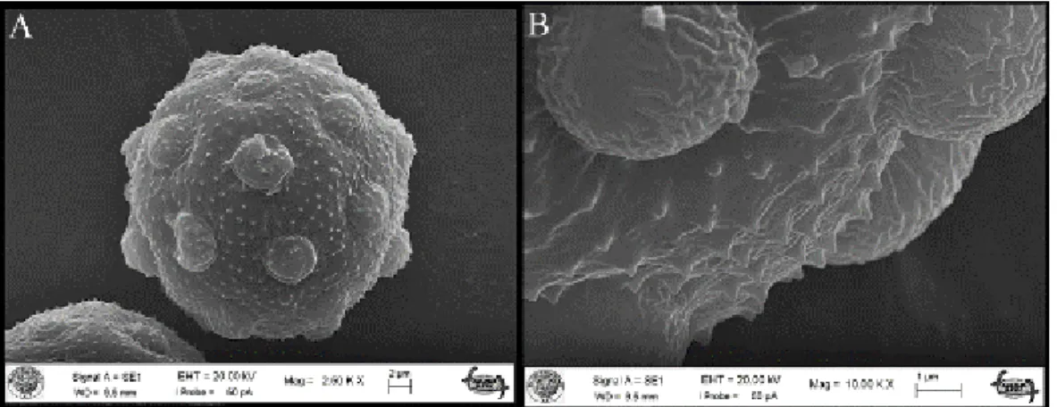
The pollen grains of S. tunicoides are monad, periporate, and spheroidal. The pollen diameter is 26.24-30.7 µm. The pore diameter is 5.1 × 5.6 µm with orbicular-shaped. The numbers of pore are 20-25, the distance between two pore is 1.92-2.12 µm. The pori is operculate with 14-17 microechinae which is triangular shaped. The microechinae dimensions 1-1.14 × 1.35-1.76 μm. The micro perforation is 1.74-2.17 μm. The exine structure is semitectate and the sculptruing is microechinate-microperforate. The exine and intine are 2.18 μm and 0.82 μm thick, respectively (Figs. 10-11).
Figure 10. The light microscope (LM) photographs of Silene tunicoides A. The general view of pollen B. The sculpturing
Figure 11. The scanning electron microscope (SEM) photographs of Silene tunicoides A. The general view of pollen B. The sculpturing of pollen
4. Conclusions and discussion
The measurements and observations of Silene tunicoides is congruent with the study of Coode and Cullen (1967) [2]. However, dimensions and properties of bracts, ovary, style, stigma, and stamen were presented for the first time as different from previous research.
In the root cross-section, S. tunicoides had filled with xylem elements in root centre. Atasagun et al. (2016) had declared that S. brevicalyx had parenchymatous cells in the root centre, whereas S. ozyurtii had a sclerenchymatic centre of root [16]. There are reports that Silene genus had calcium oxalate crystals in the endodermis of some species and multicellular hairs on the epidermis in the stem anatomy of Silene [16, 22,23,24]. These findings were consistent with our findings. In addition, S. tunicoides have starch grains inside its pith cells. However, S. ozyurtii didn’t have any starch [16]. For the classification and identification of species in Caryophyllaceae family can use pollen and seed morphological traits [25]. The members of Caryophyllaceae family had usually medium size, ranging from 25-50 µm pollen grains [9,10,11,15,26,27]. We determined that the pollen grains of S. tunicoides has the same dimensions as above mentioned. Caryophyllaceae was included in polypantoporate group by Moore et al. (1997) [28]. Our findings are in agreement with their results. There were also some differences in pollen characters between S. tunicoides and S. ozyurtii. S. ozyurtii had the bigger pollen grains, little numbers of pore, the bigger pore diameters and the smaller exine thickness than the S. brevicalyx [16] also than S. tunicoides. Structure of pore was slightly receding in S. ozyurtii, whereas structure of pore was protuberant in S. tunicoides.
The seed morphology of the Silene species showed seed type was reniform, reniform-circular, orbicular, semi-orbicular, or flabellate [15,29,30]. S. ozyurtii and S. brevicalyx had identified as reniform seed shape [16] as the same in S. tunicoides. S. ozyurtii had serrate suture outline, coarsely medium surface, triangular teeth shape, with concave lateral surface but S. tunicoides and S. brevicalyx have sinuous suture outline, fine surface, rectangular teeth shape, with convex lateral surface.
As a conclusion, these three species can easily separate each other according to anatomic (leaf shape, presence of starch in stem), pollen (microechinae structure, pore number) and seed characteristics (shape, suture structure, teeth shape).
Acknowledgements
The authors are thankful to the Research Foundation of Selçuk University for financial support (Project number: 15201097).
References
[1] Mabberley, D. J. (2008). Mabberley’s plant-book (3th ed.). Cambridge: Cambridge University Press.
[2] Coode, M.J.E. & Cullen, J. (1967). Silene L. In P.H. Davis (Ed.), Flora of Turkey and the East Aegean Islands 2. (1st ed., pp. 179–222). Edinburgh, UK: Edinburgh University Press.
[3] Melzheimer, V. (1988). Caryophyllaceae: Silene L. In K.H. Rechinger (Ed.) Flora Iranica, Vol. 163. (pp. 341-508). Graz, Austria.
[4] Townsend C. C., Melzheimer V., Kandemir A., Ghazanfar S. A., Haloob A. (2016). 90. Caryophyllaceae A.L. de Jussieu. In S.A., Ghazanfar & J.R. Ed-mondson (Eds.), Flora of Iraq 5(1), (pp. 6-123). Bagdad.
[5] Aydın, Z., Ertekin, A.S., Långström, E. & Oxelman, B. (2014). A new section of Silene (Caryophyllaceae) including a new species from South Anatolia, Turkey. Phytotaxa, 178(2), 98-112. http://dx.doi.org/10.11646/phytotaxa.178.2.2
[6] Fırat, M. & Yıldız, K. (2016). Silene miksensis (Caryophyllaceae), a new species from eastern Anatolia. Phytotaxa, 273(4), 283-292. http://dx.doi.org/10.11646/phytotaxa.273.4.4
[7] Güner, E.D. & Duman, H. (2016). A new species from Turkey, Silene bilgilii (Caryophyllaceae). Phytotaxa, 246(4), 293-299. http://dx.doi.org/10.11646/phytotaxa.246.4.5
[8] Budak, U., Koc, M. & Hamzaoglu, E. (2018). Silene goksuensis (Caryophyllaceae), a new species from south Turkey. Phytotaxa, 345(2): 170-174. http://dx.doi.org/10.11646/phytotaxa.345.2.9
[9] Yıldız, K. (2001). Pollen morphology of Silene L. (Caryophyllaceae) from Turkey. Pakistan Journal of Botany, 33(1), 13-25.
[10] Yıldız, K. (2006). Morphological and palynological investigation of Silene gigantea L. var. gigantea and Silene behen L. (Caryophyllaceae) distributed in Western Anatolia and Northern Cyprus. Turkish Journal of Botany, 30(2), 105-119.
[11] Yıldız, K., Dadandı, M.Y., Minareci, E. & Çırpıcı, A. (2011). Pollen morphology of sections Siphonomorpha and Lasiostemones of the genus Silene from Turkey. Turkish Journal of Bot., 35(6), 631-642. doi:10.3906/bot-1010-119 [12] Şen, H., Bagci, Y. & Çıtak, B.Y. (2014). The investigation of morphological, anatomical and ecological properties
of endemic Silene anatolica and Silene lycaonica. Biological Diversity and Conservation, 7(1), 47-60.
[13] Keshavarzi, M., Mahdavinejad, M., Sheidai, M. & Gholipour, M.A. (2014). Anatomical study of some Silene L. species of Lasiostemones Boiss. section in Iran. Acta Biologica Szegediensis, 58(1),15–19.
[14] Keshavarzi, M., Mahdavinejad, M., Sheidai, M. & Gholipour, M.A. (2015). Seed and pollen morphology of some Silene species in Iran. Phytologia Balcanica, 21(1), 7-12.
[15] Dadandı, M.Y. & Yıldız, K. (2015). Seed morphology of some Silene L. (Caryophyllaceae) species collected from Turkey. Turkish Journal of Botany, 39, 280-297. doi:10.3906/bot-1307-35
[16] Atasagun, B., Aksoy, A. & Martin, E. (2016). Anatomical, palynological and karyological remarks of Silene brevicalyx and Silene ozyurtii (Caryophyllaceae). Phytotaxa, 270(2), 116-126. http://dx.doi.org/10.11646/phytotaxa.270.2.4
[17] Kuh, M., Yıldız, K. & Minareci, E. (2017). A taxonomic study of the Silene sections Behenantha and Dichotomae (Caryophyllaceae) in Turkey based on the micromorphology of their seed and pollen. Turkish Journal of Botany, 41(5), 493-504. doi:10.3906/bot-1610-23
[18] Davis, P.H., Mill, R.R. & Tan, K. (1988). Silene L. In P.H., Davis, R.R. Mill, & Tan, K. (Eds.) Flora of Turkey and the East Aegean Islands (Suppl.1), Vol. 10. (1st ed., pp. 76–81). Edinburgh, UK: Edinburgh University Press. [19] Johansen, D.A. (1940). Plant microtechnique. McGraw-Hill, New-York.
[20] Wodehouse, R.P. (1935). Pollen grains. Mc Grew Hill, New York.
[21] Punt, W., Hoen, P.P., Blackmore, S., Nilsson, S. & Le Thomas, A. (2007). Glossary of pollen and spore terminology. Review of Palaeobotany and Palynology, 143: 1-81.
[22] Metcalfe, C.R. & Chalk, L. (1950). Anatomy of the Dicotyledons. Oxford, UK: Clarendon Press.
[23] Yıldız, K., Minareci, E. (2008). Morphological, anatomical, palynological and cytological investigation on Silene urvillei Schott. (Caryophyllaceae). Journal of Applied Biological Sciences, 2(2), 41-46.
[24] Kılıç, S. (2009). Anatomical and pollen characters in the genus Silene L. (Caryophyllaceae) from Turkey. Botany Research Journal, 2 (2–4), 34-44.
[25] Sahreen, S., Khan, M.A., Meo, A.A. & Jabeen, A. (2008). Pollen morphology of the genus Silene (Sileneae Caryophyllaceae) from Pakistan. Biological Diversity and Conservation, 1–2, 74-85.
[26] Yıldız, K., Cırpıcı, A. & Dadandı, M.Y. (2010). Pollen morphology of the Silene taxa (Caryophyllaceae) in four sections from Turkey. Phytologia Balcanica,16(1), 85-95.
[27] Çıtak, B.Y. & Dural, H. (2019). The palynological traits of four endemic Silene L. (Caryophyllaceae) species in Turkey. Communications Faculty of Sciences University of Ankara Series C Biology, 28(1), 35-42.
[28] Moore, P.D., Webb, J.A. & Collinson, M.E. (1997). An Illustrated guide to pollen analysis (2nd ed.). Blackwell, Oxford.
[29] Yıldız, K. & Çırpıcı, A. (1998). Seed morphological studies in Silene L. from Turkey. Pakistan Journal of Botany, 30, 173-188.
[30] Fawzi, N.M., Fawzy, A.M. & Mohamed, A.H.A. (2010). Seed morphological studies on some species of Silene L. (Caryophyllaceae). International Journal of Botany, 6(3), 287-292.