the impact of spironolactone on the lung injury induced by concomitant
trastuzumab and thoracic radiotherapy
Article · January 2019 DOI: 10.18869/acadpub.ijrr.17.1.87 CITATIONS 2 READS 42 6 authors, including: Guler Yavas Baskent University 104PUBLICATIONS 338CITATIONS SEE PROFILE Cagdas Yavas Baskent University 75PUBLICATIONS 289CITATIONS SEE PROFILE Esin Celik Selcuk University 51PUBLICATIONS 75CITATIONS SEE PROFILE Ozlem Yavas Selcuk University 62PUBLICATIONS 338CITATIONS SEE PROFILE
International Journal of Radiation Research, January 2019 Volume 17, No 1
The impact of spironolactone on the lung injury
induced by concomitant trastuzumab and thoracic
radiotherapy
INTRODUCTION
Radiation therapy (RT) plays an important
role in the treatment of breast cancer. Radiation-induced lung injury (RILI) is a potentially life-threatening and dose-limiting
side effect of breast irradiation and thus the risk
should be minimized (1-3). In addition,
chemotherapy (CT), targeted therapy, and hormonal treatment used in conjunction with RT increase the risk of damage to normal tissue. Thus, reducing radiation-induced normal tissue damage is of critical importance in improving both tumor control and patient quality of life (1).
RILI which is also named as “radiation pneumonopathy” is a continuous process and
regarded as the result of an abnormal healing
response. Subclinical early damage in pneumocytes type I progress to an acute interstitial inflammation at 6-12 weeks after the
onset of RT and further to lung fibrosis after many months and years (1, 4). Histopathologically,
radiation pneumonopathy is characterized by the focal infiltration of inflammatory cells into the pulmonary interstitium, which is associated, right from the beginning, with the deposition of intercellular matrix material, leading finally to the destruction of the alveolar
histo-G. Yavas
1*, C. Yavas
1, E. Celik
2, E. Sen
3, O. Ata
3, R.E. Afsar
41Department of Radiation Oncology, Selcuk University, Konya, Turkey 2Department of Pathology, Selcuk University, Konya, Turkey 3Department of Medical Oncology, Selcuk University, Konya, Turkey
4Department of Nephrology, Selcuk University, Konya, Turkey
ABSTRACT
Background: To evaluate impact of spironolactone (S) on pulmonary toxicity of concomitant use of thoracic radiotherapy (RT) and trastuzumab (T). Materials and Methods: Eighty rats were divided into eight groups: group (G) 1 was control group; G2, G3 and G4 were RT, S and T groups; G5, G6, G7 and G8 were RT+T, T+S, RT+S and RT+T+S groups respectively. Rats were sacrificed at 6 hour, 21 and 100 day after RT and lung samples were retrieved. Results: By 100th days of RT inflammation score, lung fibrosis score and TGF- expression were significantly different within study groups (p values were 0.002, 0.001 and 0.043 respectively). Inflammation score of G8 was significantly lower than inflammation scores of G2 and G5 (p values: G2-G8= 0.004, and G5-G8=0.022). Inflammation score of G2 was significantly higher than G7 (p=0.028). There were significant differences regarding to fibrosis scores between G2-G8 (p=0.015), G2-G7 (p=0.017) and G5-G8 (p=0.011). TGF-β expression was higher in both G2 and G5 when compared to G8 (p = 0.038).
Conclusion: Our results suggested that S is an effective treatment option for improving radiation-induced pulmonary fibrosis. These findings should be clarified with further preclinical and clinical studies.
Keywords: Aldosterone, pulmonary fibrosis, radiotherapy, spironolactone, Trastuzumab. *Corresponding authors: Dr. G. Yavas, Fax: +90 332241 60 65 E-mail: [email protected] Revised: August 17 Accepted: October 17
Int. J. Radiat. Res., January 2019; 17(1): 87-95
►
Original article
DOI: 10.18869/acadpub.ijrr.17.1.87
architecture. Clinically RILI is typically divided into two phases: pneumonitis and fibrosis. The fibrotic phase tends to manifest >3 months after treatment. Fibrosis is part of the wound-healing process. Therefore radiation fibrosis is a form of chronic lung damage that usually evolves over 4-24 months after irradiation (4).
HER-2/neu proto-oncogene is a poor prognostic factor and is amplified in 20–30% of
patients with primary breast cancer. Trastuzumab, a monoclonal antibody directed
against HER2, improves response, time to progression, and overall survival in both the
metastatic and adjuvant settings in patients with HER2 positive breast cancer (5-7). Preclinical in
vitro and in vivo studies have shown that the cascade of events through the HER2 receptor is
involved in tumor radiosensibility (8, 9),
application of Trastuzumab concurrently with radiation thus increases the antitumor effect of radiation. There are same clinical evidences in
the literature that Trastuzumab also radiosensibilizes human healthy tissues and in
this way it could increase the toxicity of the treatment (10). A few study evaluated the side
effects of Trastuzumab and RT combination and the results are contradictory (11-13). Trastuzumab
-induced pneumonitis may present as rapidly progressive pulmonary infiltrate respiratory failure after the administration of 1 dose of Trastuzumab or after 6 weeks of therapy. The incidence of Trastuzumab-induced pneumonitis is 0.4–0.6% (14). Although infrequent; pulmonary
toxicity due to Trastuzumab may be life-threatening (15, 16).
Mineralocorticoid receptor (MR) activation is a contributing factor in the pathophysiology of a wide range of diseases (17). Aldosterone, which is
a physiological activator of MR, is partially responsible for increases in the extracellular matrix turnover, as observed in fibrosis of the cardiac, kidney and lung tissues (18, 19), and exerts
its effects primarily on lung epithelium. Elevated
levels of aldosterone are known to induce hypertension, alter inflammation and fibrosis
and exacerbate cardiovascular diseases (20, 21).
Spironolactone, an aldosterone receptor antagonist, may have the ability to ameliorate
pulmonary fibrosis (22-24).
By the light of these findings we hypothesized that Spironolactone would be effective in the treatment of both RT and Trastuzumab-induced lung injury by correcting pulmonary fibrosis. In the present study we aimed to evaluate whether the use of Spironolactone would be able to ameliorate the pulmonary toxicity induced by concomitant Trastuzumab and RT. Our second
aim was to investigate the impact of Spironolactone on either RILI or Trastuzumab-induced lung injury when these
two agents used separately.
MATERIALS AND METHODS
Study DesignThis study included 80 female Wistar-Albino rats (250-300 g); the use of which was approved by the Necmettin Erbakan University Ethical Committee. Animals were housed 4 per cage in a controlled animal holding room with a 12/12-hour light/dark cycle; temperature and relative humidity were continually monitored to provide standard laboratory conditions. Rats were divided into 8 groups (G) composed of 10 animals. Group (G) 1 was defined as control
group. G2, G3 and G4 were RT, Spironolactone and Trastuzumab groups respectively. G5, G6, G7 and G8 were RT + Trastuzumab, Trastuzumab + Spironolactone, RT + Spironolac-tone and RT + Trastuzumab + SpironolacSpironolac-tone groups respectively (Table 1). RT was applied under general anesthesia with intraperitoneally administered 90 mg/kg ketamine hydrochloride and 10 mg/kg xylazine. A single dose of 15 Gy was applied to the both lungs. Trastuzumab (6 mg/kg) was administered intraperitoneally and Spironolactone (80 mg/kg) was administered by oral gavage. Rats were sacrificed via cervical dislocation at 6th hour, 21st day and 100th day
after RT and the lung samples were taken for microscopical examination.
Radiotherapy protocol
RT was applied under general anesthesia with intraperitoneally administered 90 mg/kg ketamine hydrochloride (Ketalar®, EWL
88
Eczacibasi Warner Lambert Ilaç Sanayi ve Ticaret A.S., Istanbul, Turkey) and 10 mg/kg xylazine (Rompun® 2%, Bayer Kimya San. Ltd. Sti., Istanbul, Turkey). A single dose of 15 Gy was applied to the both lungs via a single anterior
field to 2 cm depth with SAD (source-axis distance) technique. One cm elasto-gel bolus was
used to build up the radiation dose on the lungs and to provide contour regularity. The field size was 4x4 cm and included the both lungs.
Trastuzumab protocol
Trastuzumab (Herceptin ®; Genentech Inc, South San Francisco, CA, USA) dose which was equivalent to 6 mg/kg adult dose was calculated for each rat and injected i.p. 2 h before the RT. The rats in G4, G6 and G8 were applied 0.5 cc 0.9% NaCl i.p.
Spironolactone protocol
Spironolactone (Aldakton-A®, Ali Raif I laç San. A.Ş., I stanbul, Turkey) dose which was
equivalent to 80 mg/kg adult dose was calculated for each rat and was administered by
oral gavage. Spironolactone administration was started a week before RT and continued until the animals sacrificed.
Histopathologic evaluation
Rats were sacrificed via cervical dislocation at 6th hour, 21st day and 100th day after RT and
the lung samples were taken for microscopical examination. The bilateral whole lungs of each
rat were excised and fixed in 10% neutral buffered formalin. Two lobes of each lung for
each rat were processed and embedded in
paraffin for light microscopic examination. The 4 µ thick sections obtained with microtome were stained with hematoxylin and eosin (H&E) to
evaluate the inflammation, and with histochemical Masson Trichrome staining to
identify the fibrosis in the lung. Extend of the
chronic inflammatory cells including lymphocytes on alveolar walls was graded on a
scale of 0 (normal) to 3 (severe). Fibrosis was
defined as the thickened alveolar walls with superimposed collagen. As a quantitative end
point, extend of the radiation induced fibrosis was graded on a scale of 0 (normal lung or minimal fibrous thickening) to 3 (severe fibrosis -total fibrous obliteration of the field). The pathologist was not aware of the treatment
groups at the time of the histological examination of the specimens. After examining
the whole sections for each rat, the average value of fibrosis and chronic inflammation per rat was taken as the fibrosis and inflammation scores and mean values of each group were calculated.
Immunohistochemistry staining and scoring procedure
Paraffin-embedded tissues of chosen slides were collected and 4 µm thick sections were
prepared for immunohistochemistry. The sections were deparaffinized at 37 ºC oven
overnight. Immunohistochemical staining was
performed by using an automatic staining machine (Ventana, Benchmark XT). The sections
were boiled at sodium citrate buffer at 95 ºC for
60 min and then incubated with primary antibody Anti TGF-β rabbit polyclonal antibody
(ABCAM, ab92486, Cambridge, UK) at a dilution of 1:100 for 52 minutes. The sections were incubated with the secondary antibody for 20 min at room temperature, incubated with Ultra I
-view detection kit and counterstained with hematoxylin for 8 min.
The immunohistochemical TGF-β stained slides were evaluated and scored by a single pathologist blinded to patients’ data. On light
microscopic evaluation of each 4 µm thick section, 10 different fields magnified 100X were
reviewed. Immuno-reactivity scoring system (IRS) which was previously described by Wang Yavas et al. / Radiation pneumonopathy & spironolactone
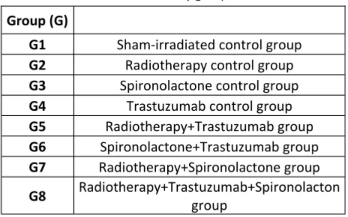
Table 1. Study groups
Group (G)
G1 Sham-irradiated control group
G2 Radiotherapy control group
G3 Spironolactone control group
G4 Trastuzumab control group
G5 Radiotherapy+Trastuzumab group G6 Spironolactone+Trastuzumab group G7 Radiotherapy+Spironolactone group G8 Radiotherapy+Trastuzumab+Spironolacton group
89
Int. J. Radiat. Res., Vol. 17 No. 1, January 2019
et al (25) was used to determine TGF-β
expression levels. This system depends on multiplication of staining intensity and TGF-βpositive alveolar cell percentage. The percentage of positive cells was scored as: 0, negative; 1, 1-25%; 2, 26-50%; 3, 51-75%; 4, 76-100% and the staining intensity as 0 (-), 1 (+), 2 (++), 3 (+++).
Statistical analysis
The Statistical Package for Social Sciences (SPSS) v. 15.0 was used for statistical analyses. As the pathological scores were ordinal in nature, the differences in pathological findings between the study groups were analyzed using
the Kruskal– Wallis test. When an overall statistically significant difference was observed,
pairwise comparisons were performed using the Mann–Whitney U test. Bonferroni correction was used for multiple comparisons. A 5 % type I
error level was used for the statistical significance cutoff for overall comparisons.
RESULTS
At 6th hour of RT; inflammation, pulmonary
fibrosis and TGF-β scores were not significantly different within the study groups (p values were
0.091, 0.082 and 0.154 respectively). The samples taken from RT groups (G2, G5, G7 and
G8) had minimal inflammation; however we did not observe any fibrosis or TGF-β expression
even in RT groups.
By 21st day of RT; the inflammation and
fibrosis score were significantly different within the study groups (p values were 0.038 and 0.038 respectively); however we could not observe any significant difference with respect to TGF-β ex-pression (p=0.072). Both the inflammation and fibrosis scores higher in RT groups (G2, G5, G7 and G8) than the control groups (G1, G3, G4 and G6); however there weren’t any significant dif-ferences between RT groups with respect to the inflammation and fibrosis scores.
By 100th days of RT the inflammation score
(table 2), lung fibrosis score (table 3) and TGF- expression (table 4) were significantly different within the study groups (p values were 0.002, 0.001 and 0.043 respectively. Pair-wise comparisons revealed that the inflammation score of the G8 was significantly lower than the inflammation scores of G2, G5 and (p values
were: G2-G8= 0.004, and G5-G8=0.022). Additionally the inflammation score of G2 was
significantly higher than that of G7 (p=0.028). When the fibrosis and scores were compared at
100th day of RT; there were significant
differences between G2-G8 (p=0.015), G2-G7 (p=0.017) and G5-G8 (p=0.011). Moreover the TGF-β expression was higher in both G2 and G5 when compared to G8 (p values were 0.038 for both).
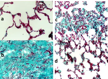
Figure 1 shows the histochemical Masson Trichrome staining of the lung samples from each group by 100th day of RT.
Figure 1. Histochemical Masson Trichrome staining (200X): Section from the lungs of the rats from a) Control group; b)
RT+Trastuzumab group; c) RT only group; d) RT+Trastuzumab+Spironolactone group in 100th day of RT. (a) Control group: Normal alveolar structures with thin alveolar walls. (b) RT+ Trastuzumab group: There were thickening in the alveolar walls with severe
fibrosis and partial lung obliteration. (c) RT only group: There were thickening in the alveolar walls with severe fibrosis.
(d) RT+Trastuzumab+Spironolactone group: There were thickening in the alveolar walls with mild fibrosis
a
b
c
d
90
Yavas et al. / Radiation pneumonopathy & spironolactone
Group (G) Mean Median Minimum Maximum P
G1 0.67 1 0 1 0.002 G2 2.25 2 2 3 G3 0.33 0 0 1 G4 1.50 1.50 1 2 G5 2.75 3 2 3 G6 1.25 1 1 2 G7 1.25 1 1 2 G8 1.25 1 1 2
Table 2. Inflammation scores of the samples taken from lung
tissue 100 days after radiotherapy.
Group (G) Mean Median Minimum Maximum P
G1 0 0 0 0 0.001 G2 2.50 2.50 2 3 G3 0.33 0 0 1 G4 1.50 1.50 1 2 G5 3 3 3 3 G6 1.25 1 1 2 G7 1.50 1.50 1 2 G8 0.75 1 0 1
Table 3. Fibrosis scores of the samples taken from lung tissue
100 days after radiotherapy
Group (G) Mean Median Minimum Maximum P
G1 0 0 0 0 0.043 G2 3 3 3 3 G3 0.67 1 0 1 G4 1.50 1.50 1 2 G5 3 3 3 3 G6 1.50 1.50 1 2 G7 1.50 1.50 1 2 G8 2 2 2 2
Table 4. TGF-β expression levels in the lung tissues 100 days after radiotherapy.
TGF-beta; Transforming growth factor beta.
DISCUSSION
In this study, we investigated the effect of MR
antagonism, via a potent MR antagonist, Spironolactone, on the use of concomitant RT
and Trastuzumab in a rat model. Our results suggested that long-term use Spironolactone improved both radiation-induced and RT + Trastuzumab induced lung toxicity. Although we observed a trend for higher scores of both in-flammation and fibrosis scores in 100th days for
G4 (Trastuzumab only group) than G6 (Trastuzumab+Spironolactone group), the difference was not statistically significant.
Therefore Trastuzumab did not cause pulmonary fibrosis when used alone and did not
deteriorate RILI. The main reason for the pulmonary fibrosis is RT. This is the first study
in the literature demonstrating that MR blockade ameliorates the radiation-induced late fibrosis in lung tissues of the rats.
In a preclinical study Barut and colleagues
investigated the effect of Spironolactone aldosterone on lung injury induced by intestinal
ischemia and reperfusion (17). Their results
suggested that Spironolactone pretreatment decreased neutrophil infiltration, nitric oxide
synthase induction, oxidative stress, and histopathological injury. Similarly, Atalay et al.
demonstrated that Spironolactone is an effective form of treatment for acute lung injury (21). Ji et
al. demonstrated the therapeutical potential of
Spironolactone, which significantly reduces the pulmonary inflammatory response induced by bleomycin (26). Spironolactone-treated lungs
exhibited fewer macrophage, lymphocyte, neutrophil and eosinophils in alveoli compared
with the untreated lungs. Lieber et al. demonstrated that Spironolactone treatment
attenuates acute pulmonary inflammation
induced by both bleomycin and lipopolysaccharide (27). In various animal
models, it was shown that Spironolactone can function as an antioxidant factor and protect
91
Int. J. Radiat. Res., Vol. 17 No. 1, January 2019
organs from oxidative damage by enhancing antioxidative defense systems while
suppressing production of free radicals (27).
However in the current study we found that
Spironolactone could not attenuate
Trastuzumab-induced pulmonary toxicity.
RILI is the reaction of the organ lung to radiation effects in various target cells. It starts as an exudative inflammation, with the clinical picture of interstitial pneumonia 6-12 weeks after irradiation, and proceeds to a productive chronic inflammation lasting several months and terminating, as other chronic inflammations do, in scar formation, called lung fibrosis. Numerous cytokines such as TNF-α, interleukin-1, platelet-derived growth factor,
fibroblast growth factor, and TGF-β have been reported to be increased within the irradiated tissue (1, 4). TGF-β levels are increased as early as
6 h after irradiation and remain elevated fibrotic lesions as long as 20 years (29, 30). In the current
study we sacrificed animals at 6th hour, 21st day
and 100th day after RT in order to observe both
pneumonitis and fibrosis phases. We found that Spironolactone was effective treatment modality for amelioration of RILI only during fibrotic phase since we observed the improvement only by 100th day.
The alveolar epithelium consists of type I and
type II epithelial cells. Type I cells cover approximately 90% of the alveolar surface and
type II cells are the precursors of type I cells. In steady state, the turnover time of the alveolar
epithelium is approximately 4-5 weeks. However after toxic injury, type I epithelial cells
are denuded and the proliferation of type II cells is stimulated up to 10-fold (31). Stimulation of
type II cells promotes the secretion of growth factors and proteases and degradation of the extracellular matrix to allow removal of dead
cells by normal processes (4). Pulmonary
irradiation also reduces microvessel density and lung perfusion and promotes hypoxia (32). All of
these injuries stimulate the recruitment of a variety of inflammatory cells to the site of the injury and it leads to establishment of chronic
inflammation and fibrosis. In this phase vascular damage and collagen deposition become apparent. Vascular injury and activation
of coagulation cascade, cellular adhesion molecules, proinflammatory and profibrotic
cytokines and oxidative stress seem to play a vital role in the development of radiation
fibrosis (33). Aldosterone is related to
extracellular matrix turnover increase, which is
associated with cardiac, and possibly lung fibrosis (18, 19). Lung cells have aldosterone
receptors, and under physiological conditions, aldosterone participates in active sodium
transport across the alveolar-capillary membrane (23, 34). It has been shown that
Aldosterone aids in maintaining the fluid-free lumen of the lung (35), and is present at high
concentrations in pulmonary fibrosis (24).
Aldosterone thus promotes interstitial fibrosis,
possibly through local dehydration. Spironolactone administration has been shown
to reduce pulmonary fibrosis (35, 36), as well as
pulmonary congestion and edema (37), by
correcting the gas diffusion capacity (38) and
renal mineral status. (39). In parallel to these
findings in the current study we also demonstrated that Spironolactone ameliorated
the RILI.
There are both preclinical and clinical
evidences showing Trastuzumab
radiosensibilizes human healthy tissues and in this way it could increase the toxicity of the treatment (9, 10, 40). The potential synergetic effect
of Trastuzumab and RT may also increase radiation morbidity when combined in the adjuvant setting. Therefore, administration of concomitant Trastuzumab and RT as be a concern. The cardiotoxic effect of Trastuzumab
especially when used with antracyclin chemotherapy has been well known, though the
mechanism is not yet fully understood (6).
However there is limited date with respect to the late toxicity of Trastuzumab administration
with RT. In a preclinical study Bese and colleagues demonstrated that, addition of
Trastuzumab to thoracic irradiation either sequentially or concomitantly did not increase radiation-induced pulmonary fibrosis in rats (41).
Similarly, in the present study we found that the use of concomitant Trastuzumab with thoracic RT did not further increase RILI.
The irradiation doses used in routine clinical
92
practice are different from the ones used in animal studies. Most patients in routine practice are treated with conventional fractionation to a total dose of 50–70 Gy. However, in recent years, stereotactic radiosurgery and intraoperative RT
that use single or two to five fractions of high-dose irradiation have become popular. It
has been postulated that the linear-quadratic
model is an appropriate methodology for determining isoeffective doses at large dose per
fraction (42). The 15 Gy single dose of RT in our
study corresponds to 48–64 Gy, the most frequent dose range used in clinical practice,
when α/β ratio of 2–4 is used.
High quality randomized trials have demonstrated that adjuvant Trastuzumab concomitant with or following chemotherapy in
node-positive and high-risk node-negative patients with HER-2 positive early stage breast
cancer improves disease free and overall survival (5, 6, 43, 44). In routine clinical practice
Trastuzumab is usually given once every week or every 3 weeks intravenously. However in experimental studies conducted on rats, a single
dose of Trastuzumab is usually preferred because of easy application (41, 45, 46). Accordingly,
in this experimental study, a single dose of Trastuzumab, which was equivalent to 6 mg/kg
adult dose was calculated for each rat and injected intraperitoneally.
Our study has some limitations that should
be mentioned as well. First of all, we could not demonstrate any difference between the RT-groups with respect to the inflammation and
fibrosis scores; and TGF-β expression before 100th day of RT. Although the inflammation
score of RT+Trastuzumab group was higher than of RT + Trastuzumab + Spironolactone group at 21st day, this difference was not at
statistically significant level. In the current study we used only light microscopy. If we used electron microscopy in addition to light microscopy we might have demonstrated some
differences at ultrastructural levels at 21st day. We think that electron microscopy may further
enlighten the possible interaction of RT and Spironolactone in this regard. Secondly, this
study is an experimental study; therefore our results should be clarified with clinical studies.
In conclusion, our results suggested that the use of concomitant Trastuzumab with thoracic RT did not further deteriorate radiation-induced
pulmonary injury. On the other hand, Spironolactone ameliorated the
radiation-induced pulmonary fibrosis. Therefore Spironolactone is an effective treatment option
for improving radiation-induced pulmonary fibrosis with long-term usage. These findings should be clarified with further preclinical and clinical studies.
Conflict of Interest
The authors declare that they have no conflict of interest.
Funding source
This work was supported by Selcuk University. There is no role of study sponsors in
the study design, in the collection, analysis and interpretation of data; in the writing of the manuscript and in the decision to submit the manuscript for publication.
Conflicts of interest: Declared none.
REFERENCES
1. Graves PR, Siddiqui F, Anscher MS, Movsas B (2010) Radia-tion pulmonary toxicity: from mechanisms to manage-ment. Semin Radiat Oncol, 20: 201–207.
2. Kocak Z, Evans ES, Zhou SM (2005) Challenges in defining radiation pneumonitis in patients with lung cancer. Int J
Radiat Oncol Biol Phys, 62: 635–638.
3. van der Veen SJ, Faber H, Ghobadi G et al. (2016) Decreas-ing irradiated rat lung volume changes dose-limitDecreas-ing toxici-ty from early to late effects. Int J Radiat Oncol Biol Phys,
94(1): 163-71.
4. Trott KR, Herrmann T, Kasper M (2004) Target cells in radi-ation pneumopathy. Int J Radiat Oncol Biol Phys, 58(2):
463–469.
5. Romond EH, Perez EA, Bryant J et al. (2005) Trastuzumab plus adjuvant chemotherapy for operable HER2-positive breast cancer. N Engl J Med, 353: 1673–1684.
6. Piccart-Gebhart MJ, Procter M, Leyland-Jones B, Gold-hirsch A, Untch M, Smith I et al. (2005) Trastuzumab after adjuvant chemotherapy in HER2-positive breast cancer. N
Engl J Med, 353: 1659–1672.
7. Slamon DJ1, Leyland-Jones B, Shak S et al. (2001) Use of chemotherapy plus a monoclonal antibody against HER2 for metastatic breast cancer that overexpresses HER2. N
Yavas et al. / Radiation pneumonopathy & spironolactone
93
Int. J. Radiat. Res., Vol. 17 No. 1, January 2019
Engl J Med, 344: 783–792.
8. Marinko T, Dolenc J, Bilban-Jakopin C (2014) Cardiotoxicity of concomitant radiotherapy and trastuzumab for early breast cancer. Radiol Oncol, 48(2): 105-112.
9. Pietras RJ, Poen JC, Gallardo D, Wongvipat PN, Lee HJ, Slamon DJ (1999) Monoclonal antibody to HER-2/neu re-ceptor modulates repair of radiation–induced DNA dam-age and enhances radiosensitivity of human breast cancer cells overexpressing this oncogene. Cancer Res, 59: 1347–
1355.
10. Law AB, Evans T, Hayward RL et al. (2009) Possible radia-tion sensitisaradia-tion by trastuzumab leading to radiaradia-tion- radiation-induced myelitis. Breast Care (Basel), 4: 40–42.
11. Belkacémi Y, Gligorov J, Ozsahin M, et al. (2008) Concur-rent trastuzumab with adjuvant radiotherapy in HER2-positive breast cancer patients: acute toxicity analyses from the French multicentric study. Ann Oncol, 19: 1110–
6.
12. Raben A, Sammons S, Hanlon A, et al. (2006) Comparison of acute breast and chest wall toxicity in women treated with external beam radiation with and without concurrent herceptin in a community cancer center. Int J Radiat Oncol
Biol Phys, 66(Suppl. 1): S541–2. 11.
13. Bellon JR, Gover MT, Burstein HJ, Harris JR, Harris LN (2005) Concurrent Trastuzumab and radiation therapy in the adjuvant treatment of breast cancer. Int J Radiat Oncol
Biol Phys, 63(Suppl. 1): S55–6.
14. Vahid B and Marik P (2008) Pulmonary complications of novel antineoplastic agents for solid tumors. Chest, 133:
528–538.
15. Bettini AC, Tondini C, Poletti P, Caremoli ER, Guerra U, Labianca R (2008) A case of interstitial pneumonitis associ-ated with Guillain-Barré syndrome during administration of adjuvant trastuzumab. Tumori, 94: 737.
16. Pepels MJ, Boomars KA, van Kimmenade R, Hupperets PS (2009) Life-threatening interstitial lung disease associated with trastuzumab: case report. Breast Cancer Res Treat,
113: 609.
17. Barut F, Ozacmak VH, Turan I, Sayan-Ozacmak H, Aktunc E (2016) Reduction of acute lung injury by administration of spironolactone after intestinal ischemia and reperfusion in rats. Clin Invest Med, 39(1): E15-24.
18. Zannad F, Alla F, Dousset B, Perez A, Pitt B (2000) Limita-tion of excessive extracellular matrix turnover may con-tribute to survival benefit of spironolactone therapy in patients with congestive heart failure: insights from the randomized aldactone evaluation study (RALES). Rales Investigators. Circulation, 102: 2700-6.
19. MacFadyen RJ, Barr CS, Struthers AD (1997) Aldosterone blockade reduces vascular collagen turnover, improves heart rate variability and reduces early morning rise in heart rate in heart failure patients. Cardiovasc Res, 35:
30-4.
20. Frieler RA, Ray JJ, Meng H, et al. (2012) Myeloid mineralo-corticoid receptor during experimental ischemic stroke: efects of model and sex. J Am Heart Assoc, 1: e002584. 21. Atalay C, Dogan N, Aykan S, Gundogdu C, Keles MS (2010)
The efficacy of spironolactone in the treatment of acute
respiratory distress syndrome-induced rats. Singapore
Med J, 51: 501-5.
22. Broillet MC, Berger A, Horisberger JD (1993) Early effects of aldosterone on the basolateral potassium conductance of A6 cells. Phlugers Arch, 424: 91-3.
23. Hirasawa G, Sasano H, Takahashi K, et al. (1997) Colocali-zation of 11b-hydroxysteroid dehydrogenase type II and mineralcorticoid receptor in human epithelia. J Clin
Endo-crinol Metab, 82: 3859-63.
24. Zhao L, Zhao M, Fang Q (1998) Spironolactone ameliorates rat pulmonary fibrosis induced by bleomycin A5.
Zhong-hua Jie He He Hu Xi Za Zhi 21: 300-2. In Chinese.
25. Wang W, Qiu J, Liu Z, Zeng Y, Fan J, Liu Y, Guo Y (2013) Overexpression of RING box protein-1 (RBX1) associated with poor prognosis of non-muscle-invasive bladder tran-sitional cell carcinoma. J Surg Oncol, 107: 758-61. 26. Ji WJ, Ma YQ, Zhou X, et al. (2013) Spironolactone
attenu-ates bleomycin-induced pulmonary injury partially via modulating mononuclear phagocyte phenotype switching in circulating and alveolar compartments. PLoS One, 19:
e81090.
27. Lieber GB, Fernandez X, Mingo GG, et al. (2013) Mineralo-corticoid receptor antagonists attenuate pulmonary in-fammation and bleomycin-evoked fibrosis in rodent mod-els. Eur J Pharmacol, 718: 290-8.
28. Yuan J, Jia R, Bao Y (2007) Benefcial efects of spironolac-tone on glomerular injury in streptozotocin-induced dia-betic rats. J Renin Angiotensin Aldosterone Syst, 8: 118-26. 29. Dent P, Yacoub A, Contessa J, et al. (2003) Stress and radi-ation-induced activation of multiple intracellular signaling pathways. Radiat Res, 159: 283–300.
30. Martin M, Lefaix J, Delanian S (2000) TGF-beta 1 and radia-tion fibrosis: a master switch and a specific therapeutic target? Int J Radiat Oncol Biol Phys, 47: 277-290.
31. Kasper M and Haroske G (1996) Alterations in the alveolar epithelium after injury leading to pulmonary fibrosis.
His-tol Histopathol, 11: 463–483.
32. Fleckenstein K, Zgonjanin L, Chen L, et al. (2007) Temporal onset of hypoxia and oxidative stress after pulmonary irradiation. Int J Radiat Oncol Biol Phys, 68: 196-204. 33. Chen Y, Williams J, Ding I, et al. (2002) Radiation
pneu-monitis and early circulatory cytokine markers. Semin
Radiat Oncol, 12: 26-33.
34. Fischer H and Clauss W (1990) Regulation of Na channels in frog lung epithelium: a target tissue for aldosterone action. Pflugers Arch, 416: 62-7.
35. Suzuki S, Tsubochi H, Suzuki T, et al. (2001) Modulation of transalveolar fluid absorption by endogenous aldosterone in adult rats. Exp Lung Res, 27: 143-55.
36. Tsukashita M, Marui A, Nishina T, et al. (2008) Spironolactone alleviates late cardiac remodeling after left ventricular restoration surgery. J Thorac Cardiovasc Surg,
136: 58-64.
37. Nishi I, Kawano S, Misaki M, et al. (2006) Addition of spiro-nolactone to an angiotensin-converting enzyme inhibitor decreases lung congestion and edema in Dahl hyperten-sive rats. Heart Vessels 21:251-5.
38. Agostoni P, Magini A, Andreini D, et al. (2005)
Spirinolac-94
Yavas et al. / Radiation pneumonopathy & spironolactone
95
Int. J. Radiat. Res., Vol. 17 No. 1, January 2019tone improves lung diffusion in chronic heart failure. Eur
Heart J, 26: 159-64.
39. Olivera WG, Ciccolella DE, Barquin N, et al. (2000) Aldoste-rone regulates Na,K-ATPase and increases lung edema clearance in rats. Am J Respir Crit Care Med, 161: 567-73. 40. Horton JK, Halle J, Ferraro M, et al. (2010)
Radiosensitiza-tion of chemotherapy-refractory, locally advanced or lo-cally recurrent breast cancer with trastuzumab: a phase II trial. Int J Radiat Oncol Biol Phys, 76(4): 998-1004. 41. Bese NS, Umay C, Serdengecti S, et al. (2010) The impact
of trastuzumab on radiation-induced pulmonary fibrosis: results of an experimental study. Med Oncol, 27(4):
1415-9.
42. Brenner DJ (2008) The linear quadratic model is an appro-priate methodology for determining isoeffective doses at large doses per fraction. Semin Radiat Oncol, 8: 234-239.
43. Smith I, Procter M, Gelber RD, et al. (2007) 2-year follow-up of trastuzumab after adjuvant chemotherapy in HER2-positive breast cancer: a randomised controlled trial, 369:
29-36.
44. Joensuu H, Kellokumpu-Lehtinen PL, Bono P, et al. (2006) Adjuvant docetaxel or vinorelbine with or without trastuzumab for breast cancer. N Engl J Med, 354:
809-820.
45. Yavas G, Yildiz F, Guler S, et al. (2011) Concomitant Trastuzumab with thoracic radiotherapy: a morphological and functional study. Ann Oncol, 22(5):1120-6.
46. Yavas G, Gultekin M, Yildiz O, et al. (2017) Assessment of concomitant versus sequential trastuzumab on radiation-induced cardiovascular toxicity. Human and Experimental
Toxicology. 36(11):1121-1130.