Case Reports
Anatol J Cardiol 2018; 20: 363-7
365
ing a hybrid thoracic endovascular aortic repair. Interact Cardiovasc Thorac Surg 2018; 26: 951-6.
2. Martin G, Riga C, Gibbs R, Jenkins M, Hamady M, Bicknell C. Short- and Long-term Results of Hybrid Arch and Proximal Descending Tho-racic Aortic Repair: A Benchmark for New Technologies. J Endovasc Ther 2016; 23: 783-90.
3. Goodney PP, Travis L, Lucas FL, Fillinger MF, Goodman DC, Cronenwett JL, et al. Survival after open versus endovascular thoracic aortic an-eurysm repair in an observational study of the Medicare population. Circulation 2011; 124: 2661-9.
4. Shimura S, Yamaguchi M, Cho Y, Aki A, Furuya H, Ueda T. External ca-rotid-to-subclavian artery bypass in stent grafting of thoracic aorta. Asian Cardiovasc Thorac Ann 2013; 21: 546-50.
Video 1. Control angiography after the procedure.
Address for Correspondence: Dr. Habib Çakır, İzmir Katip Çelebi Üniversitesi,
Atatürk Eğitim ve Araştırma Hastanesi, Kalp ve Damar Cerrahisi Kliniği, İzmir-Türkiye
Phone: +90 532 485 90 51 E-mail: [email protected]
©Copyright 2018 by Turkish Society of Cardiology - Available online at www.anatoljcardiol.com
DOI:10.14744/AnatolJCardiol.2018.00525
SA emerging from the aneurysmatic segment of the descending aorta. For this reason, both subclavian arteries had to be bypassed.
The most common anatomic variation of the supraaortic trunk is the aberrant right SA. Surgical indications for aberrant right SA are the aneurysm rupture risk, aneurysm compression symptoms, and embolic complications (4). There were no aneurysms and embolic complications in the aberrant right SA in our case. When the literature is searched, we could not find any case of descending aortic aneurysms including the aberrant right SA orifice thus far. Also, we performed an aberrant right SA ligation to avoid Type 2 endoleak when repairing a descending aortic aneurysm.
Anatomic or extra-anatomic bypass can be used when performing total or partial debranching of the aorta. The long-term patency of the extra-anatomic bypass when performing aortic arch debranching is good (2). Anatomic bypass was not performed for debranching because of the aneurysm in ascending aorta. Bilateral external carotid arteries were used for an extra-anatomic bypass. But in general, the common carotid artery is preferred for inflow of the carotid-subclavian bypass. Shimura et al. (4) performed a left ECA-left SA bypass in 15 patients who underwent TEVAR. The authors reported good patency of the graft during the follow-up period and had only one recurrent left laryngeal nerve palsy. We decided to use external carotid arteries to avoid possible neurological complications because of the presence of an aberrant right SA and aortic arch variation, and the condition of the Willis polygon was unknown and not assessed (due to emergency operation planning). Bilateral external carotid arteries were used as inflow due to the appropriate size of the external carotid artery. No major or minor neurological complications occurred in the postoperative period.
Endoleak is one of the most common complications of endovascular aneurysm repair procedures. This situation may be due to structural or progressive nature of an aneurysm. Despite, reporting patient as event-free may be early, and a CT follow-up on the 30th day did not reveal any problems.
Conclusion
In conclusion, because of the high mortality and morbidity rates with the conventional surgery, the TEVAR treatment can be conducted by performing a partial debranching operation to ob-tain sufficient neck, if there is not sufficient neck for endovascular treatment in proximal impending ruptures of the descending aortic aneurysm. An anatomic or extra-anatomic bypass can be used when performing partial debranching of the aorta. External carotid artery can be used instead of the common carotid artery as an inflow to avoid neurological complications (if the ECA diameter is appropriate).
References
1. Shuto T, Wada T, Miyamoto S, Kamei N, Hongo N, Mori H. Ten-year experience of the thoraco-abdominal aortic aneurysm treatment
us-Concomitant left main coronary artery
and prosthetic mitral valve thrombosis
treatment
Ayhan Olcay
Department of Cardiology, Faculty of Medicine, İstanbul Aydın University; İstanbul-Turkey
Introduction
Significant left main coronary artery (LMCA) thrombosis has a 40%–80% mortality, and the treatment strategy is determined from experience in LMCA stenosis in nonacute cases (1). Coro-nary revascularization guidelines lack clear Class I suggestion for the management of prosthetic valve thrombosis owing to the lack of randomized trials and leave the choice of treatment to physicians (2). Here we describe a treatment approach used in a patient with combined LMCA thrombosis and PVT.
Case Report
A 41-year-old woman with irregular warfarin use and me-chanical mitral valve replacement performed 2 years previously
Case Reports Anatol J Cardiol 2018; 20: 363-7
366
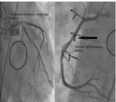
presented to the emergency department with chest pain, dys-pnea, widespread ST depression on electrocardiography, and an international normalized ratio (INR) of 1.3. She was referred for primary percutaneous coronary intervention (PCI) because she was on inotropes, was hypoxic, and in cardiogenic shock. Throm-botic occlusion of the LMCA, left anterior descending (LAD) and circumflex (CX) arteries, and normal right coronary artery (RCA) was observed upon perfroming angiography (Fig. 1).
Emergent echocardiography revealed an ejection fraction of 20% and a normal prosthetic valve; however, the obtained images were suboptimal owing to patient movement and inabil-ity for positioning. Aspirin (100 mg), Clopidogrel (600 mg), and
heparin (7500 IU) was given, and a 6-Fr JL 3.5 guide catheter was inserted through the transfemoral route. LAD and CX were easily wired with floppy guidewire. We first tried aspiration using a 6-Fr Medtronic Export aspiration catheter from LMCA to LAD and later from LMCA to CX; however, the catheter was always clogged with white dense fibrotic thrombus. The flow did not improve despite using the aspiration catheter; however, the thrombus was dispersed into the LAD and Cx. We then bal-looned the LAD and Cx with a 2.5
×
20-mm sized Simpass bal-loon, which restored the flow in both arteries. We observed that the thrombus in the LAD was very dense and not retrievable; hence, we stented the LAD with a Xcience 3.5×
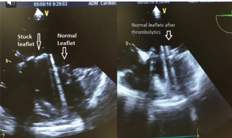
34 mm-sized stent to exclude the thrombus from the lumen. TIMI III flow was obtained in the LAD; however, distal LMCA, LAD ostium, and CX ostium had residual thrombi. We decided upon V stenting of the LMCA–LAD–CX bifurcation and advanced two stents to related vessels; however, suddenly, the thrombus disappeared, and the stents were retrieved back without implantation. The CX distal embolic material was dispersed with floppy guidewire and the procedure was completed (Fig. 2).IABP was inserted, and the patient was followed-up with high doses of dopamin, noradrenalin, and heparin infusions. One mitral leaflet was seen to be thrombosed and stuck in the echocardiography perfromed the next day. Mitral valve reop-eration was assessed as high risk; hence, tissue plasminogen activator (tPA) was given at a dose of 25 mg over 24 hours. The next day, transesophageal echocardiography (TEE) revealed mobile leaflet with residual thrombus. We continued another 25 mg tPA infusion over the next 24 hours. TEE imaging after 2 days revealed a completely functional mitral valve. Four days later, the IABP was removed and the inotropes were weaned. Trans-thoracic echocardiography was repeated, and the mitral leaf-let was found to be stuck again. Another 25 mg of tPA infusion
Figure 1. Massive thrombus in LMCA extending into the LAD and CX os-tia with normal RCA
Figure 2. TIMI III flow in LAD with residual thrombi in distal LMCA, LAD ostium, and CX ostium on the left. Disapperance of the thrombus while pre-paring for V stenting in the middle. Final angiography showing only residual distal Cx embolization at the end of the procedure on the right
Case Reports
Anatol J Cardiol 2018; 20: 363-7
367
over 24 hours was given, and the leaflet movement returned to normal again the next day (Fig. 3). Her pneumonia was treated with antibiotics and she was discharged on aspirin (100 mg), clopidogrel (75 mg), and warfarin (5 mg/day).
Discussion
Coronary embolisation is also seen in PVT, and generally, acute coronary syndrome is considered due to PVT-related embolism (3). Thrombolytic therapy may be considered for both coronary and valve thrombosis in hemodynamically stable patients (4). The superiority of surgery or thrombolytics is not clear due to the head-to-head randomized controlled trial; however, low-dose slow infusion of tPA repeated as needed without a bolus provides effective and safe thrombolysis in patients with prosthetic valve thrombosis (2, 5). In a previous case series, thrombolytic therapy was also successfull in the treatment of combined valve thrombosis and coronary embolism (6).
In our patient, we used the low dose-prolonged infusion protocol instead of the conventional tPa infusion due to bleeding risk. The TROIA study reported successful thrombolysis in 83.2% of cases in low-dose slow infusion without a significant difference between the thrombolytic protocols (5).
Conclusion
Acute thrombotic LMCA occlusion is a catastrophic event, and during intervention, the cardiologist needs to use percuta-neous transluminal coronary angioplasty, thrombus aspiration, different stenting techniques, thrombolytics, and IABP sup-port in different combinations. In LMCA thrombosis, prosthetic valves should be carefully evaluated for the presence and func-tion of thrombus with TEE and fluoroscopy.
References
1. Gupta R, Rahman MA, Uretsky BF, Schwarz ER. Left main coronary artery thrombus: a case series with different outcomes. J Thromb Thrombolysis 2005; 19: 125-31. [CrossRef]
2. Gürsoy MO, Kalçık M, Yesin M, Karakoyun S, Bayam E, Gündüz S, et al. A global perspective on mechanical prosthetic heart valve thrombosis: Diagnostic and therapeutic challenges. Anatol J Car-diol 2016; 16: 980-9.
3. Kaya H, Ertaş F, Tekbaş E, Elbey MA. Resolution of obstructive prosthetic valve thrombosis after coronary embolism. Anatol J Cardiol 2012; 12: E3-4. [CrossRef]
4. Bayar N, Arslan Ş, Köklü E, Kuş G. A case of unusual looking pros-thetic mitral valve thrombosis treated with low dose slow infusion tPA. Anatol J Cardiol 2014; 14: 297-9. [CrossRef]
5. Özkan M, Gündüz S, Biteker M, Astarcioglu MA, Çevik C, Kaynak E, et al. Comparison of different TEE-guided thrombolytic regimens for prosthetic valve thrombosis: the TROIA trial. JACC Cardiovasc Imaging 2013; 6: 206-16. [CrossRef]
6. Kalçık M, Yesin M, Gürsoy MO, Karakoyun S, Özkan M. Manage-ment of Prosthetic Valve Thrombosis Complicated with Coronary Embolism. Heart Lung Circ 2016; 25: 414-5. [CrossRef]
Address for Correspondence: Dr. Ayhan Olcay, İstanbul Aydın Üniversitesi Tıp Fakültesi, Kardiyoloji Anabilim Dalı,
İstanbul-Türkiye Phone: +90 532 527 22 07 E-mail: [email protected]
©Copyright 2018 by Turkish Society of Cardiology - Available online at www.anatoljcardiol.com
DOI:10.14744/AnatolJCardiol.2018.94220
Figure 3. Stuck mitral leaflet on the left and normal leaflet motion after thrombolytics