Acute abdomen and hemorrhagic shock caused by
spontaneous rupture of renal cyst in autosomal dominant
polycystic kidney disease
Autosomal dominant polycystic kidney disease is an important cause of end stage renal failure. Rarely, these patients may present with hemorrhagic shock caused by rupture of the renal cyst. The aim of this study was to report a rare case of a patient who arrived at the emergency department with autosomal dominant polycystic kidney disease presenting with acute abdominal pain and hemorrhagic shock. A 58-year-old male with chronic renal failure was admitted to the emer-gency department with acute abdominal pain and hemorrhagic shock. The patient was admitted to the Department of Surgery with diagnosis of acute abdomen and perirenal hematoma. Although the patient was on conservative treatment, his symptoms did not improve and the patient was operated emergently. During exploration, there was bleeding from the right polycystic kidney, which was 30x20 cm in dimension. The patient underwent nephrectomy and drainage of the he-matoma, and was discharged on the fifth postoperative day without any problems. Bleeding due to rupture of a cyst in au-tosomal dominant polycystic kidney disease occurs rarely but it may be life threatening. Although conservative methods are often preferable in treatment, surgery can be life saving for patients in whom the clinical situation does not improve. Key Words: Autosomal dominant disease, polycystic kidney disease, retroperitoneal hematoma, hemorrhagic shock, acute abdomen
INTRODUCTION
Autosomal dominant polycystic kidney disease (ADPKD) is a systemic disease associated with various renal and non-renal manifestations and is one of the most important reasons of end stage renal failure (1-3). The disease usually presents in the 4th and 5th decades (1, 4). The most common symptoms are flank
pain and hematuria, while life-threatening complications such as bleeding may rarely develop (1, 5, 6). In patients with autosomal dominant polycystic kidney disease, while hemorrhage within the cysts is frequently seen, hematoma surrounding the kidney is a rare (3%) and a dramatic complication (5-8). In patients with renal hematoma, depending on the degree and duration of bleeding hemorrhagic shock may rarely develop (3, 7, 9). Computed tomography (CT) and magnetic resonance imaging are the best diagnostic tools (6, 7). Treatment is usually conservative, invasive procedures such as renal artery embo-lization or nephrectomy is required only in complicated cases (6, 8).
In this study, we aimed to report on a rare case who presented to our clinics with renal hematoma fol-lowing spontaneous cyst rupture and hemorrhagic shock in a patient with ADPKD.
CASE PRESENTATION
A 58-year-old male patient presented to the emergency department with complaints of sudden onset abdominal pain that began 6 hours ago, nausea, vomiting, weakness, and dizziness. His past medical history revealed agenesis of the left kidney, and that he was undergoing hemodialysis for the last 12 years due to chronic renal failure caused by ADPKD. On physical examination, the patient was cachec-tic in appearance, and the abdomen was distended. His skin was pale, cold and moist with an arterial blood pressure of 70/50 mmHg, and a pulse rate of 150/min. On palpation an umbilical hernia and an immobile, irregular bordered mass was found that filled the right hypocondrium, lumbar and inguinal regions. Rebound tenderness and guarding was present in all quadrants, being most prominent in the right hypocondrium. The bladder was catheterized, an 10 cc of urine was drained.
The results of complete blood count, biochemistry and blood gas tests were within normal range except Hgb: 7.2 g/dL, Hematocrit: 21.9%, WBC: 12.06x10³ K/mm3, MCV: 99.8 fL, Urea: 136 mg/dL, Creatinine:
8.8 mg/dL, Potassium: 5.6 mmol/L. The plain abdominal x-ray showed an air-fluid level in the left up-per quadrant. The abdomen ultrasonography revealed the left kidney cannot be visualized, a 50x40 cm mass (suspicious for hematoma) which filled the entire right retroperitoneal area, within the mass the
1Department of General Surgery,
Balıkesir University Faculty of Medicine, Balıkesir, Turkey
2Clinic of General Surgery,
Manisa Merkez Efendi State Hospital, Manisa, Turkey
3Clinic of Pathology, Manisa
Merkez Efendi State Hospital, Manisa, Turkey
Address for Correspondence Dr. İsmail Yaman
Department of General Surgery, Balıkesir University Faculty of Medicine, Balıkesir, Turkey Phone.: +90 266 245 44 25 e-mail: [email protected] Received: 28.08.2011 Accepted: 18.11.2011 ©Copyright 2013 by Turkish Surgical Association Available online at www.ulusalcerrahidergisi.org
İsmail Yaman
1, İsmet Sağlam
2, Kamile Kurt
345
Ulusal Cer Derg 2013; 29: 45-7 DOI: 10.5152/UCD.2013.12
ABSTRACT
Case Report
kidney was seen as 30x20 cm in size containing multiple cysts together with cholelithiasis. Abdominal CT showed diffuse he-matoma in the right retroperitoneum and active bleeding in the perinephritic area.
The patient was hospitalized with a diagnosis of acute abdo-men -perirenal hematoma. The central venous pressure (CVP) was measured as -2 cm H2O. Three units of packed red blood
cells transfusions and fluid replacement was given, the blood pressure was still 90/60 mmHg, and the CVP +1 cm H2O, the
abdominal symptoms did not subside during follow-up. Inter-nal medicine was consulted and it was stated that the patient did not require an emergency dialysis. It was then decided to perform an emergent operation.
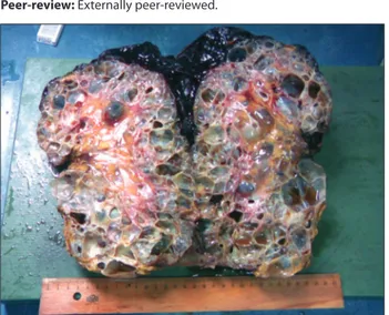
On abdominal exploration, a giant retroperitoneal hematoma of 50x40 cm in size that extended from the inferior of the liver to the pelvis, covering the right retroperitoneum completely, reaching to the transverse mesocolon and gallbladder was de-tected. The right kidney was polycystic and had a hemorrhagic and necrotic appearance with a size of 30x20 cm. The left kidney could not be palpated. The patient underwent retroperitoneal hematoma drainage, right nephrectomy, cholecystectomy, and umbilical hernia repair. In the macroscopic examination a nephrectomy specimen of 29x21x12 cm dimensions, contain-ing numerous cyst, the largest becontain-ing 6 cm. in size, filled with serosanguinous material, with areas of bleeding and necrosis was found (Figure 1, 2). On microscopic examination, benign-appearing cysts lined with single-layered epithelium, a view of thyroidisation and increase in fibrous tissue with inflammatory cell infiltration were present. The patient underwent hemodi-alysis in the postoperative period, and was discharged on the fifth day without any problems.
DISCUSSION
Autosomal dominant polycystic kidney disease is the most common hereditary renal disease and is a major cause of end stage renal failure (2, 10). In Europe and America, approximate-ly 10% of chronic diaapproximate-lysis patients have ADPKD (2, 4). As part of this disease, regarding the kidneys; asymptomatic proteinuria, chronic pain, hypertension, hematuria, cyst infection, rupture, renal failure, renal tumor can be seen together with non-renal pathologies such as liver cysts, intracranial aneurysms, heart valve abnormalities, diverticula and hernias (1, 3, 4). The
dis-ease usually manifest in the 4th and the 5th decades (4). Our
patient was 58 years old, and was undergoing hemodialysis for 12 years for chronic renal failure. In our patient, non-renal involvement except umbilical hernia was not detected. While bleeding into the cysts is frequent, rupture of the cyst is rare (5, 9). Cyst rupture may develop either secondary to trau-ma or infections or trau-may be spontaneous (9). Our patient had no history of trauma, he had a spontaneous cysts rupture that caused hemorrhagic shock.
Cyst rupture is reported to present with different clinical pic-tures depending on the degree and severity of bleeding, rang-ing from chest pain and hematuria to hemorrhagic shock (6 , 7). The treatment is usually by conservative methods including bed rest, blood transfusion and analgesic treatment (6, 8). Sur-gical intervention is reserved for patients in whom hemody-namic instability and acute abdominal findings persist despite conservative methods such as bed rest and blood transfusion (6, 8, 11).
In recent years, with advances in interventional radiology and CT technology in reference centers, conservative treatment with angiography and arterial embolization is recommended (8). The majority of patients respond well to conservative treat-ment (8, 9). In our patient, sudden onset of acute abdomen and signs of hemorrhagic shock was present. The patient was started on medical therapy. Detection of active bleeding in CT, continued hemodynamic instability, persistence of acute abdominal findings despite analgesic therapy were accepted as indications for emergent surgery. Hematoma drainage and right nephrectomy was performed. He was discharged on the fifth postoperative day without any complications.
CONCLUSION
Hemorrhage due to cyst rupture in autosomal dominant poly-cystic kidney disease is a rare but life-threatening condition. Although conservative treatment methods are preferred, surgery can be lifesaving in patients without clinical improve-ment.
Peer-review: Externally peer-reviewed.
Figure 1. Macroscopic view of the specimen Figure 2. Macroscopic view of a section of the specimen
46
Yaman et al.
Author Contributions: Study concept and design - İ.Y.; Acquisition of data - İ.Y., İ.S., K.K.; Analysis and interpretation of data - İ.Y., İ.S., K.K.; Preparation of the manuscript - İ.Y.
Conflict of Interest: No conflict of interest was declared by the authors. Financial Disclosure: The authors declared that this study has re-ceived no financial support.
REFERENCES
1. Fick GM, Johnson AM, Hammond WS, Gabow PA. Causes of death in autosomal dominant polycystic kidney disease. J Am Soc Nephrol 1995; 5: 2048-56.
2. Chijioke A, Aderibigbe A, Olarenwaju TO, Makusidi AM, Oguntoy-inbo AE. Prevalence and pattern of cystic kidney diseases in Ilorin, Nigeria. Saudi J Kidney Dis Transpl 2010; 21: 1172-8.
3. Bajwa ZH, Gupta S, Warfield CA, Steinman TI. Pain management in polycystic kidney disease. Kidney Int 2001; 60: 1631-44.
4. Şen S. Uro-oncology and cystic lesions of the kidney. Üroonkoloji Bülteni 2007; 1: 7-16.
5. Bagon JA. Hemoperitoneum originating in renal cyst in a patient with ADPKD not treated by dialysis. Nephrol Dial Transplant 2000; 15: 251-3.
6. Sirvent AE, Enríquez R, Cabezuelo JB, Ortí C, Amorós F, Reyes A. Autosomal dominant polycystic kidney disease presenting with prolonged macrohaematuria and perinephric haematoma. Nephrol Dial Transplant 1998; 13: 2422-3.
7. Reiter WJ, Haitel A, Heinz-Peer G, Pycha A, Marberger M. Spon-taneous nontraumatic rupture of a contracted kidney with sub-capsular and perirenal hematoma in a patient receiving chronic hemodialysis. Urology 1997; 50: 781-3.
8. Nishikawa Z, Kataoka A, Yuasa T, Okamoto K, Wakabayashi Y, Yo-shiki T, et al. [Renal cell carcinoma in acquired cystic disease of the kidney manifested by spontaneous renal hemorrhage]. Nihon Hinyokika Gakkai Zasshi 2000; 91: 727-30.
9. Tarrass F, Benjelloun M. Acute abdomen caused by spontaneous renal cyst rupture in an ADPKD haemodialysed patient. Nephrol-ogy 2008; 13: 177-80.
10. Altıparmak MR, Seyahi N. Advances in renoprotection in autoso-mal dominant polycystic kidney disease. Turkiye Klinikleri, J Int Med Sci 2005; 1: 40-3.
11. Ishikawa E, Kudo, M, Minami Y, Ueshima K, Chung H, Hayaishi S, et al. Intracystic hemorrhage in a patient of polycystic kidney with renocolic fistula diagnosed by contrast-enhanced ultrasonogra-phy. Inter Med 2008; 47: 1977-9.