Annals of Medical Research
DOI: 10.5455/annalsmedres.2018.06.117 2018;25(4)549-52
Original Article
Comparison of different grafting techniques in open wedge
high tibial osteotomy in terms of bone healing
and functional outcomes
Aydin Arslan
Istanbul Gelisim University, Elit Istanbul Medical Center, Orthopedics and Traumatology Clinic, Istanbul, Turkey Copyright © 2018 by authors and Annals of Medical Research Publishing Inc.
Abstract
Aim: The aim of this study was to compare bone healing and functional outcomes of open wedge high tibial osteotomy (OWHTO)
performed with different grafting techniques.
Material and Methods: From January 2009 to March 2014, 132 patients (145 knees) were included in the study. Lysholm knee scoring
scale was used in preoperative and postoperative clinical evaluation of patients. Patients were followed for at least 3 years. Clinical status of patients, time to bone union and complications were compared.
Results: Autograft was used in 31 knees (29 patients), allograft in 43 knees (39 patients), hydroxyapatite in 71 knees (64 patients),
respectively. The mean duration of bone union was 11.9 ± 3.6 (8-19) weeks in the autograft group, 13.1 ± 3.8 (10-25) weeks in the allograft group and 14.4 ± 4.9 (10-28) weeks in the hydroxyapatite group. The duration of bone union in autograft patients was significantly shorter than that in patients treated with hydroxyapatite. When the groups were evaluated in terms of delayed union, significantly high rates were found in allograft and hydroxyapatite groups when compared to the autograft group. (p<0.05) Significant improvement in clinical scores was observed in all groups postoperatively.
Conclusion: In this study, autograft use was found to be more advantageous than hydroxyapatite and did not differ from allograft in
terms of union time. The use of autograft was more advantageous than the other groups in terms of delayed union. There was no difference between groups with respect to nonunion. Functional and radiographic results were not different.
Keywords: Knee; Osteotomy; Open Wedge; Tibia;Grafting; Bone Union.
Received: 18.06.2018 Accepted: 30.07.2018 Available online: 09.08.2018
Corresponding Author: Aydın Arslan, Istanbul Gelisim University, Elit Istanbul Medical Center, Orthopedics and Traumatology Clinic, Istanbul, Turkey, E-mail: [email protected]
549
INTRODUCTION
Open wedge high tibial osteotomy is used for the treatment of medial compartmental arthritis of the knee (1). Different types of bone void filler and fixation methods have been argued by orthopedic surgeons (2). Autografts, allografts, synthetic bone substitutes (hydroxyapatite, β-tricalcium phosphate) have been used to fill osteotomy gap (2,3). However, it has been reported that osteotomy gaps could consolidate up to a 15 mm gap size without filling (4). Iliac crest autograft is accepted as the most suitable material for OWHTO due to its osteoconductive and osteoinductive features (2,4). However, harvesting autografts from the iliac crest lengthens the operation time and can cause donor site morbidity. It has been reported that minor complications such as superficial
infections, seroma, hematoma and major complications such as herniation of abdominal content, neurovascular injury, deep infections, large hematomas, iliac wing fractures could be caused by graft harvesting from iliac crest (5). Allografts have a risk of transmitting diseases and low osteoinductive features. Besides that, in-tissue degradation and compressive strength of synthetic bone substitutes have been argued by orthopedic surgeons as well (6). The present study was designed to compare bone healing and functional outcomes of OWHTO performed with different grafting techniques.
MATERIALS AND METHODS
176 knees of 161 patients underwent OWHTO operation performed by filling osteotomy gap with autograft, allograft or hydroxyapatite in our clinic from January 2009
to March 2014. 132 patients (145 knees) were included in the study due to the inability to reach 19 patients contact information, death of one patient at the 4th year, recent major surgery of 6 patients and rejection of 3 patients to participate in the study.
<65 year, >90° knee flexion, <15° varus (Hip -Knee-Ankle) can be added as explanatory angle, BMI<30, no osteochondral lesion on lateral femoral condyle and patellofemoral surface were the inclusion criteria for high tibial osteotomy.
Standing long leg roentgenogram and knee MRI were routinely performed in the preoperative period. Routine arthroscopy was performed before osteotomy in the operation. Combinations of two unlocking-wedged two whole plates or one four-hole and one two-hole plates were used. Preoperative planning was performed over standing long leg roentgenograms. Corrections were calculated and height of plate wedges were planned by adjusting HKA angles at about 3° varus. Patients were called for 45th day, 3rd month, 6th month and 1st year and yearly controls. Bone union was confirmed by trabecular and cortical continuity and the clinical disappearance of the pain and recovery of functions. Lysholm knee scoring scale was used in the preoperative and postoperative clinical evaluation of patients. Clinical evaluation was performed preoperatively, postoperatively at year 1, year 3 and at the last follow-up. Patients were followed for at least 3 years. The cases where bone union was not complete in up to 6 months were considered as delayed union. In the present study clinical status of patients, time to bone union and complications were compared.
Preoperative planning was performed over standing long leg roentgenograms. Corrections were calculated and height of plate wedges were planned by adjusting HKA angles at about 3° varus.
Infection prophylaxis was performed using 1gr cefazolin one hour before the operation. Low molecular weight heparin was used for thromboembolism prophylaxis. 40 mg/0.4 ml enoxaparin sodium (Clexane®4000 anti-Xa/ 0.4ml pre-filled syringes, Sanofi Aventis) was given subcutaneously 12 hours after the operation and continued once a day for 3 weeks. Partial weight bearing was allowed at the sixth week control visit. Full weight bearing was planned at the end of third month.
Statistical analyses
Statistical analysis was performed using SPSS for Windows 20 (SPSS, Inc.). Kolmogorov-Smirnov test was used to determine whether the data was normally distributed or not. If the data was normally distributed, differences between the three groups were evaluated using the One Way ANOVA test. And in the case of non-normally distributed data, we used Kruskal-Wallis test to evaluate the differences. Mann Whitney U test was used to compare two groups. With the assumption that α equaled 0.05 and 1-β (power) equaled 0.80 for the power analyses in terms of bone healing time, it was found out thirty
patients were enough for each group. A p-value <0.05 was considered statistically significant.
RESULTS
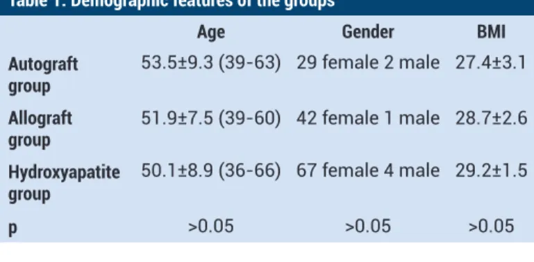
Autograft was used in 31 knees (29 patients), allograft in 43 knees (39 patients), hydroxyapatite in 71 knees (64 patients), respectively. Demographic characteristics of the patients are shown in Table 1 according to the groups. Patients were followed 58.7 ± 16.4 (36-86) months in the autograft group, 56.9 ± 11.4 (36-78) months in the allograft group and 52.3 ± 12.5 (36-70) weeks in the hydroxyapatite group. The mean duration of bone union was 11.9 ± 3.6 (8-19) weeks in the autograft group, 13.1 ± 3.8 (10-25) weeks in the allograft group and 14.4 ± 4.9 (10-28) weeks in the hydroxyapatite group. There was no difference in terms of bone union time between autograft and allograft groups. The duration of bone union in autograft patients was significantly shorter than that in patients treated with hydroxyapatite (Table 2). No delayed union was observed in patients for whom autograft is used. Delayed union was observed in 4 cases in allograft group and 8 cases in hydroxyapatite group. When the groups were evaluated in terms of delayed union, significantly high rates were found in allograft and hydroxyapatite groups when compared to the autograft group(p<0.05). Nonunion was observed in one case from the hydroxyapatite group. This case was revised using iliac crest autograft. The mean value of osteotomy gap size was 11.0 ± 1.9 mm in the autograft group, 10.9 ± 2.1 mm in the allograft group and 10.4 ± 2.3 mm in the hydroxyapatite group. There was no difference in terms of gap sizes between the groups (P> 0.05).
Table 1. Demographic features of the groups
Age Gender BMI
Autograft
group 53.5±9.3 (39-63) 29 female 2 male 27.4±3.1 Allograft
group 51.9±7.5 (39-60) 42 female 1 male 28.7±2.6 Hydroxyapatite
group 50.1±8.9 (36-66) 67 female 4 male 29.2±1.5 p >0.05 >0.05 >0.05
Table 2. p value table between groups in terms of union time
Allograft group Hydroxyapatite group Autograft group p=0.09 p=0.03
Allograft group p=0.1
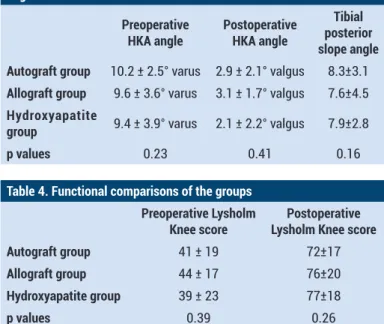
reoperative HKA angle was 10.2 ± 2.5° varus in the autograft group, 9.6 ± 3.6° varus in the allograft group, 9.4 ± 3.9° varus in the hydroxyapatite group, postoperative HKA angles ; 2.9 ± 2.1° valgus in the autograft group and 3.1 ± 1,7° valgus in allograft group, 2,1 ± 2,2° valgus in the hydroxyapatite group. There was no difference between the groups in terms of HKA angle and Tibial posterior slope angle. (Table 3)
The Lysholm Knee score was found to be 41 ± 19 in Ann Med Res 2018;25(4)549-52
autograft group, 44 ± 17 in the allograft group and 39 ± 23 in the hydroxyapatite group preoperatively. Significant improvement in clinical scores was observed in all groups postoperatively. The postoperative clinic scores of the groups are shown comparatively in Table 4. At follow up, Total Knee Arthroplasty was applied to 3 knees in autograft group, 2 knees in allograft group and 8 knees in hydroxyapatite group.
Table 3. p values of the HKA and postoperative tibial posterior slope angles
Preoperative
HKA angle Postoperative HKA angle
Tibial posterior slope angle Autograft group 10.2 ± 2.5° varus 2.9 ± 2.1° valgus 8.3±3.1
Allograft group 9.6 ± 3.6° varus 3.1 ± 1.7° valgus 7.6±4.5
Hydroxyapatite
group 9.4 ± 3.9° varus 2.1 ± 2.2° valgus 7.9±2.8
p values 0.23 0.41 0.16
Table 4. Functional comparisons of the groups
Preoperative Lysholm
Knee score Lysholm Knee score Postoperative
Autograft group 41 ± 19 72±17
Allograft group 44 ± 17 76±20
Hydroxyapatite group 39 ± 23 77±18
p values 0.39 0.26
During the operation, lateral hinge fracture was observed in 2 patients in the autograft group, 5 patients in the allograft group and 4 patients in the hydroxyapatite group. This was stabilized using long screws with the existing system without applying a new plate system. During the follow-up, none of the patients were subjected to revision because of loss of fixation. In the autograft group, 1 case had prolonged serous discharge and 2 cases had superficial infection. In the allograft group 3 cases of prolonged serous discharge and 2 cases of superficial infection occurred. In the hydroxyapatite group 7 cases of prolonged serous discharge and 4 cases of superficial infection were observed. Superficial infections were treated with antibiotic treatment and dressing. None of these groups developed a deep infection requiring revision. There was no difference between groups in terms of lateral hinge fracture and infection rates.
DISCUSSION
Although the best method for filling osteotomy gap of OWHTO is iliac crest autograft, many surgeons prefer to perform operations with allografts or synthetic materials to fill the osteotomy cavity or using techniques applied without filling the osteotomy cavity due to the morbidity in donor site (2,4). Thus, allografts or synthetic materials were used for the majority of patients in our clinic.
Osteotomy gap sizes between 11 and 15 mm were reported as cut-off points in many studies and it was reported that osteotomy cavities above these values should be filled with
autografts (7-12). Slevin et al (4). found no difference in the union rates of autograft, allograft and synthetic grafts in their review. This is what the review study has linked osteotomy cavities to be around or below these cut-off points. In the present study gap sizes in the three groups were on average 10-11 mm. There was no difference in osteotomy gap sizes between the groups. Although there was no difference in terms of union time in autograft and allograft group, there was a difference in terms of union time between hydroxyapatite and autograft groups, with the difference occurring for the autograft group.
In review studies, autografts and allografts were found to be more advantageous than synthetic grafts in terms of delayed union and nonunion (2,4). In this study, there was no delayed union or nonunion in the autograft group. Delayed union was found to be significantly higher in allograft and synthetic graft groups than in the autograft group. Nonunion was observed in one patient in hydroxyapatite group.
Various unlocking plate systems have been used in the majority of operations performed with bone void fillers. However, mostly locking plate options have been preferred in situations where the osteotomy gap is not filled (1). N. J. Lash et al (2). reported that there is no relation between plate preferences and delayed union or nonunion. In this study, the unlocked plate system was used in all patients. When the angular correction is calculated, the main purpose is to adjust the mechanical axis starting from the hip center to pass through the center of the tibiotalar joint (13). Most surgeons do this by adjusting the intersection point of mechanical axis to be 62% lateral to the knee joint surface or adjusting the HKA angle to be 3-6 degrees valgus point (14-16). In the present study, the correction amounts were calculated by adjusting the HKA angle to about 3 degrees valgus. The measured HKA values were compatible with the literature.
Lysholm Knee scoring scale was used for clinical evaluation in this study. There was a statistically significant improvement in the majority of previous studies using the same and different scoring systems (17). In this study, significant improvements were observed in mid and long term, although knee arthroplasty was applied at certain rates in each group.
In literature, lateral hinge fracture incidence is reported with a frequency of 0 to 27% (17-19). In this study, lateral hinge fracture was observed in 2 knees in the autograft group, 5 knees in allograft group and 2 knees in hydroxyapatite group during. This was stabilized using long screws with the existing system without applying a new screw plate system. At follow-up, none of the patients were subjected to revision because of loss of fixation. In the autograft group, two superficial infections and one prolonged serous discharge were observed. In the allograft group, 3 superficial infections and 4 prolonged serous discharges were observed. In the Hydroxyapatite group 5 superficial infections and 9 prolonged serous Ann Med Res 2018;25(4)549-52
discharges were observed. Superficial infections were treated with antibiotic treatment and dressing. In previous studies, rates of infection requiring revision were reported to be 0 to 9% (17-20). In this study, no infection requiring revision occurred. It has been reported that the rate of superficial infection is higher in hydroxyapatite (2). However, there was no difference between the groups in terms of infection rates in the present study.
CONCLUSION
In this study, autograft use was found to be more advantageous than hydroxyapatite and did not differ from allograft in terms of union time. The use of autograft was more advantageous when compared with the other groups in terms of delayed union. There was no difference between the groups with respect to nonunion. Functional and radiographic results were not different.
Financial Disclosure: There are no financial supports
Ethical approval: Institutional ethics committee approval and written consent from the patients were obtained for the study
REFERENCES
1. Jackson JP, Waugh W. Tibial osteotomy for osteoarthritis of the knee. J Bone Joint Surg Br. 1961;43:746-51.
2. Lash NJ, Feller JA, Batty LM, et al. Bone grafts and bone substitutes for opening-wedge osteotomies of the knee: a systematic review. Arthroscopy. 2015;31:720-30.
3. Amendola A, Bonasia DE. Results of high tibial osteotomy: review of the literature. Int Orthop. 2010;34:155-60.
4. Slevin O, Ayeni OR, Hinterwimmer S, et al. The role of bone void fillers in medial opening wedge high tibial osteotomy: a systematic review. Knee Surg Sports Traumatol Arthrosc. 2016;24:3584-98.
5. Arrington ED, Smith WJ, Chambers HG, et al. Complications of iliac crest bone graft harvesting. Clin Orthop Relat Res. 1996;300-9.
6. Aryee S, Imhoff AB, Rose T, et al. Do we need synthetic osteotomy augmentation materials for opening-wedge high tibial osteotomy. Biomaterials. 2008;29:3497-502.
7. El-Assal MA, Khalifa YE, Abdel-Hamid MM, et al.
Opening-wedge high tibial osteotomy without bone graft. Knee Surg Sports Traumatol Arthrosc. 2010;18:961-6.
8. Galla M, Lobenhoffer P. High tibial open wedge valgus osteotomy stabilized with the TomoFix™ plate fixator. Oper Orthop Traumatol 2004;16:397-417
9. Kuremsky MA, Schaller TM, Hall CC, et al. Comparison of autograft vs allograft in opening-wedge high tibial osteotomy. J Arthroplasty. 2010;25:951-7.
10. Lobenhoffer P, Agneskirchner J, Zoch W. Die öffnende valgisierende Osteotomie der proximalen Tibia mit Fixation durch einen medialen Platten fixateur. Orthopade. 2004;33:153-16.
11. Stuart M, Backstein D, Logan M, et al. Osteotomy about the knee: international roundtable discussion. Insall Scott Surg, Knee. 2012. pp. 944-51.
12. Zorzi AR, da Silva HG, Muszkat C, et al. Opening-wedge high tibial osteotomy with and without bone graft. Artif Organs. 2011;35:301-7.
13. Amis AA. Biomechanics of high tibial osteotomy. Knee Surg Sports Traumatol Arthrosc. 2013;21:197-205.
14. Hernigou P, Medevielle D, Debeyre J, et al. Proximal tibial osteotomy for osteoarthritis with varus deformity. A ten to thirteen-year follow-up study. J Bone Joint Surg Am. 1987;69:332-54.
15. Fujisawa Y, Masuhara K, Shiomi S. The effect of high tibial osteotomy on osteoarthritis of the knee. An arthroscopic study of 54 knee joints. Orthop Clin North Am. 1979;10:585-608.
16. Liu-Barba D, Hull ML, Howell SM. Coupled motions under compressive load in intact and ACL-deficient knees: a cadaveric study. J Biomech Eng. 2007;129:818-24.
17. Gouin F, Yaouanc F, Waast D, et al. Open wedge high tibial osteotomies: calcium-phosphate ceramic spacer versus autologous bonegraft. Orthop Traumatol Surg Res. 2010;96:637-45.
18. Jung WH, Chun CW, Lee JH, et al. Comparative study of medial opening-wedge high tibial osteotomy using 2 different implants. Arthroscopy. 2013;29:1063-71.
19. Schroter S, Gonser CE, Konstantinidis L, et.al. High complication rate after biplanar open wedge high tibial osteotomy stabilized with a new spacer plate (Position HTO plate) without bone substitute. Arthroscopy. 2011;27:644-52.
20. Sevimli R. Comparison of medium-period outcomes of allografts and autografts used in repair of bone defects in patients who were treated in our department due to skeletal system tumors. J Turgut Ozal Med Cent. 2017;24:187-9.
Ann Med Res 2018;25(4)549-52