Ankara Üniv Vet Fak Derg, 64, 137-139, 2017
Short Communication / Kısa Bilimsel Çalışma
First isolation of Alternaria alternata from a dog in Turkey
Meriç Lütfi AVSEVER
1, Süleyha HİLMİOĞLU POLAT
2, İlker ÇAMKERTEN
1, Adil AKSOY
11Aksaray University, Eskil Vocation of High School, Laboratory and Veterinary Sciences, Aksaray; 2Ege University, Faculty of
Medicine, Department of Medical Microbiology, İzmir, Turkey.
Summary: Alternaria alternata is a fungus species which can infect animals and people as well as being commonly found in
nature. On the other hand, animals with Alternaria infection can infect other animals and people by spreading a high amount of fungal spores. In this work, Alternaria alternata was detected for the first time in Turkey from the skin lesions of a dog, an antifungal susceptiblity testing was carried out and treatment with itracanozole to which the agent showed susceptibility was accomplished. The aim of this work was to report the Alternaria infection in dogs in Turkey for the first time, to draw attention to the zoonotic dimension of this disease and to emphasize the importance of antifungal susceptibility tests.
Keywords: Alternaria alternata, anti-fungal susceptibility test, dog.
Türkiye’deki bir köpekte ilk Alternaria alternata izolasyonu
Özet: Bir mantar türü olan Alternaria alternata doğada yaygın olarak bulunmakla birlikte insan ve hayvanları enfekte
edebilmektedir. Diğer taraftan Alternaria enfeksiyonu geçiren hayvanlar mantar sporlarını yoğun bir şekilde etrafa bulaştırıp, diğer hayvanları ve insanları enfekte edebilir. Bu çalışmada; Türkiye’de bir köpeğin deri lezyonlarından ilk kez Alternaria alternata infeksiyonu tespit edilmiş, etkene karşı antifungal duyarlılık testi uygulanmış ve duyarlı tespit edilen itraconazole etken maddesi ile tedavi gerçekleştirilmiştir. Çalışmanın amacı, köpeklerde Alternaria enfeksiyonunu ülkemizde ilk kez bildirmek, hastalığın zoonotik yönüne dikkat çekmek ve antifungal duyarlılık testlerinin önemini vurgulamaktır.
Anahtar sözcükler: Alternaria alternata, anti-fungal duyarlılık testi, köpek.
Skin lesions in dogs may develop as a result of allergic, immunologic, hormonal diseases, cancers and nutritional defects as well as parasitic, fungal and bacterial infections (13). Dermatophytes which are keratinophyllic fungi are the cause of dermatomycoses in humans and animals. Dermatophytes such as Microsporum and Trichophyton spp.; yeasts such as Candida spp., Malassezia pachydermatis are the most common causes of dermatomycoses in dogs. Also, molds such as Alternaria spp., Rhizopus spp., Trichoderma spp., Fusarium spp., Aspergillus spp. and Penicillium spp. can be found as saprophytes on skin as well as agents of dermatomycoses (13, 15, 20, 22).
There are more than 80 species in the Alternaria genus. They are commonly found in water, plants and food as well as on the skins of animals and humans as saprophytes (12). They can also be the agent for dermatomoycoses (2, 8). Although Alternariosis is commonly reported as skin Alternariosis in humans, it is also reported to be the cause of hypersensitivity, pneumonitis, bronchial asthma, allergic sinusitis, rhinitis and disseminated infection (4, 8, 21). Animals with
Alternaria infection can contaminate their surroundings with fungal spores and may infect other animals and people (16, 19).
Alternariosis is most commonly reported from cats. In cats, infection is mostly seen as slow growing painless nodules on the face, nose and less commonly on other parts of the body (11, 14, 18). Alternariosisis less commonly reported from dogs as purulant ulcerative lesions with scabs and erythrema located in different parts of the body (6, 23). Alternariosis was very rarely reported from other animals (3, 5). No reports from Turkey on Alternaria infection in dogs were found.
In this work, a ten year old dog kept in a house was presented to the laboratory with severe pruritus and redness on outer skin of auricle, ventral abdomen and joints. In the examinations, lesions with irregular borders and erythrema, scabbing and skin thickening were seen. Lesions were evaluated by parasitologists for mange but were found to be negative.
Skin scrapings and hair samples were taken from the lesions. From a portion of these samples, smears were prepared with 20% KOH and were examined under light
Meriç Lütfi Avsever - Süleyha Hilmioğlu Polat - İlker Çamkerten - Adil Aksoy 138
microscope. Thick walled, septate fungal hyphae were detected in the impression smears. From the remaining samples inoculations were made on Blood Agar (LabM) and Sabouraud Dextrose Agar (SDA) (Oxoid). Blood agar was incubated under 37oC one week and SDA plates were incubated in 25oC for 3 weeks. While there was no growth on Blood Agar, on SDA pale grey colonies with aerial mycelia were grown on the fifth day.
The surface of colonies were initially grey white and then became greyish black with a light border, the reverse of the colonies were dark brown. Lacto-phenol cotton blue mount preparations were prepared from the colonies and septate brown hyphae, and erect, brown, multicellular conidiophores, producing unbranched, club shaped, conidial chains were seen. The conidia had a round base, a short cylindrical beak and muriform septation (Figure 1). With these morphological characteristics the fungus was identified as A. alternata.
Figure 1. The appearence of slide of A. alternaria from a colony grown on SDA plate and stained with lactophenol blue under light microscopy (x40): Conidia specific to A. alternata (arrow 1), (the conidia of Alternaria alternata with a round base, a short, cylindrical beak) and muriform septation (arrow 2).
Şekil 1. SDA plaklarda üreyen A. alternaria kolonisinden laktofenol pamuk mavisi ile boyanan preparatın ışık mikrosko-pundaki görüntüsü (x40): A. alternata’ya özgü konidyumlar (ok 1) Alternaria alternata’nın enine ve boyuna bölmeli ve hife tutunan kısmı yuvarlak tabanlı, kısa, silindirik gaga şeklinde olan konidyumları (ok 2).
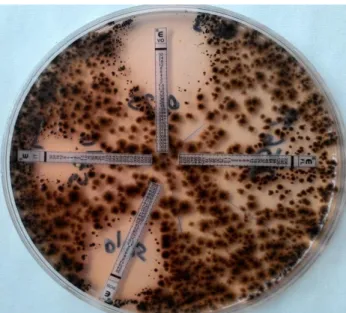
To ensure the diagnosis, skin samples were taken twice with two day intervals and A. alternata was isolated from these samples as well. Antifungal susceptibility test was performed for the isolate on RPMI 1640 medium by E test (AB biodisc) method. The MICs were as follows: Itraconazole, 0.19 μg/mL; voriconazole, 0.19 μg/mL; posaconazole, 0.125 μg/mL (Figure 2). Within the 3 antifungals (itraconazole, voriconazole, posaconazole) to which the agent was susceptible to; itraconazole was chosen for therapy due to its lesser toxicity. Treatment with itraconazole (oral solution) 10 mg/kg was daily started and given for two weeks with marked improvement of the lesions. Then itraconazole was reduced to 5 mg/kg/day and was administered for further two months. At the end of therapy control specimens were taken, and no growth was observed on the cultures.
Figure 2. Antifungal susceptibility testing, MIC values. Itraconazole, 0.19 ug/ml; Voriconazole, 0.19 ug/ ml; Posaconazole 0.125 ug/ml; Fluconazole, 256 ug/ml.
Şekil 2. Antifungal duyarlılık testi, MIK değerleri. Itraconazole, 0.19 ug/ml; Voriconazole, 0.19 ug/ ml; Posaconazole 0.125 ug/ml; Fluconazole, 256 ug/ml.
Despite the world-wide distribution of the genus Alternaria and its ubiquitous presence in the environment, its infections are rare (15). Most of the reported human cases occured either in patients with severe underlying disease or in those receiving immunosuppressive therapy (1, 2). Only rarely healthy patients are affected (24). In animals, most cases are associated with long term exposure to soil and garbage, a penetrating trauma, or subsequent to a course of steroid therapy. The infection usually manifests as a single poorly circumscribed cutaneous nodules or plaques, redness of the skin, crusts, erosions or ulcerations (3, 6, 9, 17). In the present case, the lesions detected on the skin of the auricle, ventral abdomen and on the joints, as reddness, scaling and thickening of the skin, with severe prurutis. No compatible systemic signs were detected, and there was no underlying disease or immunosuppression. Hence, the lesions could be attributed to a multiple minor traumatic injuries, during grooming or hunting outside.
For the diagnosis of fungal infections, fungal hyphae have to be seen and the fungal agent has to be cultivated in the microbiologic examinations of skin and biopsy samples. As biopsy would have required general anaesthesia, this was not carried out, as the owner of the dog rejected the biopsy. To confirm that Alternaria was the aetiologic agent in this case, three consequent specimens were taken from the same lesions and the microscopic findings were the same as for the first specimen, and A. alternata was isolated from each of the specimens.
Ankara Üniv Vet Fak Derg, 64, 2017 139
Diseases caused by melanised fungi are difficult to treat, both in humans and animals. There have been reports of many recurrences of the infections due to Alternaria, especially in cases with underlying diseases (9, 10). Azoles are the most popular treatment choice despite the necessity of surgery in some cases. Itroconazole is the preferred drug within the azoles due to its low toxicity (7, 8, 24).
In this case, itraconazole therapy was applied and was considered to be succesful as the lesions disappered after the treatment and no fungal agents were isolated from the skin scrapings from the same regions. No significant side effects were observed in the dog during treatment and this corresponds with the reports stating that itraconazole is less toxic than others.
As a result in this presentation, Alternaria alternata infection was detected for the first time from a dog in
Turkey, antifungal susceptibility testing was carried out
and treatment with itracanozole was accomplished.
References
1. Arunh DB, Zeluff B (2012): Alternaria alternata infection in a patient with acute promyelotic leukemia. J Med Cas, 3, 23-24.
2. Bras S, Sabino R, Laureano A, et al. (2015): Cutaneous infection by different Alternaria species in a liver transplant recipient. Med Mycol, 8, 1-4.
3. Cabanes FJ, Abarca L, Bragulat MR, et al. (1988): Phaeohyphomycosis caused by Alternaria alternata in a mare. J Med Vet Mycol, 26, 359-365.
4. Chabra V, Rastogi S, Barua M, et al. (2013): Alternaria alternata infection associated osteomyelitis of maxilla: A rare disease entity. Ind J Dent Res, 24, 639-641.
5. Coles BM, Stevens DR, Hunter RL (1978): Equine nodular dermatitis associated with Alternaria tenuis infection. Vet Pathol, 15, 779-780.
6. Dedola C, Stuart AP, Ridyard AE, et al. (2010): Cutaneous Alternaria infectoria infection in a dog in association with therapeutic immunosuppression for the management of immune-mediated haemolytic anaemia.Vet Dermatol, 21, 626-34.
7. Del Palacio A, Gómez-Hernando C, Revenga F, et al. (1996): Cutaneous Alternaria alternata infection successfully treated with itraconazole. Clin Exp Dermatol, 21, 241-243.
8. Demirci M, Baran N, Uzum A, et al. (2015): Cutaneous Alternariasis in a patient with renal transplant. Jundishapur J Microbiol, 8, e19082.
9. Dhein CR, Leathers CW, Padhye AA, et al. (1988): Phaeohyphomycosis caused by Alternaria alternata in a cat. J Am Vet Med Assoc, 193, 1101-1103.
10. Dye C, Johnson EM, Gruffid-Jones TJ (2009): Alternaria species infection in nine domestic cats. J Feline Med Surg, 11, 332-336.
11. Mc Kay JS, Cox CL, Foster AP (2001): Cutaneous Alternariosis in a cat. J Small Animal Practice, 42, 75-78. 12. Moriello KA, DeBoer DJ (1991): Fungal flora of the coat
of pet cats. Am J Vet Res, 52, 602-6
13. Nichita I, Marcu A (2010): The fungal microbiota isolated from cats and dogs. Spasb, 43, 411-4.
14. Outbridge CA, Myers SL, Summerbell RC (1995): Phaeohyphomycosis in a cat. Can Vet J, 36, 629-230. 15. Paixao GC, Sidrim JJC, Campos GMM, et al. (2001):
Dermatophytes and saprobe fungi isolated from dogs and cats in the city of Fortaleza, Brazil. Arg Bras Med Vet Zootec, 53, 568-573.
16. Raza H, Khan RU, Anwar K, et al. (2015): Visceral phaeohyphomycosis caused by Alternaria offering a diagnostic as well as a therapeutic challenge. Saudi J Kidney Dis Tranpl, 26, 339-343.
17. Roosje PJ, de Hoog GS, Koeman JP, et al. (1993): Phaeohyphomycosis in a cat caused by Alternaria infectoria. Mycoses, 36, 451-454.
18. Seyedmousavi S, Guillot J, de Hoog GS (2013): Phaeohyphomycoses, emerging opportunistic diseases in animals. Clin Microbiol Rev, 26, 19-35.
19. Singh B, Denning DW (2012): Allergic bronchopulmonary mycosis due to Alternaria: Case report and review. Med Mycol Case Rep, 1, 20-23.
20. Soomra IH, Shar AH, Soomra FM (2010): Fungal biota of the domestic animals in a city in Pakistan. Pak J Med Sci, 26, 964-967.
21. Stark PC, Celedon JC, Chew GL, et al. (2015): Fungal levels in the home and allergic rhinitis by 5 years of age. Environ Health Perspect, 113, 1405-1409.
22. Stojanov IM, Jaksic SM, Prodanov JZ (2007): Presence and importance of saprophyte fungal organisms on dog skin. Proc Nat Svi, Matica Sprska Novi Sad, 113, 261-265. 23. Subapriya S, Senthil NR, Vairamuthu S, et al. (2015):
Opportunistic fungi as etiologic agents of dermatitis – A case of Alternaria fungal infestation in canines. Int J Livest Res, 5, 24-28.
24. Vandeputte P, Ferrari S, Coste AT (2012): Antifungal resistance and new strategies to control fungal infections. Int J Microbiol, Article ID 713687, 26 pages. Doi:10.1155/2012/713687.
Geliş Tarihi:28.12.2015 / Kabul Tarihi: 22.06.2016
Adress for correspondence:
Dr. Meriç Lütfi AVSEVER
Aksaray University, Eskil Vocation of High School, Laboratory and Veterinary Sciences,
68800, Eskil, Aksaray, Turkey.
Gsm: +90 554 700 61 70 Email: [email protected]