Invasive Fungal Infections in Children with Acute
Lymphoblastic Leukemia: Experience from a
Reference University Hospital in Cappadocia
Ebru YILMAZ1, Arda ERDOGMUS1,2, Alper OZCAN1, Sureyya Burcu GORKEM3,
Ozgur CEYLAN4,5, Kemal DENIZ6, Ekrem UNAL1,7, Mustafa Altay ATALAY2,
Musa KARAKUKCU1, Ayse Nedret KOC2, Turkan PATIROGLU1
1 Erciyes University, Faculty of Medicine, Department of Pediatrics, Division of Pediatric Hematology Oncology, Kayseri 2 Erciyes University, Faculty of Medicine, Department of Medical Microbiology, Kayseri
3 Erciyes University, Faculty of Medicine, Department of Radiology, Division of Pediatric Radiology, Kayseri 4 Erciyes University, Faculty of Medicine, Department of Pediatrics, Division of Pediatric Infectious Disease, Kayseri
5 Baskent University, Faculty of Medicine, Department of Pediatrics, Division of Pediatric Infectious Disease, Adana 6 Erciyes University, Faculty of Medicine, Department of Pathology, Kayseri
7 Erciyes University, Gevher Nesibe Genom and Stem Cell Institution, Genome and Stem Cell Center (GENKOK),
Department of Molecular Biology and Genetic, Kayseri, TURKEY ABSTRACT
Invasive fungal infections (IFI) are an important cause of mortality and morbidity in patients with hematological malignancy. This study aims to investigate the incidence of IFI development, risk factors, the management of the infection in a pediatric patient group followed up with the diagnosis of acute lymphoblastic leukemia (ALL), and to share the experience obtained from a single center. Two hundred forty children monitored with the diagnosis of ALL in the pediatric hematology-oncology department of the Erciyes University Medical Faculty from January 2010 to September 2017 included in the study. A total of 30 (14 females and 14 males) IFI attacks were diag-nosed (12.5%) in the included patients with ALL, two of them having the attacks twice. Candida species were the dominant cause of infection (n= 17) and the rest (n= 8) had invasive Aspergillosis. Nineteen IFI attacks were assessed as proven, 6 as probable, and 5 as possible IFI. The most fungal infection was detected in blood culture (43.3%) followed by pulmonary involvement (40%). The most frequently used diagnostic methods were direct microscopic examination, histological examination, and cultures (66.6%). IFI-related mortality was 20%. IFI continues to be an important problem in pediatric patients with hematologic malignity. The 7 of the observed invasive Aspergillosis developed in non hepafiltered room. Treatment of neutropenic children in hepafiltered rooms decrease the risk of IFI. With careful assessment of the patients bearing risk factors for IFI development, early diagnosis and treatment will reduce morbidity and mortality.
Keywords: Leukemia, Children, Invasive fungal infections
INTRODUCTION
Invasive fungal infections (IFI) is an impor-tant cause of mortality and morbidity in patients with hematological malignancies. Approximately 5-10% of febrile neutropenia attacks results from proven yeast or mould infections.1 The most
im-portant reported risk factors of IFI in children with acute lymphoblastic leukemia (ALL) are the
us-age of steroids, deep and prolonged neutropenia due to the intensive chemotherapy, prolonged us-age of broad spectrum antibiotics, central venous catheters, total parenteral nutrition (TPN), severe mucositis.2-5 Although early diagnosis and
inter-vention reduce the IFI-related mortality, the use of diagnostic methods in children with ALL are lim-ited due to their invasive characteristics.
This study aims to investigate the incidence, risk factors, the management of IFI in children with ALL.
PATIENTS and METHODS
Access was gained retrospectively to the data be-longing to the patients monitored with the diagno-sis of ALL in the pediatric hematology-oncology department of the Erciyes University Medical Faculty from January 2010 to September 2017. Included in the study were the patients who were diagnosed, through clinical, laboratory, and imag-ing methods, to have proven, probable, and pos-sible IFI according to the criteria of the European Organization for Research and Treatment of Can-cer and Mycoses Study Group (EORTC/MSG).6
Patients diagnosed with Pneumocystis jirovecii were excluded from the study. The data transferred from the patients’ files and the hospital’s database and recorded into their standard forms included the patients’ ages, sexes, the risk groups according to Turkish ALL (TRALL) 2000, ALL IC Berlin-Frankfurt-Münster (BFM) 2009 and Interfant 2006 protocols, the results of the laboratory and imaging methods employed used for diagnosis and follow-up of IFI, the development period of IFI, the treat-ment given and its duration, the symptoms emerg-ing duremerg-ing the development period of IFI, absolute neutrophil count (ANC), absence or presence of central venous catheter, feeding with TPN, the state of being monitored in a single room with hepafil-ter, the use of granulocyte colony-stimulating fac-tor (GCSF) and granulocyte suspension, and IFI reactivation.7,8 Ethical permission for a review of
all records was granted by the ethics committee of Erciyes University (Approval Number: 2018/552, Date: 07.11.2018).
Statistical Analyses
The descriptive statistics and quantitative variables using Shapiro-Wilk test were expressed as mean ±standard deviation or median (minimum-maxi-mum) according to whether the distributions were normal or not. Nominal variables, however, were expressed as the number of cases and percentage (%).
RESULTS
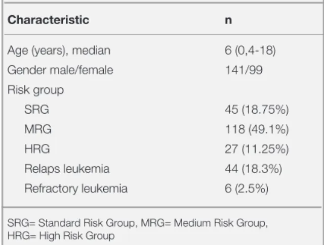
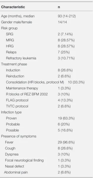
Access was achieved to the data regarding 240 patients (99 female and 141 male) monitored with the diagnosis of ALL. According to TRALL 2000, ALLIC BFM 2009, interfant 2006 treatment proto-cols, 45 of the patients were classified as standard risk group (SRG), 118 were medium risk group (MRG), 27 high risk group (HRG) and 44/6 were relaps/refractory leukemia. The baseline character-istics of the ALL patients are shown in Table 1. With IFI detected twice at different times in 2 pa-tients, a total of 30 (14 female and 14 male) IFI attacks were diagnosed. The median age of the pa-tients was 93 months (14-212 months). The char-acteristics of the 28 patients diagnosed with IFI are shown in Table 2.
While Aspergillus niger was isolated in brain biop-sy of one patient that was previously published.9,10
Aspergillus spp. was isolated in pleural fluid
cul-tures of two patients. Five patients, owing to their general condition being poor, were diagnosed to have Aspergillus spp based on serial galactoman-nan and imaging results. One patient had mucor-mycosis, one patient had Geotrichum capitatum, 4 patients had Candida albicans, and 10 patients had non-albicans Candida. While in one of the 2 patients with fungal proliferation at different times,
C. glabrata and Acremonium strictum proliferated,
in the other, C. glabrata and C. pelliculosa was generated.
Table 1. Characteristics of 240 patients with acute lymphoid
leukemia
Characteristic n
Age (years), median 6 (0,4-18)
Gender male/female 141/99 Risk group SRG 45 (18.75%) MRG 118 (49.1%) HRG 27 (11.25%) Relaps leukemia 44 (18.3%) Refractory leukemia 6 (2.5%)
SRG= Standard Risk Group, MRG= Medium Risk Group, HRG= High Risk Group
Monotherapy (L-AmB, caspofungin, fluconazole or variconazole) was applied to 15 IFI attacks and combined therapy was applied to 15 IFI. Median duration of treatment 30 (21-720) days. In addition to antifungal therapy, 12 patients received granu-locyte suspension (3 patients in addition to mon-otherapy and 9 patients in addition to combined therapy). The patients other than those in induction period and relapsing/refractory ones received 5 mcg/kg/day GCSF.
Nineteen IFI attacks were assessed as proven, 6 as probable, and 5 as possible IFI. The most fungal infection was detected blood culture (43.3%) fol-lowed by in the lungs (40%). The most frequent symptom in 30 IFI attacks was fever (96.6%), and the most frequently employed diagnostic method was conventional mycological examination (direct microscopic examination, histological examina-tion, and culture) (66.6%).
Median ANC was 100/mm3 (0-330 mm3) and the
period when ANC remained less than 500 was 45 days (11-109 days). All of the patients were taking broad spectrum antibiotics that last longer than 7 days. It was detected than only one of 8 patients diagnosed as having Aspergillus diagnosis and as-sessed as proven/probable IFI was hospitalized in the room with hepafilter. Five of the 14 IFI attacks with C. albicans/non-albicans Candida, 8 IFI at-tacks with invasive Aspergillus and 2 IFI atat-tacks with proliferated G. capitatum, A. strictum de-tected had a history of TPN use. All the patients with IFI had central venous catheter, except the one patient in whom C. kefyr was grew from blood cul-ture. The risk factors for developing invasive fun-gal infection are summarized in Table 3.
IFI deterioration occurred in 6 patients despite antifungal therapy, and 3 patients died of isolated fungal sepsis while 3 patients died of leukemia and fungal sepsis. Of the patients who died, 1 had been diagnosed as possible, 2 as probable, and 3 as prov-en IFI. Of our patiprov-ents, 8 died of refractory leuke-mia although they had recovered completely with antifungal therapy (Overall mortality was 46.6%).
Table 2. Characteristics of 28 patients with invasive fungal
infections
Characteristic n
Age (months), median 93 (14-212)
Gender male/female 14/14 Risk group SRG 2 (7.14%) MRG 8 (28.57%) HRG 8 (28.57%) Relaps 7 (25%) Refractory leukemia 3 (10.71%) Treatment phase Induction 8 (26.6%) Reinduction 2 (6.6%) Consolidation (HR blocks, protocol M) 10 (33.3%)
Maintenance therapy 1 (3.3%) R blocks of REZ BFM 2002 3 (10%) FLAG protocol 4 (13.3%) TVTC protocol 2 (6.6%) Infection type Proven 19 (63.3%) Probable 6 (20%) Possible 5 (16.6%) Presence of symptoms Fever 29 (96.6%) Cough 8 (26.6%) Dyspnea 3 (10%)
Focal neurological finding 1 (3.3%)
Nasal defect 1 (3.3%)
Abdominal pain 2 (6.6%)
SRG= Standard Risk Group, MRG= Medium Risk Group,
HRG= High Risk Group, FLAG= fludarabine, high-dose cytarabine, G-CSF, TVTC= Topotecan, Vinorelbine, Thiotepa, Clofarabine
Table 3. Risk factors for developing invasive fungal infection
Risk Factors n (%)
Steroid therapy (>14 days) 10 (33.3%) Severe neutropenia (>10 days) 30 (100%) Antibiotic use (>7 days) 30 (100%) Total parenteral nutrition 15 (50%)
DISCUSSION
Leukemia ranks the first among the cancers of pedi-atric period, and acute leukemia constitutes more than 95% of leukemic cases, and acute lympho-blastic cases constitute 75-80% of acute leukemia cases.11 In recent years, considerable progress has
been made in the treatment of pediatric ALL, and the 5-year survival rate is over % 85. Relapse oc-curs in nearly 20% of ALL cases, and the prognosis of these cases is poor.12 In our study it has been
de-termined that, consistent with the literature, 10% of our patients (25 of 240 patients) received the diag-nosis of T-ALL, and the rate of relapse was 18,3%. The incidence of IFI has recently increased in par-allel with the use of high dose chemotherapy.13
Rosen et al.14 have reported the IFI incidence to
be 4.6% in pediatric patients monitored for solid tumor and leukemia, and deprived of primary anti-fungal prophylaxis. Mor et al.15, on the other hand,
while reporting the IFI incidence in pediatric pa-tients with hematologic disease and solid tumor to be 7.2%, in the same study found the incidence in AML patients to be %39.4. In ALL patients, how-ever, it was established to be 11.2%. In a study in-cluding 125 pediatric patients diagnosed to have ALL, the incidence of IFI has been reported to be 19.2% . In our study it was 12.5%.
Long-term, severe neutropenia, steroid use, cen-tral venous catheter use, mucositis associated with high dosed methotrexate use are reported to be the risk factors in IFI development.2-4 In
addi-tion, in patients with hematologic malignity, even if their ANC is normal functional neutropenia oc-curs, which phagocytosis and pathogens being destroyed.16 Observed in all of our patients were
severe neutropenia lasting longer than 10 days, the use of central venous catheter in 29 IFI attacks, the use of TPN in 15 IFI attacks. Ten patients had to receive steroid longer than 14 days as required by the induction/re-induction protocols.
The most common observed IFI was Candida spe-cies (46.6%) in our patients as previously report-ed.17,18 In the study by Baytan et al.19 which
includ-ed 160 children with hematologic disease, IA was reported by 74% as the most common IFI agent, and hospital renovation activities are reported to have increased the risk. Some studies have argued
that IA is correlated to contaminated ventilation system, hospital construction and renovation.19 In
our study, it was found that IA developed in 1 and 7 of the 8 patients hospitalized with and without hepafilters, respectively.
In the study by Kaya et al.18 C. albicans have been
found more frequently than non-albicans Candida species. However, in our study C. albicans have been detected in 5 (35.7%) and non-albicans
Can-dida species in 9 (64.28%) of 14 patients,
consist-ent with the studies supporting the opposite of the detection in the study of Kaya et al respectively.15,16
The use of histopathologic and/or cytopathologic diagnostic methods are restricted because of their invasive characteristics. Compared with pulmo-nary graph, High-resolution computed tomogra-phy (HRCT) has recently been regarded as a more valuable diagnostic method.20 The data obtained
through radiological means in diagnosing IFI have a greater predictive value in pediatric patients, but they are not specific.21 The most frequently used
diagnostic method for the 30 IFI attacks in our study has been conventional mycological examina-tion (66.6%) followed by computed tomography/ HRCT (30%). A recent retrospective analysis of 139 pediatric invasive aspergillosis cases reported that the most frequent diagnostic radiologic finding was nodules at a rate of 34.6%.22 Nodular
appear-ance has been detected with HRCT in all of our patients.
Follow-up with galactomannan can contribute to early diagnosis and the improvement of clinical findings. However, its sensitivity has been reported to be 64.5% in diagnosing proven IA, 26.4% in probable IA, and 25.5% in possible IA. Herbert et al.23, have reported its specificity as 92.8% in adults
and 47.6% in children. In the same study, the false positivity rate has been found to be in children while it is 0.9% in adults.23 Therefore, they should
be used together with other diagnostic tools, and repetitive tests should be done. In our study it has been detected that while galactomannan positivity had been observed in 6 of the 8 patients diagnosed to have invasive aspergillus, and the diagnosis had been made histopathologically and without check-ing the galactomannan levels in two patients.
In the study by Kaya et al.18 in which 154 pediatric
patients with acute leukemia have been reported and fluconazole has been used as prophylaxis, the incidence has been found to be 13.6%. In our study no patient has received any systemic antifungal prophylaxis other than nystatin used for oral care, and the IFI incidence has been found to be 12.5%. In studies on the recipients of hematopoietic stem cell transplantation and on the patients with hema-tologic malignancy, secondary antifungal prophy-laxis has been found to be effective in preventing IFI from relapsing. However, there are no specific suggestions as to the drugs to be used for secondary prophylaxis. Nevertheless, there are publications suggesting that the pathogen causing the previous IFI and the response to previous antifungal therapy be considered when choosing the medicine.24-27 In
the study by Liu et al.25 on 164 patients, however, no
statistically significant difference in the recurrence rate has been obtained by using drugs and other broad-spectrum antifungal which has been used previously and proven to be effective. Secondary prophylaxis was not administered to patients other than who developed fungemia during the intensive chemotherapy period after complete recovery has been achieved with radiologic fungal antigen and/ or culture results. Despite this, IFI recurrence asso-ciated with the same factor was not observed in any of them. In 8 patients, however, antifungal therapy on end together with chemotherapy was continued until complete remission was achieved. With small number of the patients having been low and kept on antifungal and chemical therapy until complete remission, it would not be right to comment on sec-ondary prophylaxis in the light of this study. In empirical treatment, caspofungin and L-AmB are the first-line agents.4,25 In our center L-AmB
has been used for the first-line agent in empirical treatment. The antifungal drug used most frequent-ly by our patients in our hospital has been L-AmB (73.3%), and it has been used as monotherapy in 9 IFI attacks, and as adjuvant drug in combined therapy in 13 IFI attacks.
The rate of response to voriconazole in cancer pa-tients is over 45%.28 In our study voriconazol has
been used as monotherapy in 5, and as part of a combined therapy in 13 of 18 IFI attacks. The
com-bination of L-AmB and voriconazole was used in 11 patients, L-AmB and caspofungin in 2 patient, and combined voriconazole and caspofungin in 2 patients.
Mortality rate is still high despite advances in new antifungal drugs and supportive care. The IFI-relat-ed mortality in pIFI-relat-ediatric patients with hematologic malignity and aplastic anemia is in 40% range.19
Despite the appropriate treatment, recurrence was observed by 16% following intensive chemothera-py.30 Recurrence was observed in none of our
pa-tients. However in 6 patients progression was ob-served, despite combined antifungal therapy, and three of the patients died of isolated fungal sepsis, and the other three died of refractory leukemia and IFI (IFI-related mortality was 20%).
In this retrospective study, the data and experience that belong to a single center have been shared. Ac-cordingly, there is a need for more comprehensive prospective randomized controlled studies so that risk factors (such as mucositis and neutropenia), risk specific fungal agents and treatment modali-ties (such as combined therapy, primary and sec-ondary prophylaxis) can be identified.
IFI continues to be an important problem in pediat-ric patients with hematologic malignity. With care-ful assessment of the patients bearing risk factors for IFI development, early diagnosis and treatment will reduce morbidity and mortality. New diagnos-tic methods and antifungal treatment choices will contribute to the enhancement of the survival rate in this patient group.
Acknowledgements:
The authors thank to Prof. Dr. Mustafa Kursad OZTURK, Prof. Dr. Mehmet Akif OZDEMIR for their valuable contributions to the study.
REFERENCES
1. Karthaus M, Cornely OA. Recent developments in the man-agement of invasive fungal infections in patients with hemato-logical malignancies. Ann Hematol 84: 207-216, 2005 2. Patiroglu T, Altuner Torun Y, Yikilmaz A, et al. Invasive
Pul-monary Aspergillosis Associated With Pleural Effusion and Pneumothorax. Erciyes Med J 30: 48-51, 2008.
3. Celkan T, Kizilocak H, Evim M, et al. Hepatosplenic Fungal Infections in Children With Leukemia-Risk Factors and Out-come: A Multicentric Study. J Pediatr Hematol Oncol 41: 256-260, 2019.
4. Lehrnbecher T, Phillips R, Alexander S, et al. Guideline for the
management of fever and neutropenia in children with cancer and/or undergoing hematopoietic stem-cell transplantation. J Clin Oncol 30: 4427-4438, 2012.
5. Rüping MJ, Vehreschild JJ, Cornely OA. Patients at high risk of invasive fungal infections: when and how to treat. Drugs. 68: 1941-1962, 2008.
6. Donnelly JP, Chen SC, Kauffman CA, et al. Revision and Up-date of the Consensus Definitions of Invasive Fungal Disease From the European Organization for Research and Treatment of Cancer and the Mycoses Study Group Education and Re-search Consortium. Clin Infect Dis 71: 1367-1376, 2020.
7. Yuksel Soycan L. BFM-TR ALL 2000: first Turkish multicentric
study in the treatment of pediatric acute lymphoblastic leuke-mia. J Pediatr Hematol Oncol 29: 21, 2007.
8. Uderzo C, Dini G, Locatelli F, et al. Treatment of childhood acute lymphoblastic leukemia after the first relapse: curative strategies. Haematologica 85: 47-53, 2000.
9. Patiroglu T, Unal E, Karakukcu M, et al. Multiple fungal brain abscesses in a child with acute lymphoblastic leukemia. My-copathologia 174: 505-509, 2012.
10. Canpolat M, Ceylan O, Per H, et al. Brain abscesses in chil-dren: results of 24 children from a reference center in Central Anatolia, Turkey. J Child Neurol 30: 458-467, 2015. 11. Arora RS, Arora B. Acute leukemia in children: A review of the
current Indian data. South Asian J Cancer 5: 155-160, 2016. 12. Nguyen K, Devidas M, Cheng SC, et al. Factors influencing survival after relapse from acute lymphoblastic leukemia: a Children’s Oncology Group study. Leukemia 22: 2142-2150, 2008.
13. Auberger J, Lass-Flörl C, Ulmer H, et al. Significant alterations in the epidemiology and treatment outcome of invasive fungal infections in patients with hematological malignancies. Int J Hematol 88: 508-515, 2008.
14. Rosen GP, Nielsen K, Glenn S, et al. Invasive fungal infections in pediatric oncology patients: 11-year experience at a single institution. J Pediatr Hematol Oncol 27: 135-140, 2005. 15. Mor M, Gilad G, Kornreich L, et al. Invasive fungal infections
in pediatric oncology. Pediatr Blood Cancer 56: 1092-1097, 2011.
16. Sahbudak Bal Z, Yilmaz Karapinar D, Karadas N, et al. Proven and probable invasive fungal infections in children with acute lymphoblastic leukaemia: results from an university hospital, 2005-2013. Mycoses 58: 225-232, 2015.
17. Hazar V, Karasu GT, Uygun V, et al. Risks and outcomes of invasive fungal infections in pediatric allogeneic hemat-opoietic stem cell transplant recipients receiving fluconazole prophylaxis: a multicenter cohort study by the Turkish
Pedi-atric Bone Marrow Transplantation Study Group. Med Mycol 57: 161-170, 2019.
18. Kaya Z, Gursel T, Kocak U, et al. Invasive fungal infections in pediatric leukemia patients receiving fluconazole prophylaxis. Pediatr Blood Cancer 52: 470-475, 2009.
19. Baytan B, Gunes AM, Celebi S, Gunay Ü. Invasive fungal dis-eases in children with hematologic disorders. Turk J Haema-tol 26: 190-196, 2009.
20. Althoff Souza C, Müller NL, Marchiori E, et al. Pulmonary in-vasive aspergillosis and candidiasis in immunocompromised patients: a comparative study of the high-resolution CT find-ings. J Thorac Imaging 21: 184-189, 2006.
21. Thomas KE, Owens CM, Veys PA, et al. The radiological spectrum of invasive aspergillosis in children: a 10-year re-view. Pediatr Radiol 33: 453-460, 2003.
22. Burgos A, Zaoutis TE, Dvorak CC, et al. Pediatric invasive aspergillosis: a multicenter retrospective analysis of 139 con-temporary cases. Pediatrics 121: e1286-e1294, 2008. 23. Herbrecht R, Letscher-Bru V, Oprea C, et al. Aspergillus
ga-lactomannan detection in the diagnosis of invasive aspergil-losis in cancer patients. J Clin Oncol 20: 1898-1906, 2002. 24. Sav H, Atalay MA, Koc AN, et al. Utility of the Aspergillus
galactomannan antigen testing for neutropenic paediatric pa-tients. Infez Med. 25: 38-44, 2017.
25. Tragiannidis A, Dokos C, Lehrnbecher T, Groll AH. Antifungal chemoprophylaxis in children and adolescents with haemato-logical malignancies and following allogeneic haematopoietic stem cell transplantation: review of the literature and options for clinical practice. Drugs 72: 685-704, 2012.
26. Liu M, Li Y, Zhang Y, et al. Secondary antifungal prophy-laxis in hematological malignancy patients with previous in-vasive fungal disease: a retrospective analysis. PLoS One 9: e115461, 2014.
27. Lee JY, Jung CW, Kim K, Jang JH. Impact of previous in-vasive pulmonary aspergillosis on the outcome of allogeneic hematopoietic stem cell transplantation. Korean J Hematol 47: 255-259, 2012.
28. Walsh TJ, Lutsar I, Driscoll T, et al. Voriconazole in the treat-ment of aspergillosis, scedosporiosis and other invasive fun-gal infections in children. Pediatr Infect Dis J 21: 240-248, 2002.
29. Grigull L, Beier R, Schrauder A, et al. Invasive fungal infec-tions are responsible for one-fifth of the infectious deaths in children with ALL. Mycoses 46: 441-446, 2003.
30. Cornely OA, Böhme A, Reichert D, et al. Risk factors for breakthrough invasive fungal infection during secondary prophylaxis. J Antimicrob Chemother 61: 939-946, 2008.
Correspondence:
Dr. Ekrem UNAL
Erciyes Üniversitesi, Tip Fakültesi, Pediatri Anabilim Dali Pediatri Hematoloji-Onkoloji ve Hematopoietik Kök Hücre Nakli Merkezi
Moleküler Biyoloji ve Genetik Anabilim Dali Gevher Nesibe Genom ve Kök Hucre Enstitusu Genom ve Kök Hücre Merkezi
38010 Melikgazi, KAYSERI / TURKEY Tel: (+90-352) 207 66 66 ext 25475 Fax: (+90-352) 437 58 25 e-mail: [email protected] [email protected] ORCIDs: Ebru Yilmaz 0000-0003-4802-0986 Arda Erdogmus 0000-0003-1895-9132 Alper Ozcan 0000-0002-6100-1205
Sureyya Burcu Gorkem 0000-0001-8949-6476
Ozgur Ceylan 0000-0001-6910-7250
Kemal Deniz 0000-0001-7749-2152
Ekrem Unal 0000-0002-2691-4826
Mustafa Altay Atalay 0000-0003-4169-0637
Musa Karakukcu 0000-0003-2015-3541
Ayse Nedret Koc 0000-0002-1736-9707