Address for correspondence: Dr. Erdem Özel, İzmir Tepecik Eğitim ve Araştırma Hastanesi, Kardiyoloji Kliniği, Güney Mahallesi 1140/1 Sokak No: 1 Yenişehir-Konak 35540, İzmir-Türkiye
Phone: +90 232 469 69 69 E-mail: [email protected] Accepted Date: 26.07.2019 Available Online Date: 15.08.2019
©Copyright 2019 by Turkish Society of Cardiology - Available online at www.anatoljcardiol.com DOI:10.14744/AnatolJCardiol.2019.09633
AUTHORS: Erdem Özel
1, Mustafa Feridun Koşar
2, Emin Evren Özcan
3, Burak Hünük
4,
Taner Ulus
5, Vedat Aytekin
6REVIEWERS: Aylin Yıldırır
7, Bülent Özin
7, İzzet Erdinler
8, Ömer Akyürek
91Department of Cardiology, İzmir Tepecik Training and Research Hospital; İzmir-Turkey
2Department of Cardiology, İstanbul Bakırköy Dr. Sadi Konuk Training and Research Hospital; İstanbul-Turkey 3Department of Cardiology, Faculty of Medicine, İzmir Dokuz Eylül University; İzmir-Turkey
4Department of Cardiology, Faculty of Medicine, İstanbul Yeditepe University; İstanbul-Turkey 5Department of Cardiology, Faculty of Medicine, Eskişehir Osmangazi University; Eskişehir-Turkey
6Department of Cardiology, İstanbul Koç University Hospital; İstanbul-Turkey 7Department of Cardiology, Faculty of Medicine, Başkent University; Ankara-Turkey
8Department of Cardiology, Memorial Ataşehir Hospital; İstanbul-Turkey 9Department of Cardiology, Faculty of Medicine, Ankara University; Ankara-Turkey
Turkish Society of Cardiology consensus report on recommendations
for athletes with high-risk genetic cardiovascular diseases or
implanted cardiac devices
Introduction
Regular exercise has many favorable effects on the cardio-vascular system. However, intense physical activity, especially in a competitive fashion, may become hazardous to genetically vulnerable individuals.
A competitive athlete is one who participates in an orga-nized team or individual sport that requires regular competition against others as a central component, places a high premium on excellence and achievement, and requires some form of sys-tematic (and usually intense) training. Soccer, basketball, run-ning, cycling, and swimming are some examples of competitive sports with high dynamic activity, whereas gymnastics, weight lifting, and sailing are examples of competitive sports with high static activity.
Sudden cardiac death (SCD) in sports and exercise is defined as cardiac arrest occurring during or within 1 hour of exercise or sports-related activity, and genetically transmitted cardiovascu-lar diseases are among the most common pathologies leading to SCD in competitive athletes (1).
Sudden death of a competitive athlete has a mass and unde-sirable effect on the community. Properly diagnosing inherited cardiovascular pathologies, making appropriate recommenda-tions on physical activity in athletic population, and providing close supervision to diseased athletes are the main duties of cardiovascular specialists.
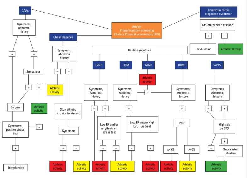
The aim of our document is to summarize the various as-pects of the most common genetic cardiovascular diseases in athletes and to make recommendations for diseased athletes and athletes with implanted cardiac pacemakers/Implantable Cardioverter Defibrillators (ICDs) based on the current literature. Our general recommendations are summarized in Figure 1 and discussed below in detail.
The incidence of SCD in athletic population varies between 0.5 and 13 deaths per 100.000 athletes according to current lit-erature (2, 3).
The most common genetic cardiovascular diseases which leads to SCD varies among different regions of the world. In the United States (US), hypertrophic cardiomyopathy (HCM) is re-sponsible for one-third of the mortality in the athletic population
and congenital coronary anomalies is the second in frequency (4). In the Veneto region of Italy, arrhytmogenic right ventricular cardiomyopathy (ARVC) has been reported as the most common cause of SCD in young athletes (5). Another study which investi-gated the causative pathologies of SCD in athletes with detailed post mortem evaluation found that idiopathic left ventricular hy-pertrophy/fibrosis and ARVC were the most common causes of SCD in athletes (6). The etiology of SCD in competitive athletes involves a wide range of pathologies as listed in Table 1 and dis-cussed below.
Cardiomyopathies
Hypertrophic cardiomyopathy
HCM is an inherited disorder defined by the presence of in-creased left ventricular (LV) wall thickness that cannot be ex-plained by abnormal loading conditions of a cardiac or a system-ic disease. LV wall thsystem-ickness of ≥15 mm in at least one segment measured by an imaging technique is required for the definite
diagnosis of HCM (7). Since HCM has been accepted as one of the major pathology that leads to SCD in young athletes (4, 8), it is of paramount importance to detect the disease before athletic participation to make the distinction between HCM and physi-ological hypertrophy and manage patients during the sporting activity.
Main abnormalities on electrocardiography (ECG) of athletes with HCM are large QRS voltages, T wave inversion, ST depres-sion, and pathological Q waves. On echocardiographic examina-tion, most athletes with HCM had larger LV cavity dimensions with lesser LV hypertrophy and better indices of diastolic func-tion compared to non-athletic HCM patients. The pattern of LV hypertrophy in athletes with HCM is mostly asymmetric and fo-cal in contrast to the symetrifo-cal pattern of physiologifo-cal LV hy-pertrophy of the athlete’s heart. Apical hyhy-pertrophy is also seen in a substantial number of athletes with HCM. Healthy athletes show 10%–20% increase in LV wall thickness. LV wall thickness of 13–16 mm falls into a “gray zone”, and more clinical and
imag-CAAs Symptoms, Abnormal history Symptoms, Abnormal history Symptoms, Abnormal history Symptoms, Abnormal history Symptoms, Abnormal history Symptoms, Abnormal history Stress test Athletic activity Athletic activity Athletic activity Athletic activity Surgery Symptoms, positive stress test Reevaluation Reevaluation Stop athletic activity, treatment Symptoms Athletic
activity Athletic activity Athletic activity Athletic activity Athletic activity Athletic activity Athletic activity Athletic activity Low EF and/or
arrythmia on stress test
Low EF and/or High
LVOT gradient LVEF High risk on EPS
Successfull ablation Channelopaties
Athlete Preparticipation screening (History, Physical examination, ECG)
Cardiomyopathies
Commotio cordis diagnostic evaluation Structural heart disease
LVNC HCM ARVC DCM WPW ≤40% >40% + + + + + + + + + + + + – – – – – – – – – – – – –
Figure 1. Athletic activity recommendations for athletes with genetic cardiovascular pathologies and commotio cordis
For Athletic activity boxes; Red box: Athletic activity prohibited. Yellow box: Athletic activity allowed selectively according to the level of activity and athlete’s condition. Green box: All athletic activities allowed. ARVC - arrythmogenic right ventricular cardiomyopathy, CAAs - coronary artery anomalies, DCM - dilated cardiomyopathy, HCM - hypertrophic cardiomyopathy, LVNC - left ventricular non compaction, WPW - Wolf Parkinson White syndrome
ing data is required to differentiate an athlete’s heart from HCM. Non-sustained ventricular tachycardia (VT) on Holter ECG, late gadolinium enhancement (LGE) in cardiac magnetic resonance imaging (MRI), and genetic testing further help establish the di-agnosis of HCM and discriminate the pathologic condition from the athlete’s heart (9).
Although vigorious physical activity is restricted, the rela-tionship between athletic activities and SCD in HCM is not fully understood. Population based studies showed that the majority of SCDs of HCM patients occurred during routine daily activities and rest or sleep (10, 11). A recently published study examined 35 athletes with a definite diagnosis of HCM. The majority of the athletes had a low risk profile. During 9 years of follow-up, the in-cidence of events and symptoms was not different between ath-letes who continued to exercise and who quit to exercise (12).
Exercise recommendations for athletes with HCM could be challenging. In recent years, trials which investigate the effect of supervised exercise training on patients with HCM found posi-tive effects of exercise on peak oxygen consumption without serious adverse effects (13). In addition, observational studies conducted on athletes with HCM found that high intensity ex-ercise had positive effects on structural and functional cardiac parameters (9, 14).
Patients with HCM are advised to restrict participation in athletic activities according to the AHA/ACC task force report which was published in 2015 (10). The latest ESC report stated that participation in intensive exercise programs and competi-tive sport should be considered on an individual basis. After full evaluation of the disease characteristics and risk determinants,
patients with HCM who have high-risk clinical characteristics must be withheld from athletic activities (15). It is of paramount importance to discriminate the ones who have high-risk features of HCM when restricting athletes from competitive activities.
Non-compaction cardiomyopathy
Left ventricular non-compaction (LVNC) is a genetic car-diomyopathy which is characterized by prominent myocardial trabeculations and deep intertrabecular recesses associated with LV dysfunction. Heart failure symptoms, syncope, systemic thromboembolism, and VT are the main clinical presentations of the patients with LVNC (16). The main symptom was found to be syncope without prodromal symptoms during activity in athletes with LVNC (17). Although LVNC is rarely seen in athletes and accounts for the minority of SCDs in athletic cohorts, differen-tiating the morphological alterations of LVNC from the adaptive changes of an athlete’s heart is an important issue. There is no specific ECG finding for LVNC. LV hypertrophy, repolarization ab-normalities, and QT prolongation were found to be the most com-mon abnormalities on ECG (18). Sustained VT can also be seen in athletes with LVNC (17). Several echocardiographic criteria were identified for making the diagnosis of LVNC. The main findings on echocardiography are ≥2 ratio of non-compacted/compacted layer, presence of deep intertrabecular recesses filling with ven-tricular blood and numerous trabeculations protruding from LV wall with reduced ejection fraction (<50%) (16).
A study which compared the echocardiographic results of elite athletes with normal population and LVNC patients found that 20% of the athlete group expressed an increased number of LV trabeculation and 10% of the athletes had fullfilled the con-ventional echocardiographic criteria of LVNC. Authors stated that this may not be the true incidence of the disease and more stringent criteria for the diagnostic consideration of LVNC are required in this special population (19). According to another study, prominent LV trabeculation was found only in 1.4% of a large athlete population. Although 66% of the athletes who had LV hypertrabeculation match the echocardiographic criteria for LVNC, none of them had LV dysfunction, positive family history, pathologic cardiac MRI findings, and symptoms (20). Thus, ath-letes with hypertrabeculation have to be evaluated carefully and should not be diagnosed directly as LVNC. However, athletes with symptoms and/or high-risk clinical features should be re-stricted from the athletic activities.
Dilated cardiomyopathy
Dilated cardiomyopathy (DCM) is a myocardial disease char-acterized by dilated and hypokinetic LV with or without right ven-tricle dysfunction. Dilated cardiomyopathy may be idiopathic or originating from infection, inflammation, toxic agents or ischemia (21). Occasionally, DCM is a known cause of SCD in athletes (2, 3).
The ECG may be normal or exhibit similar changes to those of athletic individuals such as atrial dilatation, axis deviation or large QRS voltages, and T wave inversion in lateral leads.
Table 1. Etiology of sudden cardiac death in competitive athletes • Cardiomyopathies ¡ Hypertrophic cardiomyopathy ¡ Non-compaction cardiomyopathy ¡ Dilated cardiomyopathy Myocarditis
¡ Arrythmogenic right ventricular cardiomyopathy
• Commotio cordis
• Coronary artery anomalies
• Channelopaties
¡ Long QT syndrome
¡ Short QT syndrome
¡ Brugada syndrome
¡ Catecholaminergic polymorphic ventricular tachycardia
¡ Early repolarization syndrome
• Wolf Parkinson White syndrome
• Idiopathic left ventricular hypertrophy/fibrosis
Consensus statements
Recommendations References Hypertrophic cardiomyopathy
Athletes with HCM who are asymptomatic and do not have significant LVOT gradient could be
supervised closely and may selectively participate in athletic activities. 10, 15
Athletes with HCM who have a history of aborted SCD, exercise-induced ventricular tachycardia, unexplained syncope, significant LVOT gradient, and abnormal blood pressure response to
exercise have to be restricted from athletic acitivities. 10, 15
Genotype positive phenotype negative asymptomatic HCM patients without evidence of LV
hypertrophy by imaging methods may participate in athletic activities. 10, 15
Genotype positive phenotype negative HCM patients should supervise closely to monitor the
progression to hypertrophic phenotype. 10, 15
Non-compaction cardiomyopathy
Athletes who have a diagnosis of LVNC with normal EF, without symptoms and ventricular tachycardias on ambulatory monitoring and stress testing may not be restricted from athletic
activities but close supervision needed. 15, 19, 20
Athletes with LVNC who have symptoms (especially syncope), reduced EF, thromboembolic events, and ventricular tachycardias on ambulatory monitoring or stress testing should be restricted from
the athletic activities. 15, 19, 20
Asymptomatic athletes with hypertrabeculation and without a diagnosis of LVNC can participate in
all competitive sports. 15, 19, 20
Dilated Cardiomyopathy
Asymptomatic athletes with DCM and mildly decreased LV systolic function (EF> 40%), may
selectively participate in athletic activities. 15, 22
In DCM, athletes with symptoms or reduced LV ejection fraction (<40%) or frequent and complex ventricular tachyarrhythmia in ambulatory ECG monitoring or exercise tests or history of
unexplained syncope should not be recommended to deal with athletic acitivities. 15, 22
Myocarditis
If LV function and serum biomarkers of myocardial injury are normalized and no clinically
releveant aryythmia detected on 24 h ECG monitoring, it is reasonable for athletes with myocarditis
to return athletic activities under close supervision after a healing period of 3 to 6 months. 10, 24 Athletes with myocarditis should be followed regularly in case of risk of recurrence and silent
progression of the disease especially during the first 2 years. 10, 24
Athletes with myocarditis should be restricted from athletic activities for a period of 3 to 6 months. 10, 15, 24 Arrhythmogenic right ventricular cardiomyopathy
ARVC patients should not participate in high intensity athletic activities. 15, 27
Genotype positive phenotype negative ARVC patients should not participate in high intensity
athletic activities. 15, 27
ICD implantation in an athlete with ARVC for the sole purpose of participation in high intensity
athletic activity is not recommended. 15, 27
Commotio cordis
Commotio cordis survivors should undergo a complete cardiac study to exclude structural heart
disease and underlying arrythmic condition. 29, 30
After comprehensive evaluation of commotio cordis survivors, athletes without any underlying
Healthy athletes show a 10%–15% increase in both left and right ventricular cavity size. In Olympic athletes, 45% have LV cavity size over the upper limits of normal. Family history and additional ECG changes help establish the diagnosis of DCM. Diastolic dysfunction
and failure of improvement in LV systolic function on exercise echo-cardiography suggests DCM. Additionally, low peak VO2 on cardio-pulmonary testing, non-sustained VT on Holter ECG, LGE on cardiac MRI, and positive genetic testing favor a diagnosis of DCM (22). Consensus statements
Recommendations References Coronary artery anomalies
Athletes with anomalous origin of a coronary artery without either symptoms or positive stress test may be selectively participate in athletic acitivities after counseling with the athletes and/or
parents of the athlete. 15, 36, 43
After successful surgical repair; operated athletes with CAAs may consider to return athletic activities 3 months after surgery if the athlete is asymptomatic and a stress test shows no
evidence of ischemia. 15, 36, 43
Athletes with anomalous origin of a coronary artery which shows an interarterial course should
be restricted from athletic acitivities before surgical repair. 15, 36, 43
Athletes with anomalous origin of a coronary artery who exhibits symptoms or arrythmias or signs of myocardial ischemia in stress tests should be restricted from athletic acitivities before surgical
repair. 15, 36, 43
Channelopathies
Athletes with a suspected cardiac channelopathy should be evaluated by an experienced heart
rhytm specialist. 46, 47, 52
It is advised to perform sports in places with on-board automated-external-defibrillator and near
people who are already informed about the disease for athletes with channelopathy. 46, 47, 52
Asymptomatic athletes with genotype positive phenotype negative channelopathy might be
allowed to participate in all sports with appropriate precautionary measures. 46, 47, 52
It is recommended that symptomatic athletes with any suspected or diagnosed channelopathy should be restricted from all competitive sports until a detailed evaluation has been completed, appropriate treatment has been applied and asymptomatic status on therapy has been provided for 3 months.
46, 47, 52 Drugs which induce a Brugada-pattern on ECGand drugs which prolongs QT interval should be
avoided in athletes with BrS and LQTS respectively. 46, 47, 52
Dehydration, excessive sweating, electrolyte disturbances, and hyperthermia should be avoided
for athletes with channelopathy. 46, 47, 52
Wolff Parkinson White syndrome
Athletes with high-risk characteristics during EP study should undergo RF ablation to retain
athletic eligibility. 52, 69
An athlete may return to athletic acvities after 3 months of successfull ablation procedure in case
of no recurrence of arrythmia. 52, 69
Athletes with implanted cardiac devices
Among athletes with ICD, performing an exercise test to determine the athlete’s upper heart rate
for tachycardia zone programming is recommended. 15, 73
Among the athletes with permanent pacemaker or ICD, only low–moderate intensity athletic
activities except those with risk of bodily collision are recommended. 15, 74
In asymptomatic athletes with Mobitz type 2 or complete AV block without structural heart disease, a deconditioning period up to 2 months is recommended. Persisting or recurring of
symptoms after deconditioning may indicate pacemaker implantation. 15, 73
Asymptomatic athletes with sinus bradycardia or sinus pauses that are secondary to elevated
Presence of symptoms, ejection fraction, and arrhythmic status are the main determinants of an athlete with DCM who desires to participate in athletic activities.
Myocarditis
Myocarditis usually presents with symptoms of exertional dyspnea, chest pain, and arrhythmia. It easily mimics acute coronary syndrome, and coronary angiography is needed for the definite diagnosis (23). Autopsy studies in the series of sud-den deaths in athletes indicate that myocarditis is a significant cause. Among the US military recruits, myocarditis-associated sudden death was found to be the most significant etiology (24).
Acute myocarditis can lead to DCM and can be resolved af-ter a period of time with myocardial scar formation. This condi-tion can lead to arrythmias both during the acute and chronic phases. All physical activities should be restricted during acute myocarditis and exercise recommendations should be tailored after acute phase according to the clinical condition, laboratory, and imaging parameters of an athlete.
Arrhythmogenic right ventricular cardiomyopathy
ARVC is a hereditary myocardial disease caused by muta-tions, especially in genes encoding desmosomic proteins. ARVC is histologically characterized by the loss of myocytes in the right ventricular myocardium and/or LV myocardium with fibrofatty re-placement which results in segmental or diffuse wall thinning (25). The ECG findings of ARVC include most commonly T wave inversion in precordial leads V1 through V3 with a rare finding of Epsilon waves. The left bundle branch patterned ventricular tachyarrhythmia can be observed in ARVC patients (25).
Echocardiography and cardiac MRI may show right ventricu-lar dilation or segmental wall motion abnormalities with morpho-logical alterations. Fatty deposition in the right ventricular wall can be identified with cardiac MRI.
Both ventricular cavities can dilate physiologically in re-sponse to chronic exercise and this situation must be differenti-ated from the pathological conditions of ARVC. Generally, no seg-mental wall motion defects are seen in the physiological state. A positive family history along with ECG and echocardiographic findings may help identify ARVC, and typical findings on cardiac MRI confirms the diagnosis (26).
The most important risk factors in athletes with ARVC include prior history of SCD, sustained VT, or syncope (27).
Most of the SCDs in ARVC patients occur during exercise. Exer-cise itself can both facilitate the natural progression of the disease and lead to lethal arrhytmias. Reducing the intensity of the exer-cise in ARVC patients is associated with lower risk. Since exerexer-cise and SCDs have a definite causative relationship in ARVC, competi-tive athletic activities should be restricted in these patients.
Commotio cordis
Commotio cordis is defined as SCD due to ventricular fibril-lation (VF) triggered by a blunt, non-penetrating blow to the
pre-cordium. Although initially considered extremely rare, it is now accepted as one of the most common causes of SCD in young athletes. Commotio cordis predominantly affects young male athletes (28).
Blows occur on the left chest wall and usually involve im-pact from a hard spherical object, such as a baseball, hockey ball, football, or volleyball. Strong body impacts which can occur during sports like karate and boxing may also lead to commotio cordis. Using soft balls or chest protection instruments during athletic activities can be useful but not absolutely protective (29).
More recently, the Commotio Cordis registry suggested that survival rates have increased steadily over the past 15 years, at >50%. Survival can be improved by earlier recognition of commo-tio cordis, shortening of the time interval from collapse to cardio-pulmonary resuscitation, increased use of automated external defibrillators in the community, and increased number of people receiving training in cardiopulmonary resuscitation (30).
Coronary artery anomalies
The frequency of Coronary Artery Anomalies (CAAs) in the community is mostly learned by autopsies, angiography, and oth-er imaging techniques (31, 32). Even though diffoth-erent results woth-ere also found in a number of angiography series, prevalence of CAAs have been reported between 0.21% and 5.79% in older surviving subjects (33, 34). Autopsy evaluation among military recruits and young athletes who experienced sudden death shows that CAAs are responsible for 17%–44% of cardiovascular mortality (31, 35).
In order of frequency among the general population, CAAs with an abnormal origin might present as the left circumflex ar-tery originating from the right sinus valsalva, a single coronary artery originating from the left sinus valsalva, all coronary arter-ies originating from the right coronary sinus, or the left anterior descending coronary artery (LAD) originating from the right coronary sinus (36). Among athletes who have died suddenly, anomalous origin of LMCA and LAD from the right sinus valsalva is more prevalent. The main clinical problem is the interarterial course of the left main coronary artery, LAD, and right coronary artery. The course of the vessels between the aorta and the pul-monary artery may cause compression of vessels, which in turn could lead to myocardial ischemia, ischemic arrhythmias, and sudden death (37-39).
Anomalous origin of left coronary artery from the pulmonary artery (ALCAPA) is usually an isolated anomaly. ALCAPA consti-tutes 0.22%–0.4% of congenital cardiac anomalies. Most patients die within the first year after delivery, so it is rarely seen among the athletes who have died suddenly (40).
The importance of preparticipation screening, especially in competitive young athletes, is evident in order to reduce mor-tality and morbidity caused by CAAs. Unfortunately, in contrast to congenital arrhythmia syndromes and cardiomyopathies, the diagnostic power of ECG in asymptomatic CAAs is not very high.
Echocardiography is superior to detect the concomitant struc-tural congenital heart disease in athletes with CAAs. An
experi-enced echocardiographer can inform us about the origin of coro-nary ostia with 98% certainity. Transesophageal echocardiography can be performed if the images are not clear enough (41, 42).
Since our aim is to protect a young person from the negative consequences of sport activity, a maximal stress test must be per-formed in the third line after routine ECG and echocardiography in athlete with a possible diagnosis of CAA. If the test result is nega-tive, it will carry him to a better level in terms of risk. If the stress test is suspicious, myocardial perfusion scan or stress echocar-diography will be the best method to evaluate ischemia (43).
Recently, Multi Detector Computerized Tomography (MDCT) data became recognized as an appropriate method to obtain the closest factual results. However, it is not practical to use MDCT as a routine control test due to restrictions related to the usage of X-ray and contrast agent (44, 45). Coronary angiography is indi-cated for definite diagnosis, and intracoronary functional asses-ment may be required.
The only treatment method for CAAs in athletes is surgery. Absence of myocardial ischemia must be demonstrated before returning to athletic activities in operated athletes.
Channelopathies Long QT syndrome
Cardiac ion-channel disorders, also known as “Channelopa-thies”, are inherited primary electrical disorders without “gross” cardiac structural abnormalities. Long QT syndrome (LQTS) is the most frequent within this group of suggestively pure “electrical” diseases, with an approximate prevalence of 1: 2000 (46, 47). The disease is characterized by a prolongation of the QTc interval (heart rate corrected QT) due to the mutations in genes encod-ing for subunits of potassium, sodium, or calcium voltage-depen-dent-ion-channels in the absence of secondary causes (48, 49). It is associated with syncope or SCD due to lethal ventricular arrhytmias (VAs), especially Torsades de Pointes, mainly trig-gered by adrenergic activation. The conditions associated with arrhythmic events are mostly gene-specific, with most arrhyth-mic events occurring during physical or emotional stress in LQT1, at rest or in association with sudden noises in LQT2 patients, and at rest or during sleeping in LQT3 patients (46, 47).
Restriction from virtually all competitive sports has formerly been the guideline-based recommendation since 2005 for ath-letes with any cardiac channelopathy (50, 51). However, starting from the “EHRA Expert Consensus Statement on the Inherited Primary Arrhythmia Syndromes” by Priori et al. (47) and 2015 AHA statement paper (52), expert panels tended to publish more liberal yet evidence-based recommendations (46, 47). ICD im-plantation is recommended in patients with previous SCD and in patients with syncope and/or for sustained VT occurring while receiving
β
blockers (46, 48).In their milestone paper, Johnson et al. (53) sought for the outcomes of the global conventional exercise restriction rules suggested in LQTS. 70 athletes (54%) were competing contrary to European guidelines but within Bethesda guidelines (51, 54).
However, none had a sport-related event. Of the 60 LQTS ath-letes (46%) continuing in sports contrary to both guidelines, only 1 experienced sporting-related events being equal to “1” event in “331 athlete-years”. In a large Italian registry young athletes with LQTS (0.6% of all non-eligible) were disqualified according to the contemporary guideline. During follow-up, no cardiac events in the disqualified athletes were reported. After more than 30 years of screening, the authors observed that only two of the LQTS cases had died suddenly (55). These results revealed that inci-dence of serious cardiac events in athletes with LQTS is lower than expected.
Athletes with LQTS should be supervised closely, and pre-cautionary measures including treatment options should be implemented effectively.
Short QT syndrome
Short QT syndrome (SQTS) is a rare channelopathy charac-terized by a reduced duration of cardiac repolarization building the substrate for the development of lethal arrhythmias (46, 48). Five genes have been linked to SQTS (KCNH2, KCNQ1, KCNJ2, CACNA1C and CACNB2b), but the yield of genetic screening re-mains low (about 20% overall) (56). Resuscitated-SCD might be the first manifestation of the disease with a peak incidence in the first year of life (49, 56). SCD-survivors have a high recurrence rate; therefore, implantation of ICD is strongly recommended in this group of patients with/without quinidine or sotalol (46, 48). SQTS is diagnosed in the presence of a QTc ≤340 ms or QTc ≤360 ms and one or more of the clinical disease features (46). Limited data are available to quantify arrhythmic risk during competitive physical activity as-well-as genotype-phenotype relations in SQTS patients while even “syncope” seems to fail in predicting future events (46, 47). Individuals with SQTS should avoid dehy-dration, protein-supplements, excessive sweating, and hyper-thermia during exercise.
Brugada syndrome
Brugada syndrome (BrS) is characterized by SCD and/or syn-copal events due to VT/VF in young and apparently healthy indi-viduals without significant medical history and with classical ST-segment-elevation-patterns in right precordial ECG leads (46, 47). BrS is inherited as an autosomal-dominant trait, which is more fre-quent in young adults and in men (57). The prevalence ranges from 1/1000 to 1/10 000 (46, 58). Either a decrease in the inward-sodium or calcium current or an increase in the outward-potassium-cur-rents has been shown to be associated with the BrS phenotype. Ventricular-arrhythmia/SCD occurs at a mean 41±15 years, but it usually gets manifest during rest or sleep. ICD implantation is the definitive therapy in BrS patients with aborted SCD or with a history of cardiac-syncope and spontaneous type-1-pattern. The pattern should carefully be distinguished from Brugada-phenocopies, which is challenging in an athlete’s ECG (59).
Data about the probable relation of exercise physiology and BrS is limited and mostly mechanistic rather than prognostic (60, 61).
In a meta-analysis (62) which included anecdotal cases; ST augmentation was observed during the early-recovery-phase of exercise in 57% of patients. There are insufficient data on the risks of exercise in BrS to make a recommendation. According to observations which suggest exercise might worsen the ST ab-normalities in BrS and produce VA, patients with BrS might be restricted from vigorous exercise (60, 61). There is a risk of acti-vation of temperature-dependent mutations at the climax of the exercise and sympathetic withdrawal in BrS patients. However, SCD in BrS occurs most often during sleep (60, 61). Alltogether, this limited evidence might imply an already high-risk subgroup of BrS individuals manifesting their own poor-prognostic fea-tures instead of the detrimental effects of the “exercise physiol-ogy” itself.
Randomized and prospective data is certainly needed in or-der to reveal mechanistic relations and provide firm recommen-dations on cessation of sport participation. Until then, we lack crude evidence to expel all BrS population from exercising, apart from the high-risk-subgroup.
Catecholaminergic polymorphic VT
Catecholaminergic polymorphic VT (CPVT) is a rare, poten-tially life-threatening inherited arrhythmia with an estimated prevalence of 1:10.000 (63). It is diagnosed in the presence of a structurally normal heart, normal ECG, and unexplained exercise- or catecholamine-induced bidirectional VT or poly-morphic ventricular premature beats or VT in an individual <40 years and/or in carriers of a pathogenic mutation in a CPVT-associated gene (47). Patients with CPVT often present with symptoms during the first decade of their life (63). Abnormal storage and release of calcium from the sarcoplasmic reticu-lum are the suggested mechanisms (64).
Intense physical activity has been implicated as a trigger for life-threatening cardiac arrhythmias in patients with CPVT. Exercise at a very low level should be allowed after the approv-al of CPVT experts (46). Appropriate precautionary measures should be taken during the physical activity of an athlete with CPVT including initation of beta blocker therapy, electrolyte/ liquid replacement, avoidance of dehydration, acquisition of a personal automatic external defibrillator, and establishment of an emergency action plan with the appropriate school or team officials (52).
Early repolarization syndrome
Early repolarization (ER) is a common ECG finding character-ized by J-point elevation ≥1 mm ≥2 contiguous leads (47). The ER pattern in the precordial leads has been considered a benign phenomenon, but its presence in the inferior and/or lateral leads has been associated with idiopathic VF and/or polymorphic VT (ER syndrome) (47, 65). Non-anterior ER pattern including the in-ferior subtype is commonly seen in young competitive athletes (ranging from 14% to 44%) (66, 67). Noseworthy et al. (66) showed that both ER and the inferior subtype increased in prevalence
with intense physical training. These data suggest that the ER pattern is a direct result of exercise training. ST-segment mor-phology variants associated with ER may help separate subjects with and without an increased risk of arrhythmic death in middle-aged subjects. Rapidly ascending ST segments after the J-point, which is the dominant ST pattern in healthy athletes, are not as-sociated with an increased risk for arrhythmic death (68).
In conclusion, there is no evidence of an increased risk of SCD in healthy athletes with an ER pattern (63, 67, 68). Competi-tive sports may be allowed for a previously symptomatic athlete with ER syndrome with appropriate precautionary measures (52).
Wolff Parkinson White syndrome
The prevalence of Wolff Parkinson White (WPW) pattern is estimated to be 1–3 in 1000 individuals (69). Approximately 65% of adolescents and 40% of individuals over 30 years with a WPW pattern are estimated to be asymptomatic. Ventricular pre-exci-tation accounts for approximately 1% of SCD in athletes (4). SCD may occur due to the development of atrial fibrillation (AF) with a rapid ventricular response that degenerates to VF (70). The main risk factor for SCD is the presence of an accessory pathway (AP) with short antegrade refractoriness (7).
Radiofrequency (RF) ablation should be performed in patients with WPW syndrome resuscitated from aborted cardiac arrest due to AF and rapid conduction over the AP causing VF (46, 69). If an athlete is symptomatic with syncope or palpitation, an elec-trophysiologic (EP) study is recommended. RF ablation is recom-mended if the refractory period of the AP pathway is ≤240 ms or the shortest preexcited RR interval is <220 ms during induced AF (46, 69). After 3 months of successfull ablation procedure, an asymptomatic athlete may return to athletic activities.
Approximately 65% of adolescents and 40% of individuals over 30 years with a WPW pattern are estimated to be asymp-tomatic (69). According to the 36th Bethesda Conference, an EP
study is recommended for asymptomatic athletes if they par-ticipate in moderate- or high-level competitive sports (54). The ESC mandates that all athletes with WPW undergo an EP study for risk assesment (52). Asymptomatic patients with a short preexcited RR interval ≤250 ms in AF or <220 ms during stress or isoproterenol, the refractory period of the AP ≤240 ms, pres-ence of multiple APs, or easily induced AF are at increased risk for SCD (47, 69). Athletes with high-risk characteristics men-tioned above during EP study should undergo RF ablation to re-tain athletic eligibility. In addition, asymptomatic patients with WPW and structural heart disease or ventricular dysfunction secondary to dyssynchoronous contractions may be consid-ered for ablation regardless of the antegrade characteristics of the AP (69).
Athletes with implanted cardiac devices Athletes with permanent cardiac pacemakers
Highly-trained endurance athletes have dominant parasym-pathetic tone at rest associated with marked sinus bradycardia,
first degree and Mobitz type I atrioventricular block. Mostly these findings are physiological events that do not require intervention (71). Underlying structural heart disease should be excluded in case of Mobitz type 2 atrioventricular block or third-degree atrio-ventricular block (72).
Potential risk for device and lead damage may limit the profes-sional life of athletes. The 36th Bethesda Recommendations state
that PM dependent athletes should not participate in sports that can involve bodily trauma (1). Although not addressed in formal recommendations, protection with padding might be considered. Programing upper tracking rates at higher levels is important in athletes with complete heart block. Heart rates of the patient during vigorous exercise should be considered. Myopotential inhibition may lead to inhibition of pacing, which is of concern in pacemaker-dependent patients. Therefore, bipolar leads should be selected in athletes.
Athletes with ICD
It should be kept in mind that ICDs do not prevent the occur-rence of ventricular arrhythmias and do not effect the progres-sion of the underlying disease. Therefore, careful consideration of the underlying disease is mandatory before participation in competitive sports (15).
However, a long-term prospective multinational registry pro-vides promising data about this topic. After a mean follow-up period of 44 months of 440 patients, there were no arrhythmic deaths, externally resuscitated tachyarrhythmias during sports participation, or injury resulting from arrhythmia-related syncope or shock during sports (73). 31 definite and 13 possible lead mal-functions were reported. The estimated lead survival free of defi-nite plus possible malfunction was 94% at 5 years and 85% at 10 years. No generator malfunction was reported.
On the other hand, approximately one in five received both appropriate and inappropriate shocks, which mainly occurred during competition or physical activity. Therefore, programing an ICD of an athlete is always challenging due to the risk of inap-propriate shocks from high heart rates during exercise. Recent-ly, the role of ICD programming characteristics on occurrence of shocks, transient loss of consciousness, and death among athletes was assessed by prospective, observational, interna-tional registry (74). High-rate cutoff and long-detection duration programming in athletes was associated with reduction in total and inappropriate ICD shocks without affecting survival or the incidence of transient loss of consciousness. Since it is not easy to recommend certain tachycardia detection zones, patient-tai-lored programming seems a better approach.
The potential risks associated with mechanical trauma are possible for ICDs. The athlete’s ability to participate in sports should be discussed individually. Underlying cardiovascular dis-ease, type and the programing of the device, type of the sport, risk for trauma, and risks related to potential syncope or shock should be considered.
Conclusion
Specific return-to-play protocols should be developed for competitive athletes following treatment of various cardiovascu-lar conditions like cathater ablation, cardiac device implantation, and corrective surgeries.
Athletic activities performed in a competitive fashion could have hazardous effects on the cardiovascular health of the athletes. Athletes who have high-risk genetic cardiovascular diseases and implanted cardiac devices should be closely su-pervised by sports cardiologists, exercise specialists, and their personal trainers in a deep collaboration with the guidance of professional and scientific recommendations.
Conflict of interest: None declared.
Peer-review: Externally and internally peer-reviewed.
Authorship contributions: Concept – E.Ö., M.F.K.; Design – E.Ö., M.F.K.; Supervision – E.E.Ö., B.H.; Fundings – E.E.Ö., B.H., T.U.; Materi-als – E.Ö., E.E.Ö., B.H.; Data collection &/or processing – E.E.Ö., B.H., V.A.; Analysis &/or interpretation – T.U., V.A.; Literature search – E.Ö., V.A.; Writing – E.Ö., M.F.K., E.E.Ö., B.H., T.U., V.A.; Critical review – E.Ö., T.U., V.A.
References
1. Maron BJ, Zipes DP. Introduction: eligibility recommendations for competitive athletes with cardiovascular abnormalities-general considerations. J Am Coll Cardiol 2005; 45: 1318–21. [CrossRef]
2. Harmon KG, Asif IM, Klossner D, Drezner JA. Incidence of sudden cardiac death in National Collegiate Athletic Association athletes. Circulation 2011; 123: 1594-600. [CrossRef]
3. Malhotra A, Dhutia H, Finocchiaro G, Gati S, Beasley I, Clift P, et al. Outcomes of Cardiac Screening in Adolescent Soccer Players. N Engl J Med 2018; 379: 524-34. [CrossRef]
4. Maron BJ, Doerer JJ, Haas TS, Tierney DM, Mueller FO. Sudden deaths in young competitive athletes: analysis of 1866 deaths in the United States, 1980-2006. Circulation 2009; 119: 1085-92. [CrossRef]
5. Corrado D, Basso C, Rizzoli G, Schiavon M, Thiene G. Does sports activity enhance the risk of sudden death in adolescents and young adults? J Am Coll Cardiol 2003; 42: 1959-63. [CrossRef]
6. Finocchiaro G, Papadakis M, Robertus JL, Dhutia H, Steriotis AK, Tome M, et al. Etiology of Sudden Death in Sports: Insights From a United Kingdom Regional Registry. J Am Coll Cardiol 2016; 67: 2108-15. 7. Authors/Task Force members, Elliott PM, Anastasakis A, Borger
MA, Borggrefe M, Cecchi F, et al. 2014 ESC Guidelines on diagnosis and management of hypertrophic cardiomyopathy: the Task Force for the Diagnosis and Management of Hypertrophic Cardiomyopa-thy of the European Society of Cardiology (ESC). Eur Heart J 2014; 35: 2733-79. [CrossRef]
8. Peterson DF, Siebert DM, Kucera KL, Thomas LC, Maleszewski JJ, Lopez-Anderson M, et al. Etiology of Sudden Cardiac Arrest and Death in US Competitive Athletes: A 2-Year Prospective Surveillance Study. Clin J Sport Med 2018; doi: 10.1097/JSM.0000000000000598. [Epub ahead of print] [CrossRef]
9. Sheikh N, Papadakis M, Schnell F, Panoulas V, Malhotra A, Wilson M, et al. Clinical Profile of Athletes With Hypertrophic Cardiomy-opathy. Circ Cardiovasc Imaging 2015; 8: e003454. [CrossRef]
10. Maron BJ, Udelson JE, Bonow RO, Nishimura RA, Ackerman MJ, Estes NA 3rd, et al.; American Heart Association Electrocardiogra-phy and Arrhythmias Committee of Council on Clinical Cardiology, Council on Cardiovascular Disease in Young, Council on Cardio-vascular and Stroke Nursing, Council on Functional Genomics and Translational Biology, and American College of Cardiology. Eligibil-ity and Disqualification Recommendations for Competitive Athletes with Cardiovascular Abnormalities: Task Force 3: Hypertrophic Car-diomyopathy, Arrhythmogenic Right Ventricular Cardiomyopathy and Other Cardiomyopathies, and Myocarditis: A Scientific State-ment From the American Heart Association and American College of Cardiology. Circulation 2015; 132: e273-80. [CrossRef]
11. Aro AL, Nair SG, Reinier K, Jayaraman R, Stecker EC, Uy-Evanado A et al. Population Burden of Sudden Death Associated With Hyper-trophic Cardiomyopathy. Circulation 2017; 136: 1665-7. [CrossRef]
12. Pelliccia A, Lemme E, Maestrini V, Di Paolo FM, Pisicchio C, Di Gioia G, et al. Does Sport Participation Worsen the Clinical Course of Hypertrophic Cardiomyopathy? Clinical Outcome of Hypertrophic Cardiomyopathy in Athletes. Circulation 2018; 137: 531-3. [CrossRef]
13. Saberi S, Wheeler M, Bragg-Gresham J, Hornsby W, Agarwal PP, Attili A, et al. Effect of Moderate-Intensity Exercise Training onPeak Oxygen Consumption in Patients With Hypertrophic Cardiomyopa-thy: A Randomized Clinical Trial. JAMA 2017; 317: 1349-57. [CrossRef]
14. Dejgaard LA, Haland TF, Lie OH, Ribe M, Bjune T, Leren IS, et al. Vig-orous exercise in patients with hypertrophic cardiomyopathy. Int J Cardiol 2018; 250: 157-63. [CrossRef]
15. Pelliccia A, Solberg EE, Papadakis M, Adami PE, Biffi A, Caselli S, et al. Recommendations for participation in competitive and lei-sure time sport in athletes with cardiomyopathies, myocarditis, and pericarditis:position statement of the Sport Cardiology Section of the European Association of Preventive Cardiology (EAPC). Eur Heart J 2019; 40: 19-33. [CrossRef]
16. Towbin JA, Lorts A, Jefferies JL. Left ventricular non-compaction cardiomyopathy. Lancet 2015; 386: 813-25. [CrossRef]
17. Ganga HV, Thompson PD. Sports Participation in non-compaction cardiomyopathy: a systematic review. Br J Sports Med 2014; 48: 1466-71. [CrossRef]
18. Steffel J, Kobza R, Oechslin E, Jenni R, Duru F. Electrocardiographic characteristics at initial diagnosis in patients with isolated left ven-tricular non compaction. Am J Cardiol 2009; 104: 984-9. [CrossRef]
19. Gati S, Chandra N, Bennett RL, Reed M, Kervio G, Panoulas VF, et al. Increased left ventricular trabeculation in highly trained athletes: do we need more stringent criteria for the diagnosis of left ventricu-lar non-compaction in athletes? Heart 2013; 99: 401-8. [CrossRef]
20. Caselli S, Ferreira D, Kanawati E, Di Paolo F, Pisicchio C, Attenhofer Jost C, et al. Prominent left ventricular trabeculations in competitive athletes: A proposal for risk stratification and management. Int J Cardiol 2016; 223: 590-5. [CrossRef]
21. Pinto YM, Elliott PM, Arbustini E, Adler Y, Anastasakis A, Böhm M, et al. Proposal for a revised definition of dilated cardiomyopathy, hypokinetic non-dilated cardiomyopathy, and its implications for clinical practice: a position statement of the ESC working group on myocardial and pericardial diseases. Eur Heart J 2016; 37: 1850-8. [CrossRef]
22. Pelliccia A, Culasso F, Di Paolo FM, Maron BJ. Physiologic left ven-tricular cavity dilatation in elite athletes. Ann Intern Med 1999; 130: 23-31. [CrossRef]
23. Sinagra G, Anzini M, Pereira NL, Bussani R, Finocchiaro G, Bartunek J, et al. Myocarditis in Clinical Practice. Mayo Clin Proc 2016; 91: 1256-66. [CrossRef]
24. Phillips M, Robinowitz M, Higgins JR, Boran KJ, Reed T, Virmani R. Sudden cardiac death in Air Force recruits. A 20-year review. JAMA 1986; 256: 2696-9. [CrossRef]
25. Corrado D, Link MS, Calkins H. Arrhythmogenic Right Ventricular Cardiomyopathy. N Engl J Med 2017; 376: 61-72. [CrossRef]
26. Bauce B, Frigo G, Benini G, Michieli P, Basso C, Folino AF, et al. Dif-ferences and similarities between arrhythmogenic right ventricular cardiomyopathy and athlete’s heart adaptations. Br J Sports Med 2010; 44: 148-54. [CrossRef]
27. Calkins H, Corrado D, Marcus F. Risk Stratification in Arrhythmo-genic Right Ventricular Cardiomyopathy. Circulation 2017; 136: 2068-82. [CrossRef]
28. Maron BJ, Estes NA 3rd. Commotio cordis. N Engl J Med 2010; 362: 917-27. [CrossRef]
29. Maron BJ, Gohman TE, Kyle SB, Estes NA 3rd, Link MS. Clinical pro-file and spectrum of commotio cordis. JAMA 2002; 287: 1142-6. 30. Maron BJ, Haas TS, Ahluwalia A, Garberich RF, Estes NA 3rd, Link
MS. Increasing survival rate from commotio cordis. Heart Rhythm 2013; 10: 219-23. [CrossRef]
31. Alexander RW, Griffith GC. Anomalies of the coronary arteries and their clinical significance. Circulation 1956; 14: 800-5. [CrossRef]
32. Eckart RE, Scoville SL, Campbell CL, Shry EA, Stajduhar KC, Potter RN, et al. Sudden death in young adults: a 25-year review of autop-sies in military recruits. Ann Intern Med 2004; 141: 829-34. [CrossRef]
33. Temel MT, Coşkun ME, Başpınar O, Demiryürek AT. Prevalence and characteristics of coronary artery anomalies in children with con-genital heart disease diagnosed with coronary angiography. Turk Kardiyol Dern Ars 2017; 45: 527-32. [CrossRef]
34. Tuo G, Marasini M, Brunelli C, Zannini L, Balbi M. Incidence and clini-cal relevance of primary congenital anomalies of the coronary arter-ies in children and adults. Cardiol Young 2013; 23: 381-6. [CrossRef]
35. Thompson PD. Preparticipation screening of competitive athletes: seeking simple solutions to a complex problem. Circulation 2009; 119: 1072-4. [CrossRef]
36. Brothers J, Gaynor JW, Paridon S, Lorber R, Jacobs M. Anomalous aortic origin of a coronary artery with an inter arterial course: un-derstanding current management strategies in children and young adults. Pediatr Cardiol 2009; 30: 911-21. [CrossRef]
37. Cheitlin MD, De Castro CM, McAllister HA. Sudden death as a com-plication of anomalous left coronary origin from the anterior sinus of Valsalva, A not-so-minor congenital anomaly. Circulation 1974; 50: 780-7. [CrossRef]
38. Barth CW 3rd, Roberts WC. Left main coronary artery originating from the right sinus of Valsalva and coursing between the aorta and pulmonary trunk. J Am Coll Cardiol 1986; 7: 366-73. [CrossRef]
39. Yildiz O, Karabay KO, Akman C, Aytekin V. Anomalous origin of the left main coronary artery from the right coronary artery with a preaortic course. Tex Heart Inst J 2015; 42: 243-5. [CrossRef]
40. Fendrychová V, Ondrášek J, Zatočil T, Poloczek M, Bohatá S. Anom-alous left coronary artery from the pulmonary artery: A rare case diagnosed in an adult. Cor et Vasa 2018; 60: e412-7. [CrossRef]
41. Pelliccia A, Spataro A, Maron BJ. Prospective echocardiographic screening for coronary artery anomalies in 1,360 elite competitive athletes. Am J Cardiol 1993; 72: 978-9. [CrossRef]
42. Cohen MS, Herlong RJ, Silverman NH. Echocardiographic imaging of anomalous origin of the coronary arteries. Cardiol Young 2010; 20 (Suppl 3): 26-34. [CrossRef]
43. Mery CM, Lawrence SM, Krishnamurthy R, Sexson-Tejtel SK, Car-berry KE, McKenzie ED, et al. Anomalous aortic origin of a coronary artery: toward a standardized approach. Semin Thorac Cardiovasc Surg 2014; 26: 110-22. [CrossRef]
44. Bluemke DA, Achenbach S, Budoff M, Gerber TC, Gersh B, Hillis LD, et al. Noninvasive coronary artery imaging: magnetic resonance angiography and multidetector computed tomography angiography: a scientific statement from the American Heart Association com-mittee on cardiovascular imaging and intervention of the council on cardiovascular radiology and intervention, and the councils on clinical cardiology and cardiovascular disease in the young. Circu-lation 2008; 118: 586-606. [CrossRef]
45. Srinivasan KG, Gaikwad A, Kannan BR, Ritesh K, Ushanandini KP. Congenital coronary artery anomalies: diagnosis with 64 slice multidetector row computed tomography coronary angiography: a single-centre study. J Med Imaging Radiat Oncol 2008; 52: 148-54. 46. Priori SG, Blomström-Lundqvist C, Mazzanti A, Blom N, Borggrefe
M, Camm J, et al.; ESC Scientific Document Group. 2015 ESC Guide-lines for the management of patients with ventricular arrhythmias and the prevention of sudden cardiac death: The Task Force for the Management of Patients with Ventricular Arrhythmias and the Pre-vention of Sudden Cardiac Death of the European Society of Cardi-ology (ESC). Endorsed by: Association for European Paediatric and Congenital Cardiology (AEPC). Eur Heart J 2015; 36: 2793-867. 47. Priori SG, Wilde AA, Horie M, Cho Y, Behr ER, Berul C, et al. HRS/
EHRA/APHRS expert consensus statement on the diagnosis and management of patients with inherited primary arrhythmia syn-dromes: document endorsed by HRS, EHRA, and APHRS in May 2013 and by ACCF, AHA, PACES, and AEPC in June 2013. Hear Rhythm 2013; 10: 1932-63. [CrossRef]
48. Al-Khatib SM, Stevenson WG, Ackerman MJ, Bryant WJ, Callans DJ, Curtis AB, et al. 2017 AHA/ACC/HRS guideline for management of patients with ventricular arrhythmias and the prevention of sud-den cardiac death: Executive summary: A Report of the American College of Cardiology/American Heart Association Task Force on Clinical Practice Guidelines and the Heart Rhythm Society. Heart Rhythm 2018; 15: e190-252.
49. Priori SG, Schwartz PJ, Napolitano C, Bloise R, Ronchetti E, Grillo M, et al. Risk stratification in the long-QT syndrome. N Engl J Med 2003; 348: 1866-74. [CrossRef]
50. Zipes DP, Ackerman MJ, Estes NAM, Grant AO, Myerburg RJ, Van Hare G. Task Force 7: Arrhythmias. J Am Coll Cardiol 2005; 45: 1354-63. 51. Pelliccia A, Fagard R, Bjørnstad HH, Anastassakis A, Arbustini E,
Assanelli D, et al.; Study Group of Sports Cardiology of the ing Group of Cardiac Rehabilitation and Exercise Physiology; Work-ing Group of Myocardial and Pericardial Diseases of the European Society of Cardiology. Recommendations for competitive sports participation in athletes with cardiovascular disease: a consensus document from the Study Group of Sports Cardiology of the Work-ing Group of Cardiac Rehabilitation and Exercise Physiology and the Working Group of Myocardial and Pericardial Diseases of the European Society of Cardiology. Eur Heart J 2005; 26: 1422-45. 52. Ackerman MJ, Zipes DP, Kovacs RJ, Maron BJ; American Heart
Association Electrocardiography and Arrhythmias Committee of Council on Clinical Cardiology, Council on Cardiovascular Disease in Young, Council on Cardiovascular and Stroke Nursing, Council on Functional Genomics and Translational Biology, and American College of Cardiology. Eligibility and Disqualification Recommenda-tions for Competitive Athletes With Cardiovascular Abnormalities: Task Force 10: The Cardiac Channelopathies: A Scientific
State-ment From the American Heart Association and American College of Cardiology. Circulation 2015; 132: e326-9. [CrossRef]
53. Johnson JN, Ackerman MJ. Competitive sports participation in ath-letes with congenital long QT syndrome. JAMA 2012; 308: 764-5. 54. Pelliccia A, Zipes DP, Maron BJ. Bethesda Conference #36 and the
European Society of Cardiology Consensus Recommendations re-visited a comparison of U.S. and European criteria for eligibility and disqualification of competitive athletes with cardiovascular abnor-malities. J Am Coll Cardiol 2008; 52: 1990-6. [CrossRef]
55. Corrado D, Basso C, Pavei A, Michieli P, Schiavon M, Thiene G. Trends in sudden cardiovascular death in young competitive ath-letes after implementation of a preparticipation screening program. JAMA 2006; 296: 1593-601. [CrossRef]
56. Mazzanti A, Kanthan A, Monteforte N, Memmi M, Bloise R, Novelli V, et al. Novel insight into the natural history of short QT syndrome. J Am Coll Cardiol 2014; 63: 1300-8. [CrossRef]
57. Gehi AK, Duong TD, Metz LD, Gomes JA, Mehta D. Risk stratification of individuals with the Brugada electrocardiogram: a meta-analy-sis. J Cardiovasc Electrophysiol 2006; 17: 577-83. [CrossRef]
58. Antzelevitch C, Brugada P, Borggrefe M, Brugada J, Brugada R, Cor-rado D, et al. Brugada syndrome: report of the second consensus conference: endorsed by the Heart Rhythm Society and the Euro-pean Heart Rhythm Association. Circulation 2005; 111: 659-70. 59. Gottschalk BH, Anselm DD, Baranchuk A. Brugada phenocopies are
the leading differential diagnosis of Brugada syndrome. Clin Med (Lond) 2015; 15: 308-9. [CrossRef]
60. Antzelevitch C. J wave syndromes: molecular and cellular mecha-nisms. J Electrocardiol 2013; 46: 510-8. [CrossRef]
61. Antzelevitch C, Brugada P, Borggrefe M, Brugada J, Brugada R, Cor-rado D, et al. Brugada syndrome: report of the second consensus conference. Heart Rhythm 2005; 2: 429-40. [CrossRef]
62. Papadakis M, Petzer E, Sharma S. Unmasking of the Brugada phe-notype during exercise testing and its association with ventricular arrhythmia on the recovery phase. Heart 2009; 95: 2022. [CrossRef]
63. Cheung CC, Laksman ZW, Mellor G, Sanatani S, Krahn AD. Exercise and Inherited Arrhythmias. Can J Cardiol 2016; 32: 452-8. [CrossRef]
64. Priori SG, Napolitano C, Memmi M, Colombi B, Drago F, Gasparini M, et al. Clinical and molecular characterization of patients with catecholaminergic polymorphic ventricular tachycardia. Circula-tion 2002; 106: 69-74. [CrossRef]
65. Haïssaguerre M, Derval N, Sacher F, Jesel L, Deisenhofer I, de Roy L, et al. Sudden cardiac arrest associated with early repolarization. N Engl J Med 2008; 358: 2016-23. [CrossRef]
66. Noseworthy PA, Weiner R, Kim J, Keelara V, Wang F, Berkstresser B, et al. Early repolarization pattern in competitive athletes: clinical correlates and the effects of exercise training. Circ Arrhythm Elec-trophysiol 2011; 4: 432-40. [CrossRef]
67. Pelliccia A, Quattrini FM. Clinical significance of J-wave in elite ath-letes. J Electrocardiol 2015; 48: 385-9. [CrossRef]
68. Tikkanen JT, Junttila MJ, Anttonen O, Aro AL, Luttinen S, Kerola T, et al. Early repolarization: electrocardiographic phenotypes associated with favorable long-term outcome. Circulation 2011; 123: 2666-73. 69. Pediatric and Congenital Electrophysiology Society (PACES); Heart
Rhythm Society (HRS); American College of Cardiology Foundation (ACCF); American Heart Association (AHA); American Academy of Pediatrics (AAP); Canadian Heart Rhythm Society (CHRS), Cohen MI, Triedman JK, Cannon BC, Davis AM, Drago F, Janousek J, et al. PACES/HRS expert consensus statement on the management of the asymptomatic young patient with a Wolff-Parkinson-White (WPW, ventricular preexcitation) electrocardiographic pattern: developed in
partnership between the Pediatric and Congenital Electrophysiology Society (PACES) and the Heart Rhythm Society (HRS). Endorsed by the governing bodies of PACES, HRS, the American College of Car-diology Foundation (ACCF), the American Heart Association (AHA), the American Academy of Pediatrics (AAP), and the Canadian Heart Rhythm Society (CHRS). Heart Rhythm 2012; 9: 1006-24. [CrossRef]
70. Munger TM, Packer DL, Hammill SC, Feldman BJ, Bailey KR, Ballard DJ, et al. A population study of the natural history of Wolff–Par-kinson–White syndrome in Olmsted County, Minnesota, 1953–1989. Circulation 1993; 87: 866–73. [CrossRef]
71. Kusumoto FM, Schoenfeld MH, Barrett C, Edgerton JR, Ellenbogen KA, Gold MR, et al. 2018 ACC/AHA/HRS Guideline on the Evaluation and Management of Patients With Bradycardia and Cardiac Con-duction Delay. Circulation 2018: CIR0000000000000628. [CrossRef]
72. Heidbuchel H, Panhuyzen-Goedkoop N, Corrado D, Hoffmann E, Biffi A, Delise P, et al. Recommendations for participation in leisure-time physical activity and competitive sports in patients with arrhyth-mias and potentially arrhythmogenic conditions Part I: Supraven-tricular arrhythmias and pacemakers. Eur J Cardiovasc Prev Reha-bil 2006; 13: 475-84. [CrossRef]
73. Lampert R, Olshansky B, Heidbuchel H, Lawless C, Saarel E, Acker-man M, et al. Safety of Sports for Athletes With Implantable Cardio-verter-Defibrillators: Long-Term Results of a Prospective Multina-tional Registry. Circulation 2017; 135: 2310-2. [CrossRef]
74. Olshansky B, Atteya G, Cannom D, Heidbuchel H, Saarel E, Anfinsen OG, et al. Competitive athletes with implantable cardioverter-defi-brillators-How to program? Data from the Implantable Cardioverter-Defibrillator Sports Registry. Heart Rhythm 2019; 16: 581-7. [CrossRef]