Journal of Neurological Sciences [Turkish] 29:(4)# 33; 877-890, 2012 http://www.jns.dergisi.org/text.php3?id=609
Review
Diagnosis and Management of The Congenital Cranial Teratomas: Report of Four Cases and Review
Nejat IŞIK1, Gökalp SİLAV3, Berrin GÜÇLÜER2, İlhan ELMACI3
1SB. İstanbul Medeniyet University Goztepe Education and Research Hospital, Department of
Neurosurgery, İstanbul Turkey 2SB. İstanbul Medeniyet University Goztepe Education and Research Hospital, Department of Pathology, İstanbul Turkey 3Medipol University School of
Medicine, Department of Neurosurgery, İstanbul Turkey
Abstract
Congenital cranial teratomas are a well-recognized but infrequent entity and are usually characterized by complete loss of the normal intracranial architecture. They contain tissues from all three germ layers. This review presents the clinical features, diagnostic methods and management of cranial teratomas based on our experience and the literature.
Material and methods: Four pathologically proven cases of congenital cranial teratomas
were reviewed. One case was diagnosed prenatally, and the others were diagnosed postnatally. While CT and MRI had been performed in all patients, ultrasonography was only performed in one patient. Craniofacial mass, macrocephalus and/or hydrocephalus were the prominent findings. Localized neurological deficits were not observed even with very large tumors. In all patients, the mass was totally removed, and a ventriculoperitoneal shunt was performed in two patients who had hydrocephalus. Histological examination demonstrated a variety of tissues from all three germ layers. All three forms of teratomas were observed. There was no operative mortality and morbidity, but two patients died at 2 and 3 years after the surgery.
Conclusion: Teratomas can be diagnosed early with antenatal ultrasound. They are detected
postnatally in the setting of hydrocephalus and macrocephaly. CT and MRI show a multiloculated, heterogeneous solid-cystic mass with both lipids and calcification. The prognosis is poor, with death usually occurring shortly after birth, although there are rare reports of prolonged survival up to 3.5 years following tumor resection. Early diagnosis is important, and it is necessary for appropriate family counseling regarding the morbidity and mortality of fetuses diagnosed with tumors.
Keywords: Cranial, craniofacial, Congenital, CT, MRI, surgery, teratoma
Konjenital Kraniyal Teratomlarin Tanı ve Tedavi Yönetimi: Dört Olgu Sunumu ve Review
Özet
Konjenital intrakraniyal teratomlar iyi bilinen ancak ancak nadir görülen bir patolojidir, ve genellikle normal intrakraniyal yapının tam kaybı ile karakterizedirler. Her üç germ yaprağından da dokular içerirler. Bu yazıda literatür ve deneyimlerimiz ışığında kranyal teratomlarda klinik bulgular, tanı yöntemleri ve tedavi yöntemleri sunulacaktır.
Materyal ve metod: Patolojik olarak kanıtlanmış dört Konjenital Kraniyal teratom olgusu
gözden geçirildi. Biri doğum öncesi, diğerleri postnatal olarak tespit edilmişti. BT ve MRG tüm hastalarda yapılmış iken, ultrasonografi sadece bir hastada yapıldı. Kraniyofasiyal kitle, macrocephalus, ve / veya hidrosefali en belirgin bulgulardı. Lokalize nörolojik defisit çok büyük tümörlerde bile görülmedi. Tüm hastalarda, kitle total olarak çıkarıldı ve ventriküloperitoneal şant hidrosefali olan iki hastada uygulandı. Histolojik incelemede her üç germ yaprağından dokular içeren çeşitli yapılar gözlendi. Teratomların her üç formuda vardı. Operatif mortalite ve morbidite görülmedi, ancak iki hasta ameliyat sonrası 2 ve 3 yaşında hayatını kaybetti.
Sonuçlar: Teratomlar erken antenatal ultrason ile teşhis edilebilir. Postnatal makrosefali
ve/veya hidrosefali varlığı ile saptanırlar. BT ve MRG ile multiloküle, heterojen solid-kistik, hemde lipid ve kalsifikasyon içeren kitle gösterilmektedir. Tümörlerin rezeksiyonunu takiben 3.5 yıl kadar uzun sağ kalımlı nadir olgular yayınlanmış olsa da, prognoz, doğumdan kısa süre sonra meydana gelen ölüm şeklinde genellikle kötüdür. Erken tanı önemlidir ve fetal dönemde tanı konan olguların ailelerinin mortalite ve morbidite yönünden bilgilendirilmeleri gereklidir.
Anahtar Kelimeler: BT, Cerrahi, Konjenital, Kranial, Kraniofasial, MRG, Teratom
INTRODUCTION
Intracranial tumors are among the most common neoplasms in pediatric patients, second to neuroblastoma in infancy and to
lymphoid-hematopoietic system malignancies in childhood(3,9). Brain
tumors that are diagnosed in the neonatal period are possibly congenital in origin. Congenital tumors are defined as those presenting within the first 60 days of life, representing approximately 0.5%–1.9% of all childhood tumors(3,9,12,36). Of these tumors, 10% arise from the central nervous system and approximately 30-50% are teratomas(8,17,24,28,33).
The word teratoma comes from the classical Greek and essentially means "monstrous tumor." Teratomas are neoplasms that are composed of elements from all three germ layers and occur rarely in the craniofacial and neck regions. They are reported in many different sites including the nasal tip, nasopharynx, cervical and cranial cavity. Since the first report of an intracranial teratoma by Maier in 1861, which was in a 10-week-old infant, sporadic cases and small series have been reported. These reports have described several forms of congenital intracranial teratomas, including massive tumors replacing the intracranial contents,
smaller tumors producing hydrocephalus, large intracranial tumors extending into the orbit or neck, and extracranial teratomas
extending into the cranium(1,2,3,6,7,10,11,12,13,14,17,20,22,27,30,31,33).
The aim of this study is to present and discuss the findings and management of congenital intracranial teratomas based on our experience and the literature.
MATERIAL AND METHODS
Between January 2001 and December 2011, 11 patients with congenital intracranial tumors underwent surgery at our clinic. The study included 2 male and 2 female patients with congenital intracranial teratomas (2 craniofacial teratomas, one intraventricular, and one pineal). One case had been reported previously(10). All of the patients were less than 1 month old. All diagnoses were confirmed with neuroimaging, surgery, and histological examination. Follow-up time was between 24 and 40 months (mean: 30.2 months). A written informed consent was taken from the parent of the patients for the pictures.
RESULTS
Table I summarizes the demographics and clinical features of the patients. Symptoms at presentation included a craniofacial mass, craniofacial distortion,
macrocephaly, and skull protrusion in two patients. The mass was an immobile, lobulated mass displacing and disturbing the normal anatomic structures. Split cranial sutures and prominent scalp veins were observed. The overlying skin appeared thin (Figure 1a). Three patients had hydrocephalus and its clinical sequelae. Other systems were normal, and there were no neurological deficits. The head circumference was between 36 cm and 47 cm. In two patients, the anterior fontanels were noted to be open and bulging. The serum α-feto protein (AFP) level was pathologic (>5; normal 0–5) in all cases, while the β-human chorionic gonadotropin (β-HCG) level was pathologic in only one patient. In one case, an intracranial mass was diagnosed prenatally with ultrasound (US). There was polyhydramnios in this case. Plain radiography of the cranium showed a defect on the skull and a large soft tissue mass in two cases. Computed tomography (CT) showed a heterogeneous mass with cystic components. CT also demonstrated regions of calcification. There were deflections and thinning of the skull, and in some areas, there was no bone between and the mass. The ventricles and brain were partially decompressed, and the mass had significantly increased in size. All of the masses were supratentorial (Figure 1b,2a). Magnetic resonance imaging (MRI) showed a large, heterogeneous mass with cystic components. MRI demonstrated mixed signals derived from different tissues (fluid, fat, and soft tissue). Heterogeneous enhancement was observed after infusion of contrast medium (Figure 1c,2b,2c,3a,3b)
Surgery: After the risks and benefits of surgery had been explained and
informed consent had been obtained, surgery was performed in each patient.
In the patients who had craniofacial masses, a multi-lobulated, well-circumscribed extracranial cystic tumor was observed after the skin incision. The surface was reddish and smooth, and the skull was deformed. The tumors were easily dissected and removed entirely. In the other patients, the tumors were dissected and removed entirely via craniotomy. There was no operative mortality, but there was transient respiratory failure for 1 week in one case. In one patient who had a choroid plexus papilloma, a second surgery was required for mass after 3 months.
Histopathological examination showed a predominantly solid mass, and sectioning showed areas of sub capsular cysts and hemorrhage. Microscopic sections demonstrated a variety of tissues from all three germ layers (Figure 4a, 4b). The infants were discharged when a normal cry and spontaneous movement were observed. A chemotherapy regimen with vincristine, dactinomycin, and cyclophosphamide was prescribed only for the patient with a malignant teratoma. On follow-up evaluation, neurological examination was normal in all patients. Postoperatively, CT and MRI revealed no residual or recurrent tumor (Figure 1d, 2d, 3c), while laboratory examination demonstrated decreased AFP levels. Neural tissues had filled into defect from the mass (Fig 1d, 2d). In two patients a ventriculoperitoneal shunt was inserted for hydrocephalus. One patient died after 2 years, while another patient died 36 months later at home. Two patients are still being followed.
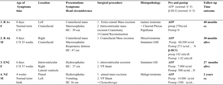
Table I: The demographics and clinical features of the patients who surgically treated in our clinic for
congenital cranial teratoma
Age of Symptoms time Location Presentations Symptoms Head circumference
Surgical procedure Histopathology Pre and postop AFP (normal: 0–5) β-HCG (normal: 0–5) Follow-up Time outcome 1. B Av F 0 days Normal term C/S Left Craniofacial Craniofacial mass Macrocephalus HC: 39 cm
1. Extra cranial Mass excision 2. Intraventricular mass excision Craniotomy 3.Cranial Reconstruction mature teratoma + Choroid Plexus Papilloma AFP preop 270iu/ml Postop N 40 months ex 2. B Ak M 0 days C/S 35 weeks Right Craniofacial Craniofacial mass Macrocephalus Respiratory distress HC: 47 cm
1. Craniofacial Mass excision Mixed teratoma Immature GIII AFP Preop. 60,500 iu/ml Postop 273 iu/ml... N β-HCG preop 182 mlu/dl. Postop 1.82 mlu/dl 30 months alive 3. ENÇ F 6 days C/S 37 weeks Intraventricular Right Lateral ventricle Hydrocephalus HC: 37 cm 1. intraventricular excision 2. VP shunt
Immature GII AFP
Preop. 7.000 iu/ml Postop 500 ıu/ml....N 27 months alive 4. NZ M 4 weeks Normal term-birth Pineal Left Hydrocephalus Vomiting HC 36 cm
1. pineal mass excision 2. VP Shunt
+Chemotherapy
Malign teratoma AFP
Preop. 14.000 iu/ml Postop 1500 ıu/ml... 2 years ex DISCUSSION Incidence:
Teratomas occur with an incidence of 1:4000 live births. Brain tumors occurring in the neonatal period have been estimated at 0.34 per million live births(3,7,9,24,33). Intracranial teratomas represent approximately 0.5–1.9% of all childhood tumors. Their incidence is 0.5% to 2.2% of intracranial tumors and is much lower in the spinal cord. These lesions account for only 2–4% of all teratomas, but they represent nearly 50% of brain tumors in the first 2 months of life. An intracranial teratoma is an uncommon intracranial neoplasm; however, it is the most common type of fetal intracranial tumor(2,3,6,15,19,26,32).
Embryology:
Teratomas appear to arise from diploid cells that have a mixture of chromosomes from both parents, suggesting that they have an alternate derivation. According to one theory, some of the embryonic germ cells fail to reach the gonadal ridge, become misplaced, or migrate aberrantly. These ectopic germ cells are usually embedded in or near midline structures and may be related to the head, mediastinum, or sacrococcygeal region. Without the
normal cues for maturation, these ectopic germ cells retain their pluripotent capabilities and may develop into teratomas or any other type of germ cell neoplasm that might otherwise arise within a gonad(19,28). Another hypothesis suggests that because every cell contains the full genetic code, theoretically any somatic cell, without being a “germ” cell, could produce any other type of cell. Although this premise is one of the principles of cloning, it is a principle that is expressed more often in science fiction than in science fact; however, it allows an alternate theory of teratoma formation from ordinary somatic cells, rather than from uncommitted, pluripotent germ cells(2,12,24,28,30). The third theory postulates that teratomas are an incomplete twinning. The genetic basis for teratomas is not well understood, and the clinical usefulness of chromosome mapping for teratomas is unclear. Deletions on chromosomes 1 and 6 were reported in children, but noted to be on chromosome 12 in adults. N-myc gene expression was noted in immature teratomas. No specific genetic risk factors have been identified for intracranial teratomas, and recurrence is unlikely(26,32). Teratomas are mainly isolated lesions but may form part of the Currarino triad
(anorectal malformation, sacral anomaly, and a presacral mass) as the presacral mass.
Age and sex distribution
In 1964, Solitare and Krigman(29) classified tumors occurring in the immature brain with congenital factors into three groups according to the age of symptom onset: 1. definitely congenital (symptoms present at birth); 2. probably congenital (most likely) (within the 1st week of life); and 3. possibly congenital (within the 1st month of life)(29). This entity (congenital tumor) may be extended to include brain tumors diagnosed in the 1st year of life(19). Wakai et al.(32) experienced two cases of neonatal brain tumor and collected 200 cases reported in the literature presenting or producing symptoms or found incidentally at autopsy within 2 months of birth. They found that 113 of those cases were definitely congenital tumors, and 12 were probably congenital according to Solitare and Krigman's classification(29,32). In this series, there were 2 probable, one possible, and one definite congenital tumors.
The prevalence of congenital intracranial teratomas is reported to be nearly equal in males and females in most series as in this series, but some series show a gender bias towards males(3,23,26).
Location:
The most common sites for congenital teratomas are the sacrococcygeal region (40%) and gonads (40%), followed by the oropharynx, orbit, and rarely the cranial cavity(10,11,13,17,28,33). The pineal gland is the most common site of origin, but intracranial teratomas may be found in the hypothalamus, ventricles, cavernous sinus, cerebellum, and suprasellar region. Brain tumors that are most likely congenital tend to develop in the supratentorial region of the immature brain in contrast to their infratentorial location in older children. Although it is often impossible to determine the exact site of origin, prenatally diagnosed intracranial teratomas
are also predominantly supratentorial as in our cases(5,9,19,26).
Classification:
Teratomas are anatomically classified as gonadal (testis or ovary) or extra gonadal (brain, face, neck, mediastinum, retroperitoneal, or sacrococcygeal). Three forms of congenital intracranial teratomas have been described in the literature and were observed in this series:
1. Massive intracranial teratoma replacing the intracranial contents of a neonate. 2. Smaller, more localized, less extensive intracranial teratomas producing hydrocephalus,
3. Intracranial teratomas with extension into the extracranial structures (orbit or neck) and a massive variant extending through the cranial base into the face and
neck region (“Epignathus”)(1,2,3,5,6,7,10,11,12,13,22,28,32,33).
The term ‘epignathus tumor' refers to teratomas of the oropharyngeal cavity in neonates without specifying any site of origin. They are called craniofacial teratomas. They are not known to be malignant but have the potential to extend into the cranium and involve the brain. Few cases have an extracranial extension along the craniopharyngeal canal. These more rare forms and massive intracranial teratomas involving the cranial base are usually diagnosed at autopsy(1,2,11,12,28). Nanda described another extremely rare form that should be mentioned as an extracranial congenital teratoma on the scalp(20). These tumors are also called skull/facial or craniofacial teratomas. Of teratomas on the skull sutures, approximately 50% are found in or adjacent to the orbit(3,10,13,20,22,27,30).
Clinical features
Intracranial teratomas generally present with symptoms of space-occupying lesions. These teratomas can appear in utero and cause massive hydrocephalus. Fetal intracranial teratoma is often
associated with an abnormally increasing head circumference (more than half of patients), bulging anterior fontanel, raised intracranial pressure, hydrocephaly and polyhydramnios. These teratomas are detected postnatally in the setting of
hydrocephalus and macrocephaly(1,2,7,11,13,17) (Figure 1a, 3a).
Typically, the skin overlying craniofacial teratomas seems to be normal, and the masses are mobile; however, the overlying skin appeared thin and there were
prominent scalp veins(1,28). The
macrocephaly is due to the size of the tumor, and the hydrocephalus is often associated with either the blockage of the ventricles, resulting in a buildup of cerebrospinal fluid, or hemorrhage from the tumor. As in our cases, localized neurological deficits usually are not observed in neonates, even in very large tumors. This phenomenon is attributed to immature brain development, lack of nerve myelination, and/or expansion of the cranial bones(2,7). The prevalence of tumor hemorrhage is much higher in children than in adults. This phenomenon has been attributed to the rapid growth of neonatal tumors(8,18,28,33,35,36).
Neuroradiological studies
Teratomas can be detected early with antenatal ultrasound (US) and MRI, and the combined use of these methods and computed tomography (CT) improves the diagnostic accuracy(16,18,23,25,31,35). General imaging features of teratomas include a heterogeneous appearance, because of their extremely variable histological components, and enhancing soft tissues (enhancement of the capsule and heterogeneous enhancement of soft tissue components). Teratomas show cystic and solid areas with fat density on CT and MRI studies(16,18,23,25,28,35,36) (Figure 1b,1c, 2b,2c,3a).
Ultrasound
Modern high-resolution ultrasound scanners facilitate examination of the cranial contents, allowing earlier diagnosis.
The diagnosis with antenatal ultrasound is made between 20 and 40 weeks of gestation and is usually suspected because of a sudden increase in uterine size, resulting from rapid tumor growth, and polyhydramnios due to impaired fetal swallowing. Only 10% of reported fetal intracranial tumors have been detected before 24 weeks of gestation. Intracranial teratomas should be suspected in the presence of mass-occupying solid or cystic lesions and a change in shape or size of the normal anatomic structures (such as a midline shift). Cystic tumors and teratomas are usually characterized by a complete loss of the normal intracranial architecture(21,24). In some cases, the lesion appears as a low echogenic structure, and it may be difficult to recognize(21,24). At prenatal US, the diagnosis of teratoma should be considered for a complex intracranial mass with calcifications. It is generally very vascular. The most common initial sonographic finding in the fetus is macrocephaly, and additional features typically include gross distortion or replacement of normal brain tissue by an echogenic disorganized mass with multiple solid and cystic components. Hydrocephalus secondary to obstruction and hydramnios can also be found(2,7,23). Polyhydramnios due to impaired fetal swallowing is a common sign in cases of massive intracranial teratomas. Polyhydramnios might have been caused by nearly complete replacement of the brain parenchyma with destruction of the brainstem or diabetes insipidus from hypothalamic destruction. The ultrasound appearances of all intracranial tumors are similar, and, therefore, prenatal detection of the precise histological type of tumor is almost impossible(21,24).
MRI
MRI has the advantage of not utilizing ionizing radiation, which may be a useful modality for diagnosis when the mass is revealed by antenatal ultrasound examination. Ultrafast MRI techniques for
antenatal diagnosis of teratoma have been described to minimize fetal motion and breathing artifacts(16,18,23,25,31). Several recent case reports have described the use of fetal MR imaging between 25 and 36 weeks gestation in helping confirm the diagnosis of an intracranial teratoma(18,25,35). Teratomas are often seen as large lesions at presentation.
Similar features also have been described with postnatal MR imaging of mature and immature intracranial teratomas (Figure 1c,2b,2c,3a-c). In the neonatal period, both CT and MRI have been described as being useful for the complete assessment of the tumor, to determine its relationship with surrounding structures, its extension and any complications(8,10,13,18,28,35,36). The typical appearance of a teratoma is a large, heterogeneous mass with cystic components on T1- and T2-weighted images (presence of fat, mucus-laden cysts, calcium (bone and chondroid nodules), and soft tissues), with no apparent difference between mature and immature teratomas(8,28). There can also be enhancing soft tissues (enhancement of the capsule and heterogeneous enhancement of the soft tissue components). The high fat content of teratomas is the cause of their strong signal on T1-weighted images and allows differentiation from cystic hygromas, which are less intense on T1- weighted images but more intense on T2-weighted images(8,10,13,18,28,33,35,36). (Figure 2b,2c,3a-c)
CT
Although MR imaging is insensitive for detecting small calcifications, CT shows regions of calcification, which are present in 50% of teratomas. CT can also better demonstrate bony defects(10,12,13,17,24)
(Figure 1b,2a).
Differential Diagnosis
As the MR imaging features of teratomas are relatively nonspecific, the differential diagnosis should also include supratentorial tumors(1,18,28,33,36).
Figure 1a: Photograph of a 5-month-old child who
has a mature teratoma, showing a large left craniofacial mass distorting the external facial anatomy with displacement of the nose and left eye.
Figure 1b: CT showing a large heterogeneous cystic
mass displacing and distorting the anatomical structures (left orbit, temporal fossa) in the same patient
Figure 1c: T1 weighted sequence in an axial
plane MRI showing a left multiloculated, heterogeneous cystic mass that contains cyst-like areas of variable size extending into the right orbit and temporal lobe (Mature Teratoma)
Figure 1d: After removal of the teratoma, a T1
weighted axial plane MRI showing no recurrence and spontaneous reshaping of the compressed skull.
Figure 2a: A coronal CT demonstrates skull
destruction extending into right orbit with a large craniofacial mass (teratoma-epignathus).
Figure 2b: T1 weighted MRI in the axial plane
reveals a multiloculated, heterogeneous solid, cystic mass that was larger than the patient's head. There is involvement of the right temporal lobe of the brain (Immature Teratoma)
Figure 2c: T2 weighted MRI sequence in the
coronal plane showing a multiloculated, heterogeneous cystic mass displacing and distorting the anatomical structures, consistent with craniofacial teratoma. MRI shows the mass with calcifications, soft tissue, fat and fluid components.
Fig 2d: T1 weighted in coronal plane MRI
images six months after removal of the teratoma reveal no recurrence and spontaneous reshaping of the cranium.
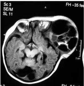
Figure 3a: T1 weighted axial MRI reveals a
midline heterogeneous cystic mass resulting in marked hydrocephalus. (Mature Teratoma)
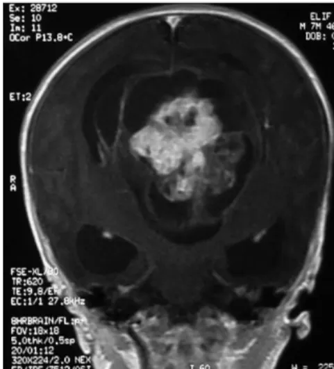
Figure 3b: T1 weighted contrast enhanced MRI
in the coronal plane showing a multiloculated heterogeneous solid-cystic mass within the lateral and third ventricle. MRI reveals calcifications, soft tissue, fat and fluid components within the mass. There is also
Figure 3c: CT two weeks after removal of
teratoma shows no recurrence but there was hydrocephalus.
Figure 4a: The external structure of a mature
teratoma is seen.
Figure 4b: Micrograph of an immature
teratoma showing tissue from all three germ layers. H&E stain at a magnification of 40x.
1. sPNET: On CT, sPNET is often seen as
a large irregular mass, typically iso- to hyper-attenuating on non-contrast imaging. Cystic components observed in approximately 65% of cases and calcification seen in approximately 70% of cases. It has heterogeneous contrast enhancement.
2. Intracranial dermoid: Typically a
well-defined, low attenuating lobulated mass is observed on CT and MRI. T1-weighted MRI typically shows a hyperintense (due to the cholesterol components) mass. On T2-weighted MRI, signals vary from hypointense to hyperintense. It may show calcification in wall, and enhancement is uncommon.
3. Choroid plexus papilloma: The CT
scan and MRI appearance of choroid plexus papillomas are distinctive. There is a heterogeneous mass showing vascularity and marked contrast enhancement with overgrowth of the choroid plexus(1,18,24,28,33,36).
4. Craniopharyngioma: Typically seen as
a heterogeneous mass in the suprasellar region. Calcification (except for the papillary type) and cysts are very common. Signal intensity varies depending on cyst contents and can appear hyperintense due to protein, blood products, and cholesterol (in the classic adamantinomatous type). Contrast enhancement is typical, with thin enhancement of the cyst wall, or diffuse heterogeneous enhancement of the solid components.
5. Atypical rhabdoid teratoid tumor (ATRT): CT shows often an isodense
mass with associated obstructive hydrocephalus. It may demonstrate heterogeneous enhancement, and calcification is common. MRI can show necrosis, multiple foci of cyst formation and sometimes hemorrhage. On T1 MRI, the mass is isointense to slightly hyperintense relative to grey matter (hemorrhagic areas can be more
hyperintense), and there is heterogeneous enhancement. On T2 MRI, the mass is generally hyperintense, although hemorrhagic areas can be hypointense.
6. Intracranial lipoma: The characteristic
finding on both CT and MRI is a mass that has appearances that are consistent with fat. CT (negative HU values) and MRI with and without fat saturation are able to provide diagnosis. Lipomas have a typical hyperechogenic, homogeneous appearance.
7. Neuroblastoma: Some helpful
distinguishing features, including frequent hemorrhage and calcification, have been reported. These features include highly vascular and lift up the periosteum, producing radial bone spiculation extending into the soft tissues. The dura often resist the spread; however, occasionally, an intratumoral hemorrhage may rupture into the brain parenchyma. Astrocytoma, ependymoma, glioma, and hemorrhage are other pathologies in the differential diagnosis that should be considered(1,10,18,24,28,33,36).
Serology:
Some teratomas contain yolk sac elements, which secrete alpha-fetoprotein (AFP). Sixty-five percent of patients with immature teratomas have elevated serum levels of AFP. This level decreases markedly after surgery. Detection of AFP may help to confirm the diagnosis and is often used as a marker for recurrence or treatment efficacy, but it is a rare method of initial diagnosis. AFP has been shown to be a reliable indicator of disease activity, and some authors advocate investigating teratoma recurrence by evaluating serial serum AFP levels(6,11,17,27,28).
Management
A multidisciplinary team, involving obstetricians, neonatologists, and neurosurgeons, is the suggested management. Over 99% of teratomas found in the fetus and newborn are
histologically benign and are diagnosed as either mature or immature teratomas. Despite the benign histopathological features of most intracranial teratomas, tumor growth is rapid and the tumor frequently replaces all normal brain tissue, resulting in massive craniomegaly(31). Because of the usually extensive involvement and destruction of normal intracranial structures, the prognosis for fetal teratoma is generally poor with death usually occurring shortly after birth. Even where surgical excision is possible, neurological handicap is not uncommon. As many cases present in the late second trimester or third trimester, termination of pregnancy is not a legal option in some countries. The timing and mode of delivery are important issues to be discussed between the management team and the patient's family. Usually, most cases are delivered by cesarean section because of an abnormally large head and difficult delivery. However, in several cases, vaginal delivery has been possible, occasionally with prior cranial decompression. Cephalocentesis, either ultrasound-guided transabdominally or intrapartum transvaginally, has been advocated as a means of facilitating vaginal delivery and thus avoiding the maternal morbidity of caesarean section in cases of severe hydrocephalus(11,24).
Therapeutic possibilities are limited due to high surgical risks(3). Of neonates with head and neck teratomas, 25% die prior to surgery, 12-80% die if the tumor is not removed in the neonatal period(1,3,31). The massive extension of congenital intracranial teratomas has made neurosurgeons hesitant to perform surgical resection. The treatment of teratomas consists of an early and complete resection of the tumor mass, and surgical
intervention can lower the risk of malignant
transformation(1,7,10,11,14,22,28,34,36). Lapras et al.(32) emphasized the importance of a radical surgical procedure, even when the teratomas is located near a functional area. Teratomas are pseudo-encapsulated and usually shell out easily, but their large size and tendency to recur make the prognosis poor(1,14,32). Large tumor size and spread do not necessarily signify that the tumor cannot be resected. Tumors involve the deeper soft tissues of the oropharynx and nasopharynx, which are less amenable to surgical excision. In the absence of intracranial extension, radical treatment of epignathus teratoma consists of early and complete surgical resection. If there is a connection between the extracranial and intracranial spaces, it should be corrected, and dural defects caused by the tumor invasion should be repaired at the same time. After removal of the mass, spontaneous reshaping of the compressed orbital cavity and skull is pursued(10,14) (Figure 4b). If reshaping is ineffective, reconstruction can be performed.
Histopathological examination and Immunohistochemical studies
Teratomas belong to a class of tumors known as nonseminomatous germ cell tumor (NSGCT). Teratomas are true congenital neoplasms derived from totipotential (pluripotent) cells and composed of tissues originating from all three germinal layers (ectoderm, mesoderm, and endoderm)(12,17).
Histopathologically, CNS teratomas are classified as either mature or immature based on the differentiation of the tumor cells into three groups (mature, immature, or malignant teratomas) and four grades (0-3)(7,17) ( Table II):
Table II : The histological teratoma classification
Mature/benign teratoma (Grade 0: all component tissues are well-differentiated) Immature teratoma
Grade 1: <10% microscopic foci contain immature tissues Grade 2: 10–50% immature tissue
Grade 3: >50% immature tissue.
Malignant (teratoma plus one or more malignant elements). These are all classified as malignant germ cell tumors.
1. Mature teratomas are composed of
well-differentiated elements; (G0) The benign lesions contain mainly mature elements, such as mature cartilage, squamous epithelium, skin appendages, columnar mucosa, and neural elements. A mature teratoma is typically benign and found more commonly in females. A dermoid cyst is a mature cystic teratoma containing hair (sometimes very abundant), and other structures are characteristic of normal skin and other tissues derived from the ectoderm. The term is most often applied to teratomas on the skull sutures and in the ovaries of females. Fetus in fetu and fetiform teratomas are rare forms of mature teratoma that include one or more components resembling a malformed fetus.
2. Immature teratomas contain primitive
elements derived from any or all of the three germinal layers. An immature teratoma is typically malignant and is more often found in males. Recurrences develop mainly in immature teratomas. A grade 1 immature teratoma that appears to be benign (e.g., because AFP is not elevated) can have a much higher risk of malignancy and requires adequate follow-up.
3. Teratomas with malignant elements (G3) are malignant teratomas that are
derived from the yolk sac or endodermal sinus, react positively for α-fetoprotein,
and are associated with poor prognoses. A teratoma with malignant transformation is a very rare form of teratoma that may contain elements of somatic (non germ cell) malignant tumors such as leukemia. Grade 0, 1 and 2 pure teratomas have the potential to become malignant (grade 3), and malignant pure teratomas have the potential to metastasize(2,5,12,17,20).
Teratomas, by nature, have varied heterogeneous histological characteristics (Fig 4b). Both mature and immature forms usually contain tissues from all three germ layers, including skeletal muscle, cartilage, bone, bronchial epithelium, gut epithelium, and neural tissue. Mature teratomas may contain many types of adult tissues with varying degrees of organoid development, whereas immature lesions may produce embryonic or extra-embryonic fetal tissue. The predominant tissue is neuroectodermal, in the form of neuroepithelial rosettes and tubules, resembling neuroblastoma. Mesodermally derived immature cartilage and primitive stroma are also common, and endodermally derived respiratory and enteric epithelium can be present in the form of cystic structures(2,5,12,24,30).
Immunohistochemistry
Tumors can be stained for p53 expression by immunohistochemistry. p53 expression
is not unusual in immature teratomas, and diffuse p53 immunopositivity is associated with recurrence or the presence of malignant elements in 50% of cases. This phenomenon may explain the poor prognosis. The finding of frequent p53-positive cells in immature teratomas should prompt a search for malignant elements within the tumor, and affected patients should be followed closely for evidence of recurrence(4).
Prognosis:
Most teratomas are benign in neonates but malignant in older children and young adults. Local recurrences are considered to be related to incomplete excision and are treated by reoperation. The prognosis is also excellent after total excision in scalp teratomas(10,11,13,20,22,28,34). Malignant degeneration of teratomas is estimated to occur in between 5% and 30%. Mature teratomas generally behave in a benign manner after complete resection(17,28). Immature teratomas have a greater propensity for recurrence. For malignant teratomas, surgery is usually followed by chemotherapy such as in our cases. Teratomas that are in surgically inaccessible locations are very complex and likely to be malignant (due to late discovery and/or treatment); these teratomas are sometimes treated first with chemotherapy(2,12,23,28). Adjuvant radiation therapy is precluded by the severe damage it would cause to the developing CNS(3).
CONCLUSION
Teratomas can be diagnosed early with antenatal ultrasound. Despite developments in prenatal diagnosis, appropriate pre- and post-operative management, the mortality associated with these tumors remains high. A multidisciplinary team is necessary for appropriate management and family counseling. Acceptable functional outcomes can be achieved by early radical resection in some forms of congenital intracranial teratomas; thus, more aggressive treatment should be attempted
for patients with resectable tumors. Serum tumor markers assist in the diagnosis of recurrent tumors.
Correspondence to:
Nejat Isik
E-mail: [email protected]
Received by: 05 October 2012 Revised by: 17 November 2012 Accepted: 23 November 2012
The Online Journal of Neurological Sciences (Turkish) 1984-2012
This e-journal is run by Ege University Faculty of Medicine,
Dept. of Neurological Surgery, Bornova, Izmir-35100TR
as part of the Ege Neurological Surgery World Wide Web service.
Comments and feedback: E-mail: [email protected] URL: http://www.jns.dergisi.org
Journal of Neurological Sciences (Turkish) Abbr: J. Neurol. Sci.[Turk]
ISSNe 1302-1664
REFERENCES
1. Arai H, Sato K, Kadota Y, Ito M, Ishimoto K, Yanai A. Skull base reconstruction in cases of intracranial teratoma extending into the extra cranial structures. Surg Neurol. 1992;Nov;38(5):383-90.
2. Canan A, Gülsevin T, Nejat A, Tezer K, Sule Y, Meryem T, Gülşen E. Neonatal intracranial teratoma. Brain Dev. 2000;Aug;22(5):340-2. 3. Carstensen. H, Juhler M, Bøgeskov L, Laursen H. A
report of nine newborns with congenital brain tumours. Childs Nerv Syst 2006;22:1427–1431. 4. Charoenkwan P, Senger C, Weıtzman S, Sexsmıth E,
Sherman Cg, Malkın D, Thorner P.. Significance of p53 Expression in Immature Teratomas. Pediatric and Developmental Pathology 2002; 5, 499–507, DOI: 10.1007/s10024-001-0268-y
890 5. Chien YH, Tsao PN, Lee WT, Peng SF, Yau KL.
Congenital intracranial teratoma. Pediatr Neurol 2000;9:72–74.
6. Desai K, Nadkarni T, Muzumdar D, Goel A. Midline posterior fossa teratoma--case report. Neurol Med Chir (Tokyo). 2001;Feb;41(2):94-6.
7. Fearon JA, Munro IR, Bruce DA, Whitaker LA. Massive teratomas involving the cranial base: treatment and outcome: a two center report. Plast Reconstr Surg 1993;91:223-228.
8. Halperin EC. Neonatal neoplasms. Int J Radiat Oncol Biol Phys 2000;47:171–178.
9. Hsieh WS, Lien RI, Lui TN, Wang CR, Jung SM. Congenital oligodendroglioma with initial manifestation of jaundice. Pediatr Neurol 2002;27:230–233.
10. Isik N, Yildirim S, Onoz M, Aras A. Surgical treatment of huge congenital extra cranial immature teratoma: a case report. Childs Nerv Syst. 2011 May;27(5):833-9. Epub 2010 Nov 16.
11. Jarrahy R, Cha ST, Mathiasen RA, Shahinian HK. Congenital teratoma of the oropharyngeal cavity with intracranial extension: case report and literature review. J Craniofac Surg. 2000;Mar;11(2):106-12.
12. Johnston JM, Vyas NA, Kane AA, Molter DW, Smyth MD. Giant intracranial teratoma with epignathus in a neonate Case report and review of the literature. J Neurosurg (3 Suppl Pediatrics) 2007;106:232–236. 13. Karaca M, Akan M, Silav G, Akoz T. Giant
craniofacial immature teratoma with primary intracranial lesions. J Craniofac Surg. 2010 May;21(3):816-8. Review.
14. Lanzino G, Kaptain GJ, Jane JA, Lin KYK. Successful excision of a large immature teratoma involving the cranial base: report of a case with long-term follow-up. Neurosurgery 1998;42:389-393.
15. Lapras C, Guilburd JN, Guyotal J, Patet JD. Brain tumors in infants: a study of 76 patients operated upon. Child\'s Nerv Syst 1988;4:100-103.
16. Liechty KW, Crombleholme TM. Management of fetal airway obstruction. Semin Perinatol 1999; 23(6):496-506.
17. Mann JR. Mature and immature extra cranial teratomas in children. The UK Children's cancer study group Experience. J Clin Oncol 2008;26:3590-3597.
18. Marden FA, Wippold FJ 2nd, Perry A. Fast magnetic resonance imaging in steady-state precession (true FISP) in the prenatal diagnosis of a congenital brain teratoma. J Comput Assist Tomogr 2003; 27:427-430.
19. Mehrotra N, Shamji FM, Vassilyadi M, Ventureyra ECG. Intracranial tumors in first year of life: the CHEO experience Childs Nerv Syst 2009;25:1563– 156.
20. Nanda A, Luis Schut and Leslie N. Sutton. Congenital forms of intracranial teratoma. Child\'s Nervous System. 1991;7(2):112-114.
21. Ng HN, Ong CL (1993) Two case reports of intracranial teratomadiagnosed antenatally [review]. Ann Acad Med Singapore 22:823–825 89 22. Onabanjo SO, Aghadiuno PU, Ogunniyi J, Adeloye
A. Congenital benign extra cranial teratoma in a nigerian neonate. Childs nerv syst 1987;3:188–190. 23. R. Douglas Wilson. Management of fetal tumors
Best Practice & Research Clinical Obstetrics and Gynaecology 2008;Vol. 22, No. 1, pp. 159–173. 24. Rickert CH, Probst-Cousin S, Louwen F, Feldt B,
Gullotta F (1997) Congenital immature teratoma of the fetal brain. Childs Nerv Syst 13:556–559 9 25. Rothschild MA, Catalano P, Urken M et al.
Evaluation and management of congenital cervical teratoma. Case report and review. Arch Otolaryngol Head Neck Surg 1994; 120(4):444-8.
26. Sano K. A statistical study of brain tumors in Japan: general features. Jpn J Clin Oncol 1987;17:19-28. 27. Seyhan T, Sener L, Refik Ozerdem O, Bal N. Mature
teratoma presenting as a scalp mass in a newborn. J Craniofac Surg. 2006;Sep;17(5):1009-11.
28. Shah Ajaz Latoo S, Ahmed I, Malik AH. Head and neck teratomas. J maxillofac Oral Surg 2009;8(1):60-63.
29. Solitare GB, Krigman MR. Congenital intracranial neoplasm. A case report and review of the literature. J Neuropathol Exp Neurol 1964;23:280-292. 30. Turgut M, ibrahım meteoglu. Mature teratoma
associated with an interparietal encephalocele Case report. J Neurosurg 2007;(4 Suppl Pediatrics) 106:305–307.
31. Ulreich S, Hanieh A, Furness ME. Positive outcome of fetal intracranial teratoma. J Ultrasound Med 1993;3:163-5.
32. Wakai S, Arai T, Nagai M. Congenital brain tumors. Surg Neurol 1984;21:597-609.
33. Ward RF, April M. Teratomas of the head and neck. Otolaryngol Clin North Am. 1989;Jun;22(3):621-9. 34. Whittle IR, Simpson DA. Surgical treatment of
neonatal cranial teratoma. Surgical Neurology 1981;15:268-273.
35. Wiatrak BJ, Myer III CM, Bratcher GO. Report of a nasopharyngeal teratoma evaluated with magnetic resonance imaging. Arch Otolaryngol Head Neck Surg 1990; 102(2):306-9
36. Yang PJ, Graham AR, Carmody RF, Seeger JF, Capp MP. Intracranial mass in a neonate. Invest Radiol. 1986;Apr;21(4):360-4.