© 2020 Hong Kong College of Radiologists. CC BY-NC-ND 4.0 e9 Hong Kong J Radiol. 2020;23:e9-11 | https://doi.org/10.12809/hkjr2017069
CASE REPORT
Radiological Findings of Falciform Ligament Necrosis:
A Case Report
C Agirgun
1, H Vehbi
2, F Agirgun
1, H Kocabas
31Department of Radiology, Bartin Government Hospital, Bartin, Turkey 2Department of Radiology, Istanbul Medipol University Hospital, Istanbul, Turkey
3Department of General Surgery, Bartin Government Hospital, Bartin, Turkey
INTRODUCTION
The falciform ligament is a sickle-shaped peritoneal ligament between the liver, the diaphragm and the anterior abdominal wall. Falciform ligament necrosis is a rare cause of right upper quadrant pain that is
often misdiagnosed.1,2 We present a case of right
upper quadrant pain caused by fatty necrosis of the falciform ligament seen during ultrasound exam and confirmed by contrast-enhanced computed tomography (CT) scan. Very few cases have been reported and correlation between CT and ultrasound findings are rarely discussed.
CASE PRESENTATION
A 35-year-old man was admitted to the emergency department of our hospital with right upper quadrant pain, epigastric tenderness, and fever. He was referred to our radiology department with suspected cholecystitis. The patient and family history were non-specific. Physical examination revealed right quadrant tenderness and positive Murphy’s sign. There was no history of drug use. Blood tests showed increased white blood cell count and C-reactive protein levels with normal liver function
parameters. Abdominal ultrasound examination showed an echogenic non-compressible lesion located between the two lobes of the liver in the falciform ligament region surrounded by fluid. The gallbladder, liver parenchyma, and other components were normal (Figure 1) as were the intrahepatic and common bile ducts.
Abdominal CT scan demonstrated a low-density oval-shaped heterogeneous mass at the falciform ligament surrounded by inflammatory changes, fat stranding and minimal free fluid consistent with falciform ligament necrosis (Figure 2). Surgical intervention and excision of the falciform ligament was performed and confirmed the radiological diagnosis (Figure 3). The patient experienced no complications and was discharged after 3 days. Histopathological examination revealed haemorrhagic areas and fatty necrosis of the falciform ligament.
DISCUSSION
The falciform ligament is a sickle-shaped area of the peritoneum that divides the right and the left subphrenic compartments. It is a remnant of the ventral fetal
Correspondence: Asst. Prof. Dr H Vehbi, Department of Radiology, Istanbul Medipol University Hospital, Istanbul, Turkey
Email: [email protected]
Submitted: 12 Jan 2019; Accepted: 13 Mar 2019.
Contributors: All authors contributed to the design of the study; acquisition and analysis of the data; drafting of the manuscript; and critical revision of the manuscript for important intellectual content. All authors had full access to the data, contributed to the study, approved the final version for publication, and take responsibility for its accuracy and integrity.
Conflicts of Interest: The authors have no conflicts of interest to declare.
Funding/Support: This case report received no specific grant from any funding agency in the public, commercial, or not-for-profit sectors. Ethics Approval: The patient was treated in accordance with the Declaration of Helsinki. The patient provided informed consent for all treatments and procedures.
Falciform Ligament Necrosis
e10 Hong Kong J Radiol. 2020;23:e9-11
mesentery that contains the paraumbilical veins, ligament teres, fatty tissue, and muscular fibres. Its arterial blood supply derives from the middle or left hepatic artery and drains into the portal or paraumbilical veins.1-3
Falciform ligament pathologies are rare. Inflammation or necrosis of this ligament can be seen in infants secondary to omphalitis. Other rare pathologies such as lipoma, cysts, leiomyosarcoma, lymphangioma,
and paraganglioma have also been recorded.4,5 Rarely
falciform ligament inflammation and abscess formation are seen as a complication of cholecystitis, or following instrumentation during laparoscopic surgery.6
Falciform ligament necrosis is a very rare cause of acute abdominal pain that is often misdiagnosed. To the best
Figure 1. A 35-year-old man presenting with right upper quadrant
pain, epigastric tenderness, and fever. Axial ultrasound image of the right upper quadrant with (a) linear and (b) convex probes showing a heterogeneous, oval, hyperechoic mass–like lesion surrounded by a hypoechoic halo at the falciform ligament region (arrows) with fluid anterior to it (arrowheads).
Figure 2. The same patient. Axial venous-phase
contrast-enhanced abdominal computed tomography scans showing (a) a heterogeneous low-attenuation mass-like lesion (arrow) at the falciform ligament region with (b) a fat strand inferior to it (arrowhead).
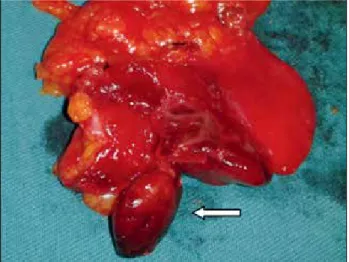
Figure 3. The same patient. Macroscopic view of the surgical
specimen showing necrotic fat tissue at the falciform ligament (arrow).
(a) (a)
C Agirgun, H Vehbi, F Agirgun, et al
Hong Kong J Radiol. 2020;23:e9-11 e11
of our knowledge, very few cases have been reported in the literature and most had no accurate preoperative diagnosis.7 Clinical presentation is similar to that of
acute cholecystitis, perforated duodenal ulcer, and pancreatitis. Infection, torsion of the ligament, venous thrombosis and arterial embolisation are the main causes of this condition, as is septic thromboembolism.8
In recent years, there has been an increase in diagnosis of intraperitoneal fat tissue and falciform ligament pathologies due to the increased availability and use of advanced imaging modalities. Ultrasound and CT play a vital role in the diagnosis of falciform ligament pathologies. Ultrasound examination accurately reveals fatty necrosis of the falciform ligament. Use of a linear probe after convex probe in suspicious cases enables a clearer view of the ligament and may reveal an oval hyperechoic mass with a hypoechoic halo in the falciform ligament region. (Figure 1). However, solitary fibrous tumours, lipoma and ligament haemorrhage should be considered as differential diagnoses.
CT scan is a very specific imaging tool for diagnosing
abdominal pathologies and their complications.5,9
Technical advances with multidetector CT allow multiplanar reformats and volume rendering. These reformats allow us to detect the falciform ligament in coronal sagittal planes and in the traditional transverse plane. Thin slices and multiple acquisitions allow more high-resolution images per tube rotation that be can be combined to create three-dimensional images and reformats, greatly increasing the diagnostic capability of CT scan. During CT scan, fatty necrosis of the falciform ligament presents as an oval fatty mass without contrast enhancement, with fluid and inflammatory changes around it.
Magnetic resonance imaging may provide an alternative imaging modality to diagnose falciform ligament pathologies. Using different sequences including the fat suppression ones enable differentiation between
fat tissue, bleeding, and effusion.7 Nonetheless its
susceptibility to artefacts of motion and organ pulsation
may limit its use in abdominal imaging.
Treatment is generally surgical excision although conservative treatment has been reported in some cases.10 Our patient underwent surgical excision with no
complications and was discharged 3 days later.
CONCLUSION
Fatty necrosis of the falciform ligament is a very rare cause of acute abdominal pain. Although it is easily diagnosed by ultrasound and CT scan, many clinicians and radiologists are unfamiliar with the condition. The falciform ligament region should be carefully examined in patients with right upper quadrant pain. Surgical excision is the treatment of choice although conservative treatment is successful in some cases.
REFERENCES
1. Standring S, editor. Gray’s Anatomy, 39th edition. Emerg Med J. 2006;23:492.
2. Ozkececı ZT, Ozsoy M, Celep B, Bal A, Polat C. A rare cause of acute abdomen: an isolated falciform ligament necrosis. Case Rep Emerg Med. 2014;2014:570751.
3. Webber CE Jr, Glanges E, Crenshaw CA. Falciform ligament. A possible twist? Arch Surg. 1977;112:1264.
4. Koca YS, Okur N, Barut İ. Isolated falciparum ligament necrosis causing right upper quadrant pain. Turk J Gastroenterol. 2017;28:531-2.
5. Lloyd T. Primary torsion of the falciform ligament: computed tomography and ultrasound findings. Australas Radiol 2006;50:252-4.
6. Lim ZS, Tan JY, Fanning S, Mitchell B. Education and imaging. Hepatobiliary and pancreatic: falciform ligament necrosis. J Gastroenterol Hepatol. 2012;27:1409.
7. Maccallum C, Eaton S, Chubb D, Franzi S. Torsion of fatty appendage of falciform ligament: acute abdomen in a child. Case Rep Radiol. 2015;2015:293491.
8. Czymek R, Bouchard R, Hollmann S, Kagel C, Frank A, Bruch HP, et al. First complete laparoscopic resection of a gangrenous falciform ligament. Eur J Gastroenterol Hepatol. 2010;22:109-11.
9. Coulier B, Cloots V, Ramboux A. US and CT diagnosis of a twisted lipomatous appendage of the falciform ligament. Eur Radiol. 2001;11:213-5.
10. Nam JG, Choi SH, Kang BS, Kim JY, Kwon WJ. Serial ultrasound and computed tomography findings of torsion of lipomatous appendage of the falciform ligament in a child treated by conservative management. J Korean Soc Radiology. 2015;72:368-71.