ORIGINAL ARTICLE
Inhibition of cell survival, viability and proliferation
by dentin adhesives after direct and indirect exposure in vitro
Safa Tuncer&Mustafa Demirci&Helmut Schweikl&
Mine Erguven&Ayhan Bilir&Aysun Kara Tuncer
Received: 14 April 2011 / Accepted: 20 December 2011 / Published online: 6 January 2012 # Springer-Verlag 2012
Abstract
Objectives The influence of dentin adhesive systems (Scotch-bond Multi-Purpose, XP Bond, Xeno V, Clearfil Protect Bond, AdheSE) on cell survival, viability and proliferation was char-acterized after direct and indirect exposure using different cell culture techniques.
Materials and methods The primers and cured bonding parts were directly exposed to cells using cell culture inserts, and complete materials were analyzed in a dentin barrier test. Cell responses were examined in 3T3 mouse fibroblasts after 24- and 72-h exposure periods by the estimation of
total cell numbers (survival), apoptosis (viability) and cell proliferation.
Results Cell numbers were effectively reduced by the primers of AdheSE, Protect Bond, and Scotchbond Multi-Purpose as well as XP bond after direct exposure in a cell culture insert test device. Likewise, Scotchbond Multi-Purpose primer in-duced a rate of apoptosis (93.9%) even higher than detected with Protect Bond primer (91.6%). Cell proliferation was entirely inhibited by primers and by Xp Bond as well. The Scotchbond Multi-Purpose was most cytotoxic in a dentin barrier test device after a 24-h indirect exposure. It also in-creased the percentage of cells in apoptosis to 15.4% com-pared to untreated controls.
Conclusion Unpolymerized primers of dentin adhesives were more cytotoxic than polymerized bonding counterparts. Moreover, total etch dentin adhesives were more cytotoxic than self-etch adhesives.
Clinical relevance When dentin adhesives are used in deep cavities without a protective dentin barrier the leachable hydrophobic and hydrophilic component of dentin adhesive systems can penetrate to the pulp and may induce cytotoxic responses in pulp tissues.
Keywords Dentin adhesive system . Biocompatibility . Cytotoxicity . Cell culture . Apoptosis
Introduction
Most of the dentin adhesives are designed to form strong bonding with dentin. The major goals of using dentin adhe-sives are to enhance the bonding strength between resin and the tooth structure, to increase the retention of restoration, to reduce the microleakage across the dentin–resin interface, and to scatter the occlusal stress [1].
S. Tuncer (*)
:
M. DemirciDepartment of Conservative Dentistry, Faculty of Dentistry, Istanbul University,
34093 Çapa Istanbul, Turkey e-mail: [email protected] H. Schweikl
Department of Operative Dentistry and Periodontology, University of Regensburg Medical Centre,
93042 Regensburg, Germany M. Erguven
Department of Clinical Biochemistry, Yeni Yüzyıl University, Faculty of Medicine,
Yılanli Ayazma Caddesi No:26 34093Cevizlibağ-Topkapı Istanbul, Turkey
A. Bilir
Department of Histology and Embryology, Faculty of Medicine, Istanbul University, 34093 Çapa Istanbul, Turkey
A. Kara Tuncer
Department of Endodontics, Medipol University, Faculty of Dentistry,
Unkapanı, Atatürk Bulvarı No. 27 34083, Fatih-İstanbul, Turkey
Current dentin adhesives can be classified based on their
underlying adhesion strategy as “etch and rinse” or
“self-etching” adhesives. The former remove the smear layer
completely via acid etching and rinsing, whilst the later incorporate the smear layer into the bonding substrate, as infiltration of resin occurs simultaneously with the self-etching process. Dental adhesives consist of methacrylates, dimethacrylates, phosphorized pentacrylates, aldehydes and organic acids. Water, acetone or alcohol is added as a solvent, and some adhesive systems also contain fillers [2]. Methacrylate-based dentin adhesives are a source of unpo-lymerized residual monomers which may be released into the oral cavity and may be active in adjacent oral tissues to cause adverse effects. Cytotoxic effects of current dentin adhesive systems have been reported from in vivo and in vitro
studies [3, 4]. The cytotoxic effects of adhesive systems
depend on the amount of unreacted resin monomers which in combination with the bonding system influences toxicity [5]. Both exposure periods and the interactions (synergism or antagonism) between the bonding components may be impor-tant parameters in determining the cytotoxicity of dentin bonding agents [6].
The common approach and principle when testing the biological behavior of materials is to start with simple in vitro tests mostly based on cell cultures. If these experiments and investigations of a material’s efficiency deliver promis-ing findpromis-ings, then more comprehensive studies on experi-mental animals and usage tests (in vivo evaluation) will be performed. Clinical studies are the final step of this evalu-ation process [7]. In early tests, materials were placed in direct contact with cells in monolayer culture and cell num-ber was used to monitor cytotoxic effects [8]. However, direct contact tests may be disadvantageous for the interpre-tation of cell reactions to dental materials that are directly placed on dentin. As a barrier dentin may markedly influ-ence the response of the adjacent target dental pulp tissue [9]. If barriers between the material and cells are incorpo-rated into an in vitro model, these models appear to be more appropriate for estimating the in vivo response. The agar diffusion test, a filter diffusion test, cell culture inserts, and dentin barrier tests are currently employed in screening assays using a physical permeable barrier between materials and target cells [10]. The main parameters for the estimation of the cytotoxicity of dental materials are to determine cell numbers, membrane permeability, intracellular metabolism or cell morphology [11]. However, when cytotoxic stimuli are severe, cells may escape from the cell cycle and undergo a programmed process of cell death called apoptosis. Apo-ptosis is considered an active process mediated by regulato-ry and effector caspases (e.g., caspase-3) and the final activation of downstream DNases [12]. Apoptotic cells can be among other means identified by flow cytometry as a sub-G1 population after being stained with propidium
iodide [13]. Viable cells are able to proliferate, synthesize new DNA and thus incorporate Bromodeoxyuridine (BrdU)
in the “S” phase of the cell cycle in an experimental
situa-tion. The measurement of the amounts of the fluorometric dye in the BrdU assay is, therefore, directly correlated to the proliferation of cells [14].
Since dentin adhesive systems remain in close contact with living dental tissues over a long period of time the biocompatibility of these materials is of particular impor-tance. Here, we used various parameters to characterize physiological cell responses [15]. The wide variety of cell types used for cytotoxicity studies of dentin bonding agents indicated the diversity of opinions on an acceptable cell culture model. Permanent mouse 3T3 fibroblasts were found sensitive and useful to test and classify the toxic effect of different dental materials. Furthermore, it was suggested that the cytotoxic effect on the mouse 3T3 cell line was some-where between that of human pulp-derived fibroblasts. 3T3 cells were used here because cytotoxicity testing of dental adhesives does not essentially require the presence of cellu-lar functions specific for target cells of the oral cavity. Furthermore, 3T3 cells can be easily amplified and are available in large numbers for testing, their behavior is well known, relatively consistent, and constant [4]. Moreover, this cell line is recommended by international standards (ISO 7405) [16]. The aim of the present study was to evaluate the in vitro effect of three self-etch and two etch and rinse bonding agents on total cell numbers as a measure of cell survival, the induction of apoptosis and the analysis of cell proliferation in exposed cell cultures. Cell morphol-ogy was assessed by scanning electron microscopy (SEM) as well. Direct exposure of cells and the use of a dentin barrier between the test materials and cell cultures for indi-rect exposure to mimic a relevant experimental parameter of a clinical situation were chosen for the testing in an established cell line in vitro.
Materials and methods Cell culture
The 3T3 mouse fibroblast cell line obtained from the Amer-ican Type Culture Collection (ATCC CRL-1658, Rockville, MD, USA) was grown in monolayers in Dulbecco’s modi-fied Eagle’s medium-F12 (DMEM F12; Biological Indus-tries, Haemek, Israel) supplemented with 10% heat-inactivated fetal calf serum (FCS; Sigma, St. Louis, MO,
USA), 50 units/ml penicillin and 50 μg/ml streptomycin
(Sigma), 1.0 mM sodium pyruvate, and 1.5 g/l sodium bicarbonate. Cells in semi-confluent cultures were harvested using trypsin (Sigma), collected by centrifugation, and resuspended in culture medium. Viability of the cells was
tested by trypan blue exclusion, and the 3T3 fibroblast cells were then plated onto 24-well culture plates in complete medium as described below.
Test materials and cell cultures Test materials
Three self-etch dentin adhesive systems including Xeno V, Clearfil Protect Bond, and AdheSE as well as two etch and rinse adhesive systems (Scotchbond Multi-Purpose and XP Bond), were tested in the present study. Manufacturers and individual components of the materials are presented in
Table1.
Sample preparation and cell exposure
Samples to be tested directly using cell culture inserts
Ad-hesives (40μl) of various dentin adhesive systems
(Scotch-bond Multi-Purpose adhesive, Clearfil PB (Scotch-bond, AdheSE bond), XP bond and Xeno V were loaded into Teflon moulds (5×2 mm) with a micropipette under sterile conditions. The specimens were light cured for 20 s with a halogen light unit (VIP; Bisco Inc., Schaumburg, IL, USA) calibrated at
600 mW/cm2. Then, the polymerized specimens were put into
cell culture inserts (24-well Millicell hanging cell culture
insert, 0.4 μm PET (polyethylene terephthalate; Millipore
Corporation, Billerica, MA, USA). The matching primers
(40μl) were also loaded into inserts with a micropipette.
HEMA (2-hydroxy ethyl methacrylate) (40 μl; 8.23 mol/l)
was used like the primers as a positive control material, and cell culture inserts without test materials were used as a nega-tive control.
3T3 fibroblasts were seeded onto 24-well plates at an initial
density of 5×10⁵cells/well in complete cell culture medium.
The cells were incubated in a humidified air atmosphere
containing 5% CO2at 37°C for 24 h to allow for cell adhesion.
Then, culture plate inserts with test materials were placed into the wells in contact to cell culture medium.
Samples to be tested indirectly in a dentin barrier test device Human third molars with almost complete root forma-tion were obtained following an informed consent protocol reviewed and approved by an appropriate institutional review board at the Istanbul University, The Institute of Medical Sciences. The teeth were stored in physiological saline at 4°C prior to the experiments. The teeth were embedded into
a methyl methacrylate resin, and dentin slices (500±50μm
thick) were cut from the coronal part of the molars under copious water cooling using a low speed saw (Isomet Saw; Buechler, Lake Bluff, IL, USA). The smear layer on the pulpal side was removed by etching with 50% citric acid for 30 s. Then, the dentin slices were sterilized by autoclaving. The test
was carried out using a stainless steel test apparatus as shown
in Fig. 1. A dentin disc with dentinal tubules open to both
sides of the apparatus was held in place between two stainless steel holders using a low-viscosity (light body) polyvinyl siloxane impression material (Zhermack Oranwash L; Zhermack Indurent Gel Badia Polesine, Rovigo, Italy). Cen-tral holes (diameter, 5 mm) in both stainless-steel holders made both sides of the dentin disc available for test materials to be applied on one side (cavity side) and cell culture medium
on the opposite (Fig.1).
Dentin adhesives were applied according to manufac-turers’ instructions and light cured for 20 s. Vitrebond was
used as a positive control [17,18]. The test apparatus with a
dentin disc alone was used as a negative control. The dentin barrier test was performed using 24 well plates. 3T3
fibro-blast cells were seeded at an initial density of 5×10⁵cells/
well in complete cell culture medium. The cells were
incu-bated in an humidified air atmosphere containing 5% CO2at
37°C for 24 h to allow for cell adhesion. Then, the dentin barrier test apparatuses with test materials were placed into separate wells that was not surrounded by medium. Such a device was held in place in the well by two small metal rods which allowed the culture medium to slightly contact the
etched side of the dentin disc (Fig. 1). In this way, a test
material was indirectly exposed to the cell culture through culture medium.
Analysis of cell responses Determination of cell survival
Cells were exposed to materials placed in cell culture inserts or a dentin barrier test device for 24 and 72 h. Then, cells from exposed cultures and untreated controls were collected by trypsinisation, and cell numbers indicating cell survival were counted with a hemocytometer [19]. These experiments were performed in triplicate with three replicates per material. Analysis of apoptosis
Cells were exposed to materials placed in cell culture inserts or a dentin barrier test device for 24 and 72 h. Cells in apoptosis were identified using an Annexin V-FITC/PI staining assay (BD Pharmingen San Diego, CA,USA). One of the manifestations of apoptosis is the translocation of phosphotidylserine (PS) from the cytosolic surface to the extracellular surface of the plasma membrane. Phospho-tidylserine was then detected by Annexin V staining. Brief-ly, cells were washed with phosphate-buffered saline (PBS) twice and resuspended in binding buffer containing 0.01 M
HEPES, 0.14 mM NaCl and 2.5 mM CaCl2. A cell
T able 1 T est materials, compounds and components Dentin adhesive Lot number Manufacturer Compound Components Scotchbond Multi-Purpose 6BF 2009 –2006 3 M ESPE St Paul, MN, USA Primer Primer: water 2-Hydroxyethyl methacrylate (HEMA) copolymer of acrylic and itaconic acids 6PN 2009 –2006 Bond Bond: bisphenol a diglycidyl ether dimethacrylate (Bis-GMA) 2-hydroxyethyl methacrylate (HEMA), Photoiniators Clearfil Protect Bond 00043A Kuraray Medical Inc., T okyo, Japan Primer Primer: 10-methacryloyloxydecyl hydrogen phosphate (MDP), 12-methacryloyloxydecyl pyridinium bromide (MDPB), 2-hydroxyethyl methacrylate (HEMA), hydrophilic dimethacrylate, water 00066A Bond Bond: 10-methacryloyloxydecyl hydrogen phosphate (MDP), bis-phenol A diglycidyl methacrylate (Bis GMA), 2-hydroxyethyl methacrylate (HEMA), hydrophobic dimethacrylate, di-camphorquinone, N ,N - diethanol-p -toluidine, silanated colloidal silica, surface treated sodium fluoride AdheSE K41839 Ivoclar V ivadent, Schaan, Liechtenstein Primer Primer: dimethacrylate, phosphonic acid acrylate, initiators and stabilizers in an aqueous solution K40597 Bond Bond: HEMA, dimethacrylate, silicon dioxide, initiators and stabilizers Xp Bond 0703002776 DENTSPL Y DeT rey GmbH Konstanz, Germany Carboxylic acid modified dimethacrylate (TCB resin); phosphoric acid modified acrylate resin (PENT A); urethane dimethacrylate (UDMA); triethylene glycol dimethacrylate (TEGDMA); 2-hydroxyethyl methacrylate (HEMA); butylated benzenediol (stabilizer); ethyl-4-dimethyl-aminobenzoate; camphorquinone; functionalized amorphous silica; t-butanol Xeno V 0801002439 DENTSPL Y DeT rey GmbH Konstanz, Germany Bifunctional acrylate; acidic acrylate; functionalized phosphoric acid ester; acrylic acid; water; tertiary butanol; initiator; stabilizer V itrebond 7JB 2010 –2012 3 M ESPE, St Paul, MN, USA Liquid Copolymer of acrylic and itaconic acids; water; 2-hydroxyethyl methacrylate 8MF 2010 –2012 Powder Glass powder; diphenyliodonium chloride
incubated with 5μl FITC-labeled Annexin V (BD Pharmin-gen) and PI (propidium iodide) for 15 min in the dark at room temperature. Then, the fluorescence of the cells for PI (Fl-3) and Annexin V-FITC (Fl-1) was measured simultaneously in a BD FACSCalibur flow cytometer and analyzed with the Cell-Quest software (BD Pharmingen). Data acquisition and anal-ysis were done with CellQuest and WinMDI programs [20, 21]. The percentages of viable cells (annexin V−; PI−; lower left quadrant), cells in apoptosis (annexin V+; PI−; lower right quadrant) and necrosis (annexin V−; PI+; annexin V+; PI−; upper left and upper right quadrants) were determined. Only the percentage of cells in apoptosis (annexin V+; PI−) is
presented in Figs.4and5. These experiments were performed
in triplicates in three independent experiments.
Analysis of cell proliferation
Cell proliferation was analyzed using a BrdU incorporation assay. After sterilization with ethanol, round cover slides were placed in 24-well plates and left under UV light overnight.
Then, 7×10⁴cells/well were seeded into each well and allowed
to adhere for 24 h. After that, cells were exposed to materials placed in cell culture inserts or a dentin barrier test device for
24 and 72 h. Next, cultured cells were labeled with 20μM
5-bromo-2-deoxyuridine (BrdU) (Sigma-Aldrich Chemie, Stein-heim, Germany) for 1 h. The slides were then fixed with
ethanol for 30 min at−20°C, and subsequently rehydrated with
PBS. Endogenous peroxidase activity was quenched with
0.5% H2O2in methanol, and double-stranded DNA was
dena-tured with 4 N HCl. The cells were washed with PBS, and a non-specific blocking reagent (Ultra-V-Block; Lab Vision Co., Westinghouse, CA, USA) was used to prevent non-specific binding of antibodies. The cells were then stained with a monoclonal mouse anti-BrdU antibody (1:50; Lab Vision Co., Fremont, CA, USA), and the primary antibody was detected by a biotinylated goat anti-mouse secondary antibody (Lab Vision Co., Fremont, CA, USA). After washing, peroxidase-conjugated streptavidin (Lab Vision) was applied and aminoethyl carbazole was used as a chromogen. The cells
were counterstained with Mayer’s hematoxylin to augment the nuclear staining. Cells were counterstained Mayers hematox-ylin in order to discriminate 3,3-diaminobenzidine tetrahydro-chloride (DAB) staining in nucleus from background and DAB unstained cells. After this, DAB staining in nucleus become more brown, DAB unstained nucleus and background became blue. BrdU labeled cells in the S-phase had red-stained nuclei. For negative controls, the cover slips of the same groups were processed with exactly the same steps, excluding the primary antibody. The BrdU-LI (the number of positively stained cells divided by the counted total number of cells) was calculated by evaluating at least 3000 cells in multiple high power fields [22]. Data were summarized from triplicates in three independent experiments.
Scanning electron microscopy
Cells grown on round cover slides as described were exposed to materials placed in cell culture inserts or a dentin barrier test device for 72 h. Then, samples were fixed and treated for evaluations using SEM. The cells were washed with PBS and fixed with 2.5% glutaraldehyde (Polysciences, Inc., Warring-ton, PA) in 0.1 mol/l phosphate buffer at pH 7.3 for 30 min, and 1% osmium tetroxide in 0.1 mol/l cacodylate buffer, pH 7.4, for 1 h. Next, the cells were washed with PBS, dehydrated in a graded series of ethanol and placed in 100% ethanol. Then, cells were critical-point dried, sputter coated with gold– palladium and analyzed by SEM (JSM 5200; Jeol, Tokyo, Japan). Photographs were taken at 10 kV using various mag-nifications and angles [23].
Statistical analysis
NCSS (Number Cruncher Statistical System) 2007&PASS 2008 Statistical Software (NCSS, Kaysville, UT, USA) was used for statistical analyses of the number of cells (cell survival), the percentage of cells in apoptosis, and cell proliferation (BrdU assay). Individual measurements were combined to mean values which were compared using the Fig. 1 Dentin barrier
one-way analysis of variance (ANOVA), follow-up compar-isons between the groups were then carried out using
Tukey’s multiple comparison test. Differences between
mean values were considered to be significant at p<0.05. The differences between mean values calculated after the two time intervals (exposure periods) were analyzed by using Student’s t-tests.
Results
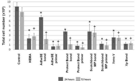
Cell survival after exposure of cell cultures towards dentin adhesives
The total number of cells exposed the various dentin adhesive systems for 24 and 72 h was determined as a measure of cell survival. In the cell culture insert test device the primers of AdheSE, Protect Bond, and Scotchbond Multi-Purpose as well as XP bond were the most effective compounds after a
24-h exposure period (Fig.2). These compounds were even
more active than HEMA. AdheSE primer, for instance, re-duced cell numbers about 9-fold compared to untreated con-trols. Similar results were observed after a 72-h exposure. Then, the lowest cell numbers were caused by Protect Bond primer. Noteworthy, for both time periods, the primer parts of the dentin adhesive systems caused significantly (p<0.05) lower cell numbers compared with the bonding counterparts except for Protect Bond after 72 h. Xeno V was as effective as
HEMA after both exposure periods (Fig.2).
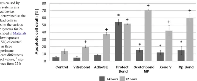
Different to the effects observed in the cell culture insert test device, the Scotchbond Multi-Purpose dentin adhesive system was the most cytotoxic material analyzed in a dentin
barrier test device after a 24-h exposure (Fig.3). The material
reduced cell numbers to about 40% compared to untreated control cultures, and was significantly more active than the positive control material (Vitrebond) included in this test system. Protect Bond, Xp Bond, Xeno V and AdheSE were in this order less cytotoxic than Scotchbond Multi-Purpose
(Fig.3). The same rank order of cytotoxic effects was detected
when cell cultures were exposed for 72 h, except that Xp Bond
was the most effective material (Fig.3).
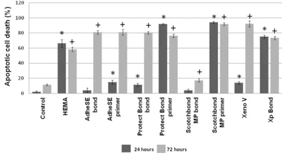
Induction of apoptosis by dentin adhesive systems
The percentage of apoptotic cells was then determined in cell cultures which survived after exposure to the various dentin adhesive systems. Scotchbond Multi-Purpose primer (93.9%) and Protect Bond Primer (91.6%) induced apoptotic cell death even higher than detected with HEMA (66.1%) in a cell
culture insert test device after a 24-h exposure (Fig.4). XP
bond (75.1%) was significantly (p<0.05) less active than Scotchbond Multi-Purpose primer and Protect Bond Primer. After a 72-h exposure period Xeno V (92.1%) and Scotch-bond Multi-Purpose primer (91.9%) induced significantly higher apoptotic cell death rate. Here, the primer parts of the dentin adhesive systems caused a significantly (p < 0.05) higher rate of apoptosis than the bonding compounds only after a 24-h exposure. Scotchbond Multi-Purpose bond was the material least effective in the induction of apoptosis (3.8%), followed by AdheSE bond (4%) and Protect Bond
bond (11.1%) after a 24-h exposure (Fig.4). The activity of
bonding counterparts of dentin adhesives, AdheSE primer and Xeno V in causing apoptosis was significantly (p<0.05) in-creased after a 72-h exposure. Yet, Scotchbond Multi-Purpose
Fig. 2 Cytotoxicity of dental adhesive systems in a cell culture insert test device. Cytotoxicity was determined as the total number of cells in cultures exposed to the various dentin adhesive systems for 24 and 72 h. Bars represent mean values (+SD) calculated from triplicates in three independent experiments (n09). *Significant differences from 24-h control values,+
sig-nificant differences from 72-h control values
bond (17.2%) was significantly less active than the other test materials. However, Protect Bond bond (80.3%) and AdheSE primer (81%), as well as AdheSE Bond (80.9%) induced apoptosis in the same range than HEMA (58.2%)
(Fig.4).
The Protect Bond system significantly (p < 0.05) in-creased the percentage of cells in apoptosis to 54.1% com-pared to 5.3% in untreated cell cultures in a dentin barrier
test device after a 24-h exposure period (Fig.5). Scotchbond
Multi-Purpose (15.4%), XP bond (15.1%) and Xeno V (12.1%) were similarly effective, but AdheSE (8.3%) was significantly (p<0.05) less effective than Scotchbond Multi-Purpose and XP Bond. Vitrebond (4.9%) caused the signif-icantly lowest rate of cells in apoptosis except for AdheSE
(Fig. 5). The efficiency of the adhesive systems to cause
apoptosis significantly increased after a 72-h exposure
except for Protect Bond (p < 0.05) (Fig. 5). After a
72-h exposure, Scotchbond Multi-Purpose induced a rate
of apoptotic cell death (70.7%) even higher than detected
with XP Bond (60.1%) (Fig.5)
Influence of dentin adhesives systems on cell proliferation Cell proliferation in a cell culture insert test device was analyzed using a BrdU incorporation assay. About 19% of the cells in untreated cultures were found to be proliferating
after a 24-h incubation period (Fig. 6). However, cell
pro-liferation was completely inhibited by AdheSE primer (1.4%), Protect Bond primer (2.4%), Scotchbond Multi-Purpose primer (1.6%), and Xp Bond (2.2%). Xeno V (6.4%) was as effective as HEMA (5%), which was used
as a control (Fig. 6). AdheSE bond (15.3%), Protect Bond
bond (7.9%), and Scotchbond Multi-Purpose bond (8%) were significantly less effective than the corresponding primers (p<0.05).
Fig. 3 Cytotoxicity of dental adhesive systems in a dentin barrier test device. Cytotoxicity was determinded as the total number of cells in cultures exposed to the various dentin adhesive systems for 24 and 72 h. Bars represent mean values (+SD) calculated from triplicates in three independent experiments (n09). *Significant differences from 24-h control values,+ sig-nificant differences from 72-h control values
Fig. 4 Apoptosis caused by dental adhesive systems in a cell culture insert test device. Apoptosis was determined as the percentage of dead cells in cultures exposed to the various dentin adhesive systems for 24 and 72 h as described in
Materials and methods. Bars represent mean values (+SD) calculated from triplicates in three independent experiments (n09). *Significant differences from 24-h control values,+ sig-nificant differences from 72-h control values
Cell proliferation in untreated cultures significantly
in-creased to 50% after a 72-h incubation period (Fig.6). Yet,
AdheSE primer (6.2%), Scotchbond Multi-Purpose primer (4.6%), and Xp Bond (6%) still significantly inhibited cell proliferation (p<0.05). Protect Bond primer (0.4%) was most
effective at that time (Fig. 6). In contrast, AdheSE bond
(14.4%) and Protect Bond bond (6.2%) even increased their inhibitory effect on cell proliferation after a 72-h exposure compared to untreated controls, and Scotchbond Multi-Purpose bond (23.4%) was as effective as after a
24-h exposure (Fig.6).
Increased cell proliferation was also detected in untreated cultures in a dentin barrier test device by 23% after a
24-h exposure period (Fig.7). Cell proliferation was again
significantly inhibited by all dentin adhesive systems. Protect Bond (8.7%) was significantly more effective than AdheSE (18%), Xeno V (14.4%), Xp Bond (11.2%) and Vitrebond (11.3%), which was used as a positive control material. Since cell proliferation in untreated cultures increased by 46% after a
72-h exposure period, the inhibitory effects of all materials tested here were very similar to those observed after 24 h except
for Xp Bond (Fig.7).
Analyses of cell cultures using scanning electron microscopy
Cells grown on cover slides and exposed to the various adhesive materials were also analyzed for morphological changes by SEM. Examples of cell cultures demonstrating
cytotoxicity of selected materials are presented in Fig.8. The
fibroblasts in untreated control cultures were well spread, and the flat cells appeared in close contact to the cover slide
surface (Fig.8a). However, the morphology of the cells
dra-matically changed after exposure to HEMA. The number of cells was considerably reduced and the few rounded cells
indicated severe cytotoxic effects of the monomer (Fig.8b).
Likewise, a similar cell response was observed with Scotch-bond Multi-Purpose Scotch-bond, while the few cells surviving Fig. 5 Apoptosis caused by
dental adhesive systems in a dentin barrier test device. Apoptosis was determined as the percentage of dead cells in cultures exposed to the various dentin adhesive systems for 24 and 72 h as described inMaterials and methods. Bars represent mean values (+SD) calculated from triplicates in three independent experiments (n09). *Significant differences from 24-h control values,+
sig-nificant differences from 72-h control values
Fig. 6 Cell proliferation in cultures exposed to dental adhesive systems in a cell culture insert test device. Cell proliferation was determined by BrdU incorporation after 24 or 72 h as described inMaterials and methods. Bars represent mean values (+ SD) calculated from triplicates in three independent experiments (n09). *Significant differences from 24-h control values,+ sig-nificant differences from 72-h control values
exposure to Scotchbond Multi-Purpose primer appeared to
increase in size (Fig.8c, d). Only very few cells were left in
cultures exposed to Xeno V, and structures indicating normal physiological cell functions were not detected in cultures
exposed to XP Bond as well (Fig.8e, f).
Discussion
Considering the clinical situation, the bioactivity of dentin adhesives in pulp tissues is of particular relevance. It has been discussed that the composition of dentin adhesive systems, clinical operative procedures, remaining dentin thickness, ad-equate lining materials and dentin permeability are factors that may influence the pulpal response to dentin adhesives [24]. The present study investigated various aspects of the cytotox-icity of currently used dental adhesive systems with different test methods.
First, this study made use of cell culture inserts which separate cells and materials. This experimental set up is suit-able for the direct testing of cell responses without the prior preparation of materials extracts. The porous membrane at the bottom of the inserts allows for the passage of leaching chemicals but not for resin particles to reach the monolayer of cells beneath it. It was reported that the use of commercially available sterile cell culture inserts was convenient for exten-sive cytotoxic screening when compared to agar overlay and Millipore filter tests [25]. Second, the dentin barrier test may help to identify compounds that repress or amplify adverse effects of substances by reducing or increasing dentin perme-ability [26]. With the importance of the principle of general-izing in vitro cytotoxicity findings to the human in vivo clinical situation, this technique is recommended for use in preference to the others (ISO 7405) [16]. Adopting the use of a
dentin barrier for indirect testing of dental materials simulates the in vivo oral environment more closely, thereby helping to identify specific components of dental filling materials which may be responsible for pulpal effects through dentin, an option not available with other barrier testing methods.
Cell responses caused by dentin adhesive systems were analyzed after two exposure periods (24 and 72 h). It was reported earlier that long periods of exposure resulted in a significant increase in the cytotoxicity of components of dentin adhesives and combinations. Likewise, longer expo-sure periods may result in a higher incidence of synergic interactions between the various adhesive components [6]. Here, the cytotoxic effects of primers were significantly higher than cell responses caused by the bonding counter-parts and other materials tested in a cell culture insert test device after a 24-h exposure. Similar results were obtained after a long exposure of 72 h except for Protect Bond. It was reported earlier that the primer part of Clearfil Protect Bond was more cytotoxic than the bonding part in a different test system. The authors suggested that the MDPB monomer in the primer part of Clearfil Protect Bond was responsible for the cytotoxic effect [27]. On the other hand, it has been discussed that this monomer has no significant deleterious effect [28]. It was assumed that cytotoxic effects of resinous dental materials depended on the concentration of leachable monomers [29]. In the present study, the primer parts of the adhesives were applied unpolymerized according to clinical situations. There is experimental evidence that polymerized dentin adhesives exhibited cytotoxicity about 2–65% lower than their unpolymerized counterparts [11]. The variation of monomer ratios in the dentin adhesive systems is probably another factor related to the different cytotoxic effects of these materials [29]. Here, the primer parts of the dentin adhesives which contain a variety of monomers were more Fig. 7 Cell proliferation in
cultures exposed to dental adhesive systems in a dentin barrier test device. Cell proliferation was determined by BrdU incorporation after 24 or 72 h as described inMaterials and methods. Bars represent mean values (+SD) calculated from triplicates in three independent experiments (n09). *Significant differences from 24-h control values,+
sig-nificant differences from 72-h control values
cytotoxic than HEMA after a 72-h exposure period. It is possible that the proportions of primers or the interaction of monomers might be responsible for the relatively severe cell responses caused by the primers in the current study [6].
In the present study, the primer parts of dentin adhesives and XP Bond exhibited the highest cytotoxic effect compared to other materials. Similar to our results, XP Bond proved more cytotoxic than Clearfil Tri-S and AdheSE than previ-ously reported [30]. This might be explained by the different ingredients of XP Bond (UDMA and TEGDMA) compared to other dentin adhesives in this study. It was reported earlier that aqueous elutes of the dentin adhesive Solobond Plus which tested severely cytotoxic contained high amounts of the monomer TEGDMA [31]. Moreover, in the present study AdheSE bond and Protect Bond bond exhibited a significant increase in cytotoxicity with increasing time periods. The differences between cytotoxic effects observed after increas-ing exposure periods were discussed controversy, and might be caused by the miscellaneous ingredients of different dentin adhesives. It is possible that cell cultures recover from damage caused by moderate cytotoxic agents more rapidly than from
exposure to severely cytotoxic materials [4,32,33].
In the dentin barrier test system Scotchbond Multi-Purpose and XP Bond were observed to be most effective after 24- or h exposure periods, respectively. After a 72-h exposure period, XP Bond caused significantly t72-he lowest cell numbers except for Scotchbond Multi-Purpose and
Vitrebond. Noteworthy, Xp Bond and Scotchbond Multi-Purpose are etch-and-rinse adhesives. In agreement with the present study, it was found earlier that etch-and-rinse adhe-sives were more effective than self-etch adheadhe-sives. It was reported that etching removes the smear layer, enlarges the dentinal tubules at the surface and, thus, increases dentin permeability [34]. Bouillaguet et al. [35] found that the cytotoxicity of dentin adhesives decreased with increasing intervals, although there was still persistent suppression of cellular metabolism even at late intervals. Furthermore, the amounts of leachable components of dentin adhesives detected in cell culture media decreased at later intervals [35]. In the present study, it is obvious that toxic constitu-ents of dentin adhesive systems accumulate at later time intervals, and more severe cell responses were induced after 72 h.
In this work, the primer parts of the adhesives were signif-icantly more effective in the induction of apoptotic cell death than the bonding counterparts after direct exposure in the cell culture insert test after a 24-h exposure. A similar effect was observed only for Scotchbond Multi-Purpose primer at the end of 72 h. At this time point, apoptosis induced by Scotch-bond Multi-Purpose Scotch-bond was significantly lower compared to the effects caused by other test materials. The proportion of apoptotic cells for bonding counterparts, AdheSE primer and Xeno V significantly increased after a 72-h exposure. More-over, in the dentin barrier test, a similar effect was observed except for Protect bond. Also, for the 72-h exposure period, apoptosis induced by Scotchbond Multi-Purpose and XP bond was higher compared to the effects caused by other test materials.
Similar to our findings, it was demonstrated that the unpo-lymerized or partially pounpo-lymerized adhesive Single Bond in-duced apoptosis very rapidly in various cell types [36]. Furthermore, it was suggested that the apoptotic potential of Clearfil SE Bond and FL Bond was material dependent [37]. The production of reactive oxygen species (ROS) was in-creased in human pulp-derived cells by various dentin adhe-sives about 5-fold in a dose-related manner. Since the production of ROS was associated with apoptosis, it was suggested that the production of ROS in the presence of adhesives may trigger apoptotic cell death [18].
The primer parts of dentin adhesives and XP Bond were most effective in the inhibition of BrdU incorporation in the cell culture insert test indicating delayed cell proliferation after 24- and 72-h exposure periods. In the dentin barrier test, however, Protect Bond adhesive significantly inhibited cell proliferation after a 24-h exposure. Similar effects on cell proliferation were observed for etch-and-rinse adhesives after 72 h. Noteworthy, cell proliferation recovered in cell cultures exposed to dentin adhesives for 72 h except for XP Bond. Yet, a significant effect on the number of cells distributed among the various phases of the cell cycle was not observed with Fig. 8 Morphology of cells in cultures treated with dentin adhesives.
Cultures treated with a negative control, b HEMA, c Scotchbond MP bond, d Scotchbond MP primer, f Xeno V, and f XP Bond. The cells were visualized by scanning electron microscopy (SEM)
AdheSE by others. Likewise, no major impact on the cell cycle was detected with self-etch adhesives, and a modest inhibition of cell proliferation might have been caused by the induction of cell death by high amounts of material. It was concluded that total etch bonding agents induced a cell cycle arrest, whereas the self-etch bonding agents did not affect the cell cycle patterns [30].
The various adverse effects of dentin adhesive systems as indicated by biochemical assays were associated with mor-phological alterations of exposed cells similar to results reported earlier [38]. These observations support our findings on the induction of apoptosis by dentin adhesives because pseudopod retraction, the rounding up of cells, and decreased cellular volume (pyknosis), or blebbing of the intact plasma membrane have long been known as morphological aspects considered typical for apoptosis [39].
In conclusion, various dentin adhesive systems were differentially active in the various test systems in the present study. Unpolymerized primer parts of dentin adhesives were more effective than their polymerized bonding counterparts after direct exposure to cells in the cell culture insert test, and similar effects were detected on apoptosis and cell proliferation. In the dentin barrier test device, total etch dentin adhesives were more cytotoxic than self-etch adhe-sives, and total etch dentin adhesives were more effective in causing cell apoptosis and inhibiting cell proliferation as well. Therefore, clinicians should consider the potential risk for pulp cells when these materials are used in deep cavities without a protective dentin barrier and when pulp cells are exposed to these materials.
Acknowledgements The current study is based on a thesis submitted to the graduate faculty, University of Istanbul, in partial fulfillment of the requirements for the degree of Doctor of Philosophy and was supported by Scientific Research Projects Coordination Unit of Istanbul University. Project number 3230.
Conflict of interest The authors declare that they have no conflict of interest.
References
1. Douglas WH (1989) Clinical status of dentine bonding agents. J Dent 17:209–215
2. Van Landuyt KL, Snauwaert J, De Munck J, Peumans M, Yoshida Y et al (2007) Systematic review of the chemical composition of contemporary dental adhesives. Biomaterials 28:3757–3785 3. Hebling J, Giro EMA, Costa CAS (1999) Human pulp response
after an adhesive system application in deep cavities. J Dent 27:557–564
4. Koliniotou-Koubia E, Dionysopoulos P, Koulaouzidou EA, Kortsaris AH, Papadogiannis Y (2001) In vitro cytotoxicity of six dentin bonding agents. J Oral Rehabil 28:971–975
5. Accorinte MLR, Loguercio AD, Reis A, Muench A, Araújo VC (2005) Adverse effects of human pulps after direct pulp capping
with the different components from a total-etch three-step adhesive system. Dent Mater 21:599–607
6. Ratanasathien S, Wataha JC, Hanks CT, Dennison JB (1995) Cytotoxic interactive effects of dentin-bonding components on mouse fibroblasts. J Dent Res 74:1602–1606
7. Schmalz G (2009) Determination of Biocompatibility. In: Schmalz G, Arenholdt-Bindslev D (eds) Biocompatibility of dental materials. Springer, Berlin, pp 13–40
8. Hanks CT, Anderson M, Craig RG (1981) Cytotoxic effects of dental cements on two cell culture systems. J Oral Pathol 10:101–112 9. Mjör IA, Hensten-Pettersen A, Skogedal O (1977) Biologic
eval-uation of filling materials. A comparison of results using cell culture techniques, implantation tests and pulp studies. Int Dent J 27:124– 129
10. Murray PE, García Godoy C, García Godoy F (2007) How is the biocompatibility of dental biomaterials evaluated? Med Oral Patol Oral Cir Bucal 12:E258–E266
11. Yasuda Y, Inuyama H, Maeda H, Akamine A, Nör JE, Saito T (2008) Cytotoxicity of one-step dentin-bonding agents toward dental pulp and odontoblast-like cells. J Oral Rehabil 35:940–946 12. Núñez G, Benedict MA, Hu Y, Inohara N (1998) Caspases: the
proteases of the apoptotic pathway. Oncogene 17:3237–3245 13. Pellicciari C, Manfredi AA, Bottone MG, Schaack V, Barni S (1993)
A single-step staining procedure for the detection and sorting of unfixed apoptotic thymocytes. Eur J Histochem 37:381–390 14. Reichl FX, Simon S, Esters M, Seiss M, Kehe K, Kleinsasser N et al
(2006) Cytotoxicity of dental composite (co)monomers and the amalgam component Hg(2+) in human gingival fibroblasts. Arch Toxicol 80:465–472
15. Schweikl H, Spagnuolo G, Schmalz G (2006) Genetic and cellular toxicology of dental resin monomers. J Dent Res 85:870–877 16. International Standard ISO 7405 (2008) Preclinical evaluation of
biocompatibility of medical devices used in dentistry—test methods for dental materials. International Organisation for Standardisation, Geneva
17. Camargo SE, Camargo CH, Hiller KA, Rode SM, Schweikl H, Schmalz G (2009) Cytotoxicity and genotoxicity of pulp capping materials in two cell lines. Int Endod J 42:227–237
18. Demirci M, Hiller KA, Bosl C, Galler K, Schmalz G, Schweikl H (2008) The induction of oxidative stress, cytotoxicity, and geno-toxicity by dental adhesives. Dent Mater 24:362–371
19. Sepet E, Pinar A, Ilhan B, Ulukapi I, Bilir A, Tuna S (2009) Cytotoxic effects of calcium hydroxide and mineral trioxide aggregate on 3T3 fibroblast cell line in vitro. Quintessence Int 40:e55–e61
20. Vermes I, Haanen C, Steffens-Nakken H, Reutelingsperger C (1995) A novel assay for apoptosis. Flow cytometric detection of phosphatidylserine expression on early apoptotic cells using fluores-cein labelled Annexin V. J Immunol Methods 184:39–51
21. Martin SJ, Reutelingsperger CP, McGahon AJ, Rader JA, van Schie RC, LaFace DM et al (1995) Early redistribution of plasma mem-brane phosphatidylserine is a general feature of apoptosis regardless of the initiating stimulus: inhibition by overexpression of Bcl-2 and Abl. J Exp Med 182:1545–1556
22. Erguven M, Akev N, Ozdemir A, Karabulut E, Bilir A (2008) The inhibitory effect of suramin on telomerase activity and spheroid growth of C6 glioma cells. Med Sci Monit 14:BR165–BR173 23. Bilir A, Erguven M, Oktem G, Ozdemir A, Uslu A, Aktas E et al
(2008) Potentiation of cytotoxicity by combination of imatinib and chlorimipramine in glioma. Int J Oncol 32:829–839
24. Soderholm KJ (1991) Correlation of in vivo and in vitro performance of adhesive restorative materials: a report of the ASC MD 156 task group on test methods for the adhesion of restorative materials. Dent Mater 7:74–83
25. Tang AT, Liu Y, Björkman L, Ekstrand J (1999) In vitro cytotoxicity of orthodontic bonding resins on human oral fibroblasts. Am J Orthod Dentofacial Orthop 116:132–138
26. Hanks CT, Watacha JC, Parsell RR, Strawn SE, Fat JC (1994) Permeability of biological and synthetic molecules through dentine. J Oral Rehabil 21:475–484
27. Grobler SR, Oliver A, Moodley D, Van Wyk Kotze TJ (2008) Cytotoxicity of recent dentin bonding agents on mouse fibroblast cells. Quintessence Int 39:511–516
28. Imazato S, Tarumi H, Ebi N, Ebisu S (2000) Cytotoxic effects of composite restorations employing self-etching primers or experimen-tal antibacterial primers. J Dent 28:61–67
29. Chen RS, Liu CC, Tseng WY, Jeng JH, Lin CP (2003) Cytotox-icity of three dentin bonding agents on human dental pulp cells. J Dent 31:223–229
30. Koulaouzidou EA, Papazisis KT, Yiannaki E, Palaghias G, Helvatjoglu-Antoniades M (2009) Effects of dentin bonding agents on the cell cycle of fibroblasts. J Endod 35:275–279
31. Geurtsen W, Spahl W, Müller K, Leyhausen G (1999) Aqueous extracts from dentin adhesives contain cytotoxic chemicals. J Biomed Mater Res 48:772–777
32. Vahid A, Hadjati J, Kermanshah H, Ghabraei S (2004) Effects of cured dentin bonding materials on human monocyte viabil-ity. Oral Surg Oral Med Oral Pathol Oral Radiol Endod 98: 619–621
33. Schweikl H, Hiller KA, Bolay C, Kreissl M, Kreismann W, Nusser A et al (2005) Cytotoxic and mutagenic effects of dental composite materials. Biomaterials 26:1713–1719
34. Vajrabhaya LO, Pasasuk A, Harnirattisai C (2003) Cytotoxicity evaluation of single component dentin bonding agents. Oper Dent 28:440–444
35. Bouillaguet S, Virgillito M, Wataha J, Ciucchi B, Holz J (1998) The influence of dentine permeability on cytotoxicity of four dentine bonding systems, in vitro. J Oral Rehabil 25:45–51 36. Mantellini MG, Botero TM, Yaman P, Dennison JB, Hanks CT,
Nör JE (2003) Adhesive resin induces apoptosis and cell-cycle arrest of pulp cells. J Dent Res 82:592–596
37. Gürpinar OA, Beklen A, Hukkanen M, Cehreli ZC, Onur MA, Konttinen YT (2006) Effects of two multi-step self-etch primer/ adhesives on apoptosis in human gingival fibroblasts in vitro. J Biomed Mater Res B Appl Biomater 79:435–440
38. Lanza CR, de Souza Costa CA, Furlan M, Alécio A, Hebling J (2009) Transdentinal diffusion and cytotoxicity of self-etching adhesive systems. Cell Biol Toxicol 25:533–543
39. Vandenabeele P, Galluzzi L, Vanden Berghe T, Kroemer G (2010) Molecular mechanisms of necroptosis: an ordered cellular explosion. Nat Rev Mol Cell Biol 11:700–714