Res Cardiovasc Med. 2016 February; 5(1): e30890. doi: 10.5812/cardiovascmed.30890
Published online 2016 January 16. Case Report
Percutaneous Retrieval of an Embolized Catheter Tip With the Balloon
Dilatation Technique
Oguz Karaca,
1,*Beytullah Cakal,
1Onur Omaygenc,
1and Muhsin Turkmen
21Cardiology Department, Faculty of Medicine, Medipol University, Istanbul, Turkey 2Kartal Kosuyolu Heart and Research Hospital, Cardiology Department, Istanbul, Turkey
*Corresponding author: Oguz Karaca, Cardiology Department, Faculty of Medicine, Medipol University, Istanbul, Turkey. Tel: +90-5053557600, Fax: +90-2124607070, E-mail: [email protected]
Received 2015 June 20; Revised 2015 July 14; Accepted 2015 August 2. Abstract
Introduction: Increasing numbers of complex percutaneous coronary interventions have been accompanied by various intra-procedural
complications. The fracture and embolization of devices or their fragments are potentially life-threatening situations, depending on the site of embolization. Different non-surgical methods to handle embolic complications have been proposed for different clinical situations.
Case Presentation: We present a case of a distally embolized catheter fragment that was percutaneously retrieved. The catheter fragment
was tightly held by the inflated balloon, moved together with the system, and successfully retrieved out of the circulation via the femoral sheath. Considerable distal embolization of the foreign body and retrieval with the balloon dilatation technique are the unique features of this case.
Conclusions: The present case appears to offer a safe and relatively simple method of balloon dilatation inside the lumen of the embolized
fragment when the foreign body is too distal to retrieve with conventional snare systems.
Keywords: Angioplasty, Transluminal, Percutaneous Coronary, Complication, Balloon Dilatation
Copyright © 2016, Rajaie Cardiovascular Medical and Research Center, Iran University of Medical Sciences. This is an open-access article distributed under the terms of the Creative Commons Attribution-NonCommercial 4.0 International License (http://creativecommons.org/licenses/by-nc/4.0/) which permits copy and redis-tribute the material just in noncommercial usages, provided the original work is properly cited.
1. Introduction
Increasing numbers of complex percutaneous coro-nary interventions are accompanied by various compli-cations (1). Fracture and embolization of devices or their fragments are potentially life-threatening situations, de-pending on the site of embolization (2, 3). Different non-surgical methods to handle embolic complications have been proposed for different clinical situations (4-7).
2. Case Presentation
A 64-year-old man with a previous history of CABG was admitted with a typical angina pectoris chest pain lasting for two hours. On the electrocardiogram (ECG), marked ST depression was noted on the inferolateral leads. Tro-ponin I was slightly elevated, and heart rate and arterial blood pressure were both within normal limits. On the transthoracic echocardiography (TTE), the ejection frac-tion was calculated as 42%, with mild aortic and mitral re-gurgitation. The patient was diagnosed with NSTEMI, and an early interventional strategy was planned.
Coronary angiography revealed subtotal occlusion of a saphenous vein graft, with the ostial and mid portions as the culprit lesions. A percutaneous intervention was planned, with pre-medication using aspirin, prasugrel, and heparin. Since the lesion was highly thrombotic, a
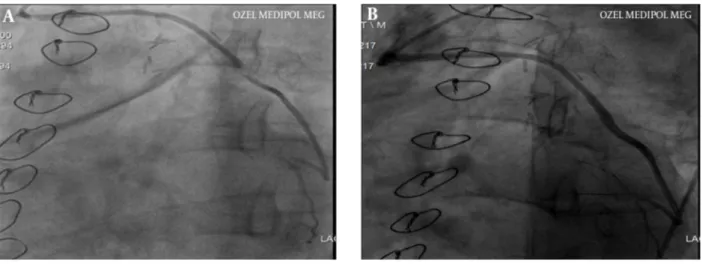
dis-tal embolic protection device (Emboshield Nav, Abbott Vas-cular, USA) was used during the procedure. After engage-ment of the saphenous vein ostium with the 7f JR guiding catheter and crossing of the lesions with a hydrophilic guidewire (Fielder J, ASAHI, Japan), the embolic protection device was located distally. The lesions were pre-dilated with 2.5 × 20 mm balloons (Sprinter Legend, Medtronic, USA), followed by implantation of a 3.0 × 28 mm Xience Pro stent (Abbott Vascular, USA) at 14 atm for the mid lesion and a 3.0 × 18 mm Xience Pro stent (Abbott Vascular, USA) at 16 atm for the ostial lesion (Figure 1A and B).
Several attempts at advancing the retrieval catheter of the embolic protection device were unsuccessful because of the struts of the ostial stent. We chose a smaller-sized hydrophilic catheter (4f, Head Hunter 1, Merit Medical, USA) to pass through the 7f JR guiding catheter in order to retrieve the protection device. The hydrophilic cath-eter was advanced on the preexisting coronary guidewire. However, during manipulations of the hydrophilic cath-eter at the ostium of the saphenous vein graft, the distal tip of the catheter was seen to break off and be lost in the ascending aorta, while the coronary guidewire and the protection device were seen to be properly localized in the saphenous graft. Fluoroscopic scanning of the entire body demonstrated that the embolized portion of the catheter
Karaca O et al.
Res Cardiovasc Med. 2016;5(1):e30890
2
was located in the distal segment of the left posterior tibial artery. In order to retrieve the foreign body percutaneous-ly, a left femoral sheath was inserted in the antegrade di-rection and a hydrophilic guidewire was advanced to the distal portion of the left foot. Since the calibration of the artery was too small at the distal posterior tibial artery, the snare catheter (En Snare, Merit Medical, USA) could not be advanced distally. We introduced a 5f JR diagnostic cath-eter beyond the popliteal artery, and used the hydrophilic wire to cross the lumen of the longitudinally-oriented catheter fragment, which had a lumen of 4f and a length of 2 cm (Figure 2A). Over the wire, a small-sized balloon (1.0 × 10 mm, Sprinter Legend, Medtronic, USA) was advanced inside the lumen of the catheter fragment and inflated at 10 atm of pressure to ensure that the balloon tightly held the inner wall of the catheter (Figure 2B). As a whole unit, the diagnostic catheter, the inflated balloon, and the guidewire were pulled out through the femoral sheath.
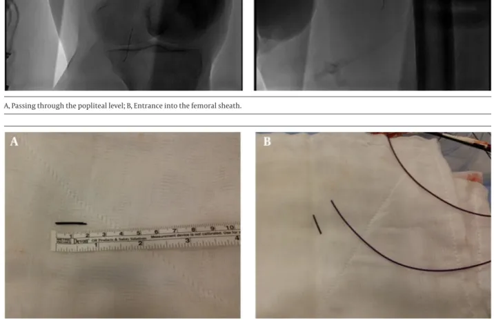
The catheter fragment was seen to be tightly held by the inflated balloon, and it was moved together with the sys-tem and successfully retrieved out of the circulation via the femoral sheath (Figure 3A and B). The broken catheter tip after removal is demonstrated in Figure 4A and B. Dur-ing the procedure, the patient was free of ischemic lower-limb symptoms, although the posterior tibial artery pulse was absent due to the embolized material.
The patient was safely transferred to the coronary care unit, where he remained hemodynamically stable after the procedure. Post-operative ECG did not show any signs of myocardial ischemia. Moreover, the distal arterial puls-es were palpable, without symptoms of limb ischemia. The patient was discharged with medications, including aspirin, prasugrel, bisoprolol, ramipril, and atorvastatin. Clinical control visits two weeks and three months after the procedure showed that the patient remained symp-tom-free in terms of angina and claudication.
Figure 1. A, Angiographic View of the Saphenous Vein Graft Lesions at the Ostial and Mid Portion; B, Successful Stenting of the Saphenous Graft Lesions
Figure 2. A, Yellow Arrow, Fluoroscopic Appearance of the Embolized Catheter Tip Found at the Left Ankle Level; Red Arrow, Hydrophilic Guidewire Near the Embolized Catheter; B, Yellow Arrow, Catheter Fragment; Red Arrow, Guidewire; Blue Arrow, Single Marker of the 1.0 × 10 mm Balloon Before Advancing into the Catheter Fragment; Green Arrow, 5f JR Diagnostic Catheter.
Karaca O et al.
3
Res Cardiovasc Med. 2016;5(1):e30890
Figure 3. Fluoroscopic Views Showing the Successful Retrieval Through the Femoral Sheath of the Catheter Tip as a Whole Unit With the Diagnostic Catheter, Inflated Balloon, and Guidewire
A, Passing through the popliteal level; B, Entrance into the femoral sheath.
Figure 4. Catheter Fragment After Removal
3. Discussion
In the present case, we demonstrated a percutaneous procedure in which a hydrophilic catheter was broken and embolized to a distal segment of the posterior tib-ial artery. After carefully examining the patient for the development of ischemic lower-limb symptoms; we de-cided to retrieve the foreign body percutaneously, which was a relatively safer and non-invasive procedure, since the patient remained asymptomatic after embolization. Practically, the patient would have been referred to a vas-cular surgeon if: 1) severe ischemic symptoms developed during the procedure, 2) the foreign body could not be retrieved percutaneously, or 3) the lumen of the catheter fragment could not be successfully wired by a coronary guidewire. However, conservative follow-up was not a
re-liable option in this case, since the posterior tibial artery pulse was absent during the procedure. In addition, in case of failure of the percutaneous procedure, the surgi-cal alternative would also have been easy, with a low risk of adverse events.
Due to the increasing numbers of complex percuta-neous coronary interventions, physicians in catheter-ization laboratories have begun to encounter various procedure-related complications (1). Depending on the level of expertise, many of the ‘unexpected’ fractures or embolic events can be managed percutaneously (2, 3). Patients who experience embolization of foreign bodies in the vasculature have a considerably increased risk for adverse events, depending on the site of embolization.
Karaca O et al.
Res Cardiovasc Med. 2016;5(1):e30890
4
Different surgical and non-surgical methods to handle embolic complications have been proposed for different clinic situations (4-9). Our study appears to offer a safe and relatively simple method of balloon dilatation inside the lumen of the embolized fragment, to be used when the foreign body is too distal to retrieve with convention-al snare systems (10).
Footnote
Authors’ Contribution:Oguz Karaca, performed the
case and drafted the manuscript; Beytullah Cakal, per-formed the case; Onur Omaygenc, perper-formed critical re-vision of the manuscript for important intellectual con-tent; Muhsin Turkmen, provided technical and material support.
References
1. Kim M, Chu A, Khan Y, Malik S. Predicting and preventing vascu-lar complications following percutaneous coronary interven-tion in women. Expert Rev Cardiovasc Ther. 2015;13(2):163–72. doi: 10.1586/14779072.2015.995635. [PubMed: 25553577]
2. Cantarelli MJ, de Paola Ade A, Alves CM, Souza JA, Castello Junior HJ, Hermann JL, et al. [Percutaneous retrieval of intravascular for-eign bodies]. Arq Bras Cardiol. 1993;60(3):171–5. [PubMed: 8250746]
3. Mainar V, Pico Aracil F, Bordes P, Ruiz Ros JA, Campos Peris JV, Marin F. [Percutaneous extraction of intravascular foreign bod-ies: a series of 38 cases]. Rev Esp Cardiol. 1996;49(1):41–7. [PubMed: 8685511]
4. Tai S, Zhou SH, Tang L, Zhu ZW, Hu XQ, Fang ZF. "Jailed-wire" tech-nique is useful for percutaneous retrieval of entrapped guide-wire. Int J Cardiol. 2015;189:223–4. doi: 10.1016/j.ijcard.2015.04.062. [PubMed: 25897910]
5. Woodhouse JB, Uberoi R. Techniques for intravascular foreign body retrieval. Cardiovasc Intervent Radiol. 2013;36(4):888–97. doi: 10.1007/s00270-012-0488-8. [PubMed: 23073559]
6. Yang FS, Ohta I, Chiang HJ, Lin JC, Shih SL, Ma YC. Non-surgical retrieval of intravascular foreign body: experience of 12 cases. Eur J Radiol. 1994;18(1):1–5. [PubMed: 8168572]
7. Sowinski H, Kobayashi D, Turner DR. Transcutaneous removal of an intravenous catheter fragment using a spider FX Embolic Pro-tection device. Catheter Cardiovasc Interv. 2015;86(3):467–71. doi: 10.1002/ccd.25839. [PubMed: 25601639]
8. Singh J, Thingnam SK, Das D, Singh H, Sharma R, Vijayvergia R. Surgical removal of entrapped and broken percutaneous trans-luminal coronary angioplasty balloon catheter. Interact Cardio-vasc Thorac Surg. 2007;6(1):94–6. doi: 10.1510/icvts.2006.140194. [PubMed: 17669780]
9. Wani SP, Rha SW, Park JY, Poddar KL, Wang L, Ramasamy S, et al. A novel technique for retrieval of a drug-eluting stent after cath-eter break and stent loss. Korean Circ J. 2010;40(8):405–9. doi: 10.4070/kcj.2010.40.8.405. [PubMed: 20830255]
10. Kharge J, Sreekumar P, Swamy K, Bharatha A, Ramegowda RT, Nanjappa MC. Balloon-assisted retrieval of a broken stent-deliv-ery system. Tex Heart Inst J. 2012;39(5):644–6. [PubMed: 23109758]